
Introduction
Colorectal cancer (CRC) is the third-most common malignant cancer worldwide, which is one of the most common causes of cancer-related deaths in America and Europe.[Reference Thomaidis, Maderer, Kornmann, Bauer, Trautmann, Schwarz, Neumann, Formentini, Lyros, Schad, Galle and Moehler1] Approximately 30% of patients are diagnosed with metastatic disease at present, which results in poor prognosis and markedly shortens their lifespan.[Reference Thomaidis, Maderer, Formentini, Bauer, Trautmann, Schwarz, Neumann, Kittner, Schad, Link, Rey, Weinmann, Hoffman, Galle, Kornmann and Moehler2] The most important reason for this is the lack of a noninvasive and efficient screening tool for the early detection of CRC. Additionally, usual radiology examinations are not specific enough and intestinal endoscopy is unfavorable for the patient. Thus, the early diagnosis of CRC and even precancerous CRC lesions will revolutionize diagnosis and treatment guidelines. A novel “cross-talk” strategy that enhances the targeting capability by combining MRI and near-infrared fluorescence (NIRF) could make the early diagnosis of CRC possible.
CRC is a hypervascular tumor showing prominent enhanced permeability and retention (EPR) effects, resulting in nanoparticles (NPs) being easily retained in tumor tissues; the full advantage of this fact can be taken for nano-imaging and while using nanocarriers.[Reference Khan3] Magnetic iron oxide NPs have been demonstrated to be an irreplaceable choice as novel agents for cancer diagnosis using magnetic resonance imaging (MRI), because of their delicate surface modification and controllable particle size. Especially for CRC, magnetic iron oxide NPs can be used as T 2 contrast agents that can generate a negative signal and produce a prominent contrast with soft intestinal tissue[Reference Lee and Lee4]; these advantages are more pronounced in patients with rectal cancer or colon cancer. Iron oxide NPs can trigger the local relaxation change of the nearby water protons and vastly reduce transverse (T 2) relaxation time, resulting in a negative contrast (low signal) on the T 2-weighted MR images.[Reference Rezayan, Mousavi, Kheirjou, Amoabediny, Ardestani and Mohammadnejad5–Reference Qiao, Wang, Zhang, Gao, Liang, Gao, Jiang, Qiao, Han, Zhang, Qiu, Tian, Gao and Cao8] Meanwhile, biocompatible polymers coated with NPs,[Reference Jiao, Sun, Tang, Ren and Yang9–Reference Chen, Shao, Li, Wang, Qu, Gu, Yu and Ye12] such as dextran, polyethylene glycol (PEG), polydopamine, and ferritin, have been widely used to coat NPs, for various biomedical applications, because of their excellent biocompatibility and biodegradability. In addition, PEG is rich in activated carboxyl groups, which can provide lots of combining sites in PEG-based NPs with targeted-biomolecules, improve their stability, prolong their half-life in blood, and reduce their non-specific uptake by the mononuclear phagocytotic system (MPS).[Reference Yan, Zhao, Shang, Liu, Xie, Qiang, Xiong, Zhang, Li, Sun and Kang13] Nevertheless, the NIRF-labeled magnetic Fe3O4 NPs have been found to be able to enhance tumor imaging sensitivity, while ensuring specificity. Furthermore, the optical modality is highly desirable to offer specific insights into cellular events and the real-time monitoring of the targeting ability of NPs in vitro and their biodistribution in vivo.[Reference Hu, Zhang, Shukla, Gu, Yu and Steinmetz14, Reference Liu, Tan, Xie, Yang, Zhao, Bai, Huang, Zhan, Wan, Zou, Han and Wang15]
Recently, coating versatile tumor-specific ligands like antibodies, polypeptides, and folic acid on the surface of iron oxide particles have been another major strategy for enhancing the tumor imaging sensitivity of magnetic Fe3O4 NPs. A series of reports have shown that biomolecules loaded onto the NP surfaces could improve their targeting capacity and curative effects.[Reference Wang, Qu, Li, Yin, Shen, Chen, Yang, Bian and Fang16–Reference Yan, Shang, Sun, Zhao, Wang, Xiong, Yuan, Zhang, Huang, Wang, Li, Tian, Kang and Feng18] However, one of the main difficulties to establish efficient cancer therapies and targeted imaging for cancer diagnosis and treatment is the high tumor heterogeneity and complicated molecular subtyping. The key to active tumor targeting using molecular imaging is the use of genetic and epigenetic alterations unique to cancer tissues as highly specific and efficient markers. This approach would allow tumor recognition, regardless of the presence of cancer cells or other components in the tumor microenvironment. For CRC, the vascular endothelial growth factor receptor (VEGFR) and the epidermal growth factor receptor (EGFR) pathway cross-talk in CRC survival and angiogenesis.[Reference Tonra, Deevi, Corcoran, Li, Wang, Carrick and Hicklin19–Reference Jebali and Dumaz21] The synergistic EGFR/VEGFR mechanism has been elucidated, and a series of reactions, including the MAPK kinase cascade and P13 K/ATK pathway, has been shown to be in the increased cellular proliferation, angiogenesis, and loss of apoptosis.[Reference Tonra, Deevi, Corcoran, Li, Wang, Carrick and Hicklin19] Their potential therapeutic combination has been suggested for in vitro and in vivo use, including clinical research.[Reference Thomaidis, Maderer, Formentini, Bauer, Trautmann, Schwarz, Neumann, Kittner, Schad, Link, Rey, Weinmann, Hoffman, Galle, Kornmann and Moehler2, Reference Funakoshi, Latif and Gasky22, Reference Moehler, Thomaidis, Zeifri, Barhoom, Marquardt, Ploch, Schattenberg, Maderer, Schimanski, Weinmann, Woerns, Kranich, Warnecke and Galle23] Thus, the “cross-talk” synergistic effect of EGFR/VEGFR for CRC tumors can be a promising tactic for notably enhancing the active targeting effect; it is also helpful for the early diagnosis of CRC using molecular imaging. Accordingly, it can completely reverse the current status of the diagnosis and treatment of CRC.
Herein, we prepared the Fe3O4-based NPs, combining the bispecific human EGFR/VEGFR single chain fragment variable (ScFv) with MRI/ NIRF imaging modalities. ScFv merely retained the binding domain of a full human antibody and showed the following characteristics: constant targeting, smaller molecular weight, and no immunogenicity.[Reference Abdelgawad, Bakr, Alkhoja and Mohamed24–Reference Zhou, Wei, Su, Chen, Qiu and Liu27] Carboxyl functional sites allow the conjugations with bispecific ScFv2 (EGFR/VEGFR) to Fe3O4 NPs to yield the Fe3O4-Dye800-ScFv2 NPs. The bispecific ScFv2 fragment was constructed by click chemistry. The MRI/NIRF imaging capability of Fe3O4-Dye800-ScFv2 NPs was explored in a CRC-bearing mouse model.
Materials and methods
Materials
Fe3O4 magnetic NPs, sodium hydrogen phosphate, oxoammonium hydrochloride, DSPE-PEG5000-COOH, trans-cyclooctene and tetrazine, N-hydroxysuccinimide (NHS) and ethyl (dimethylaminopropyl)-carbodiimide (EDC) and IRDye800CW were supplied by Xi'an ruixi Biological Technology Co, Ltd. (Xian, China) ScFvegfr (purity above 93.25%) and ScFvvegfr (purity above 91.66%) were gifted by Beijing Gegen Biotechnology Co, Ltd. (Beijing, China). The above materials were directly used for experiments without further purification.
Preparation of Fe3O4-Dye800-ScFv2 NPs
Fe3O4 NPs and DSPE-PEG5000-COOH were then mixed in a certain proportion in a rotary evaporator to yield a homogeneous Fe3O4@ PEG5000-COOH NPs solution. ScFvegfr and ScFvvegfr were linked via Tetrazine-PEG5-NHS and TCO-PEG4-NHS, respectively, to yield the ScFv2 fragments. The fragments were then labeled with Dye800-NHS and parallelly conjugated onto the NP surfaces.
Characterization of Fe3O4-Dye800-ScFv2 NPs
Transmission electron microscopy (TEM JEOL-1011; JEOL, Japan) was used to determine the size of the Fe3O4 NPs and to visualize the morphology of the Fe3O4@ PEG5000 NPs after negative staining. The hydrodynamic particle size distribution was characterized by dynamic light scattering (DLS) using a Malvern Zetasizer (ZEN 3600; Malvern, UK). The fluorescence excitation and emission spectra of the probe were detected by fluorescence spectrofluorometer (F-7000; Hitachi) at room temperature. The transverse relaxation rate (R 2) of the probe was determined by magnetic resonance scanning (Bruker BioSpec 70/20, Germany).
Cell culture and animal model
Human CRC cells (SW480, HT29, HCT116, DLD-1) were used for the following experiments. All the cells were cultured in complete medium in a CO2 incubator (Heracell, Germany). Male nude mice were anesthetized with 4% chloral hydrate (i.p., 0.15 mL/20 g). HCT116 cell-bearing mice were subcutaneously implanted with tumors. HCT116-luc cells (3 × 10⁶) were injected subcutaneously to establish the subcutaneous tumor model. 5 × 10⁶ cells in 15 µL phosphate buffer (PBS) were injected into the splenic capsule. The cell suspension formed a bolus slowly at a constant rate in 3 min. Tumor proliferation was monitored by an IVIS device. Three weeks later, the mouse models could be used for in vivo experiments.
All animal experiments reported in this paper were performed according to a protocol approved by the National Cancer Hospital Animal Care and Use Committee.
Western blotting
The indicated CRC cells were harvested during the exponential phase and lysed in 1 × PBS with 1% Nonidet P-40, 2 µg/mL Aprotinin, 2 µg/mL Leupeptin, and 50 µg/mL proteinase inhibitor. Cell lysates were centrifuged at 12,000g at 4 °C for 30 min. The total proteins were extracted from the supernatants and then quantified by the Pierce bicinchoninic acid assay kit. Proteins from each cell line were separated by sodium dodecyl sulfate polyacrylamide gel electrophoresis (SDS-PAGE) and electroblotted onto the polyvinylidene fluoride membrane by using the semi-dry blotting system. The membrane was blocked using 1 × PBST (polybutanediol terephthalate) buffer for 30 min and incubated with the indicated primary antibodies and anti-mouse or anti-rabbit secondary antibodies conjugated with horseradish peroxidase. Chemiluminescence signals were detected using ImageQuant LAS 4000.
Confocal imaging of ScFv binding to CRC cells
The ability of the ScFv2 fragment to target HCT116 cells was detected by confocal microscopy imaging. HCT116 cells were cultured in 24-well plates (1 × 10⁴ cells/well) for 24 h and fixed with 4% paraformaldehyde. For single ScFv fragment targeting, ScFvvegfr fragments labeled by AF488 were incubated with HCT116 cells for 4 h. The same steps were followed in the case of ScFvegfr labeled by AF647. Moreover, for bispecific targeting, the ScFv2 fragments labeled by AF488 were then incubated with HCT116 cells for 4 h. For specific binding, the cells were preincubated with 200 µL (20 µg/mL) of anti-VEGFR monoclonal antibody (mAb) and anti-EGFR mAb for 2 h for blocking. Nuclear staining with 4',6-diamidino-2-phenylindole (DAPI) was then performed for 5 min. The cells were observed using a PE fluorescence confocal microscope (PE Ultraview) and imaged using the ZEN software. Mean fluorescence intensity was analyzed using Image J 2X.
Cytotoxicity
HCT116 cells were seeded in 96-well plates (1 × 10⁴ cells/well) and cultured for 24 h. The ScFv2 fragments, Fe3O4 NPs, and Fe3O4-Dye800-ScFv2 NPs were incubated with HCT116 cells at different concentrations (0, 25, 50, 100, 250, and 500 µg/mL). The methyl thiazolyl tetrazolium (MTT) assay was used to evaluate cell viability. 3-(4,5-Dimethyl thiazole-2-base)-5-(3-carboxy methyl ester)-2-(4-sulfonated benzene)-2H-tetrazolium solution (20 µL) was mixed with fresh medium (100 µL) and added to each well and then placed into a CO2 incubator for 4 h. The OD values at 490 nm were then measured using a microplate reader (Molecular Devices, USA), and cell viability (%) was calculated by the following equation:
 $$\hbox{Cell viability}\, (\% ) = \displaystyle{{A_{{\rm Test \; sample}}} \over {A_{{\rm Control}}}} \times 100\%, $$
where
A Test sample and A Control were the absorbances of the test sample and the control, respectively.
In vivo NIRF and MR imaging
HCT116 cells-bearing model mice were anesthetized with 4% chloral hydrate (i.p., 0.15 mL/20 g). The NIRF images were acquired via an optical imaging system (IVIS Imaging Spectrum System, PerkinElmer Co, Ltd, USA) with a 750-nm filter for excitation and 800-nm filter for emission 2 h after the administration of the Fe3O4-Dye800-ScFv2 NPs through the tail vein. The fluorescence intensity of the subcutaneous tumor regions and liver metastases was measured; all measurements were repeated thrice.
For in vivo MR imaging, nude mice bearing HCT116 subcutaneous tumors were anesthetized and then injected with Fe3O4-Dye800-ScFv2 NPs in PBS solution (100 µL) through the tail vein.
The MR images were acquired on a 7.0 T MRI instrument at predetermined time points post injection (6, 24, 48, and 72 h). T 2 mapping MRI parameters were as follows: TR, 2500 ms; TE, 15, 30, 42, 52, 60, 72, 88, 100, 120 ms, and slice thickness, 4.0 mm. Three mice each were sacrificed at the designated time points for Fe quantitation in tumor tissue and other main organs by using inductively coupled plasma-atomic emission spectroscopy (ICP-AES) after the stripped tissues were ordered with HNO3.
Histological analysis
Tumor tissues and tissues from other major organs including heart, liver, spleen, lung, and kidney were collected for histological analysis. The frozen tissue slides were further stained with hematoxylin and eosin, and immunohistochemistry was performed following the standard protocol.
Statistical analysis
All the quantitative data were recorded as mean ± standard SD. Means were compared using Paired t test and two-way analysis of variance (ANOVA). P* < 0.05 were considered as statistical significance; P** < 0.01, remarkably; P*** < 0.001, very remarkably.
Results and discussion
Characterization of the nanoprobe
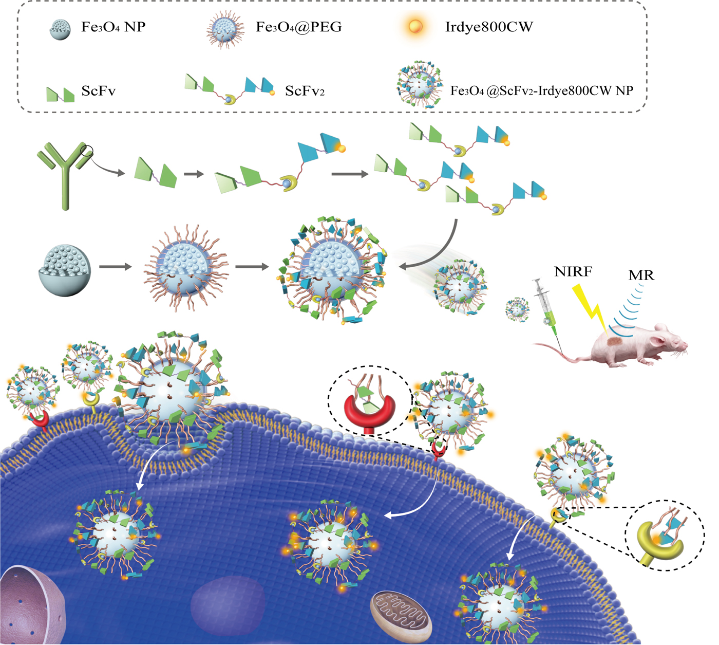
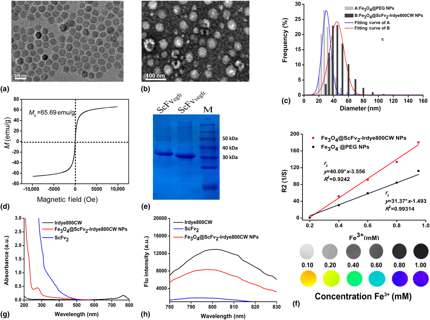
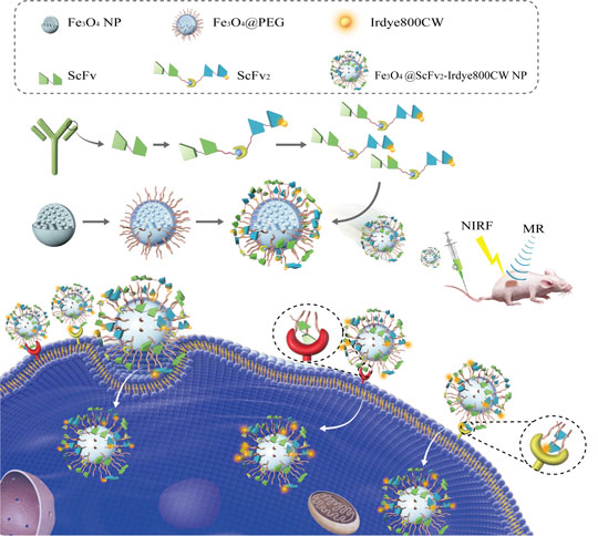
The Fe3O4-Dye800-ScFv2 NPs were prepared as shown in Fig. 1. Fe3O4 NPs were prepared by the thermal decomposition method to yield a high dispersion in the oil phase by oleate coating. The average size of the Fe3O4 NPs was 12.52 ± 1.61 nm and was verified by TEM [Fig. 2(a)] and showed a uniform and cubic microstructure, whereas the average hydrodynamic diameter, as measured by DLS in Fig. 2(c), was found to be 24.2 ± 9.17 nm. TEM images revealed a globular shape and a mean size of 33.26 ± 4.05 nm [Fig. 2(b)], indicating that nearly all of the Fe3O4 NPs were coated by PEG. The average size of the Fe3O4-Dye800-ScFv2 NPs was 46.03 ± 11.15 nm by DLS in Fig. 2(c). Furthermore, the field-dependent magnetization curve of the probe [Fig. 2(d)] at room temperature was obtained and was found to have no remanence coercivity at zero field and the magnetic property was 65.69 emu/g, indicating the ultra-paramagnetic nature of Fe3O4-Dye800-ScFv2 NPs, consistent with previous reports.[Reference Liu, Le, Mei, Wang, Chen, Lei, Cui, Chen, Cui and Shao10, Reference Mu, Zhang, Kong, Zhang, Zhang, Ge, Liu and Jiang11] And apparent Zeta potential of the probe was about −50 mV in Fig. S1, there was no significant difference between pre- and post-conjugation. Meanwhile, SDS-PAGE results for ScFvegfr and ScFvvegfr fragment are shown in Fig. 2(e). The distinct lines revealed that the ScFvegfr and ScFvvegfr fragment were successfully synthesized, and that their molecular weight was about 25,000. ScFv2@Dye800 can be easily acquired via the ScFv2 fragments and Dye800-NHS in alkaline solution (pH = 8.3) at room temperature. The carboxyl groups of PEG5000-COOH could be activated via EDC/NHS and then conjugated with ScFv2@Dye800 through covalent bonds. This parallel conjugation not only improved the combination efficiency but also economized the materials.[Reference Arami, Khandhar, Tomitaka, Yu, Goodwill, Conolly and Krishnan28]
Schematic illustrating the synthetic process of Fe3O4-Dye800-ScFv2 NPs synthesis and the proposed mechanism for targeting colorectal cancer.

Characteristics of the Fe3O4-Dye800-ScFv2 NPs. (a) The electron microscopic images of the magnetic Fe3O4 NPs. (b) The electron microscopic images of the probe. (c) The distribution of the hydrodynamic diameter of the probe at pre- and post-conjugation. (d) Magnetization loops of the Fe3O4-Dye800-ScFv2 NPs, showing good magnetic properties. (e) ELISA of the ScFvegfr and ScFvvegfr protein preparation. (f) T 2 relaxation rates (r 2) of the Fe3O4-Dye800-ScFv2 and Fe3O4@PEG NP solutions at different concentrations. (g) UV absorbance spectra of ScFv2, Dye800, and Fe3O4-Dye800-ScFv2 NPs. (h) Fluorescence emission spectra of ScFv2, Dye800, and Fe3O4-Dye800-ScFv2 NPs.
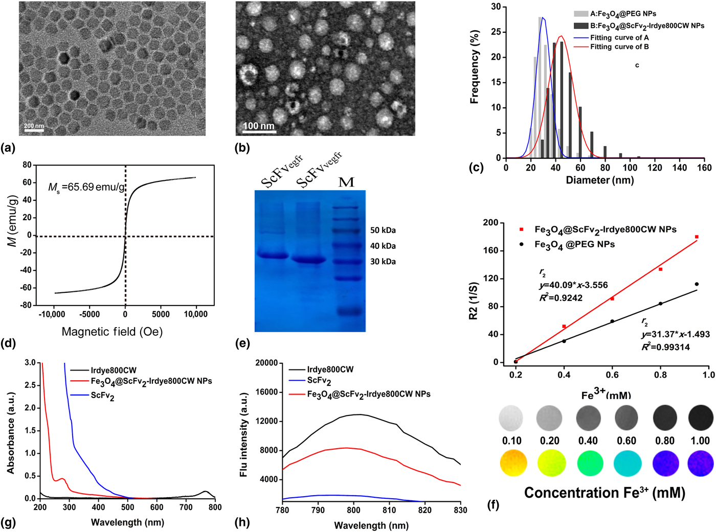
The capacity of magnetic imaging depended on the relaxation rate. Fe3O4 NPs had a prominent ability to enhance T 2 relaxation via shortening of the transverse relaxation time, showing a negative signal in T 2-weighted imaging.[Reference Yan, Zhao, Shang, Liu, Xie, Qiang, Xiong, Zhang, Li, Sun and Kang13] Essentially, a nanoprobe with high relaxivity can be detected at lower concentrations. The larger size of iron oxide NPs displayed a stronger relaxation ability.[Reference Arami, Khandhar, Tomitaka, Yu, Goodwill, Conolly and Krishnan28] Therefore, the R 2 of Fe3O4-Dye800-ScFv2 NPs and Fe3O4 NPs were measured ex vivo to evaluate the potential of Fe3O4-Dye800-ScFv2 NPs as a T 2 contrast agent. As shown in Fig. 2(f), the transverse relaxivity (r 2) of the probe was 40.09 × 10⁻³M−1S−1, which was acquired using the 7.0 T MRI device. The r 2 of the Fe3O4 NPs was 31.37 × 10⁻³M−1S−1. The phantom imaging demonstrated that the surface modification of Fe3O4 NPs via PEG-COOH changed the transverse relaxivity at a certain degree. The probe presents a significantly stronger T 2 effect than the Fe3O4 NPs.
On the other hand, the optical characteristics of Fe3O4-Dye800-ScFv2 NPs were evaluated and compared with those of the ScFv2 fragment and Fe3O4 NPs. As expected, the absorption peak at 755 nm derived from the conjugated Dye800 appeared in the UV absorption spectrum of the probe, whereas the ScFv2 fragment and Fe3O4 NPs presented no absorption, as seen in Fig. 2(g). On the other hand, the fluorescence emission spectrum of the probe showed a peak of up to 805 nm, similar to the case for free Dye800 (800 nm). In contrast, no fluorescence emission peak was observed for the Fe3O4 NPs [Fig. 2(h)]. The fluorescent characteristic of Fe3O4-Dye800-ScFv2 NPs enabled the visualization of the tumors in deeper tissues with a lower signal background.
Finally, Fe3O4 NPs were capped by PEG to improve their biocompatibility, resulting in their persistent circulation in the blood and accumulation in tumor tissues.[Reference Gref, Luck, Quellec, Marchand, Dellacherie, Harnisch, Blunk and Muller29, Reference Yan, Chen, Sun, Zhao, Zhang, Bian, Yang, Liu, Yuan, Yao and Wu30] The average hydrodynamic diameter of the probe was less than 50 nm, and thus, a notable amount of the probe was phagocytosed by the mononuclear endothelial system, as expected.[Reference Jun, Huh, Choi, Lee, Song, Kim, Yoon, Kim, Shin, Suh and Cheon31, Reference Jun, Seo and Cheon32] Therefore, Fe3O4-Dye800-ScFv2 NPs were successfully synthesized, with an excellent fluorescence and T 2 relaxivity effect. These characteristics were necessary for using the probe in the in vivo experiments.
EGFR/VEGFR expression in human CRC cells
Relative expression of both EGFR and VEGFR were evaluated in the following human CRC cell types: DLD-1, HT29, HCT116, and SW480 (Fig. S2). Actin was used as a normalized loading control for western blotting. EGFR had a high expression among these CRC cell types, and VEGFR was overexpressed in most of the CRC cell lines. As shown in Fig. S1, EGFR and VEGFR were simultaneously overexpressed in HCT116 cells, supporting that HCT116 cells were positive cells and can be used for the following in vitro and in vivo experiments.
Cytotoxicity
The cytotoxicity of Fe3O4-Dye800-ScFv2 in HCT116 cells was evaluated by using the MTT assay. As shown in Fig. S3, with the increasing concentrations of the probe, the cell viability was higher than 85% when the concentration was under 100 µg/mL. The cell viability sharply decreased to 45% when the concentration was above 250 µg/mL. The MTT assay showed that the viability of HCT116 cells was impacted by the probe concentration in a dose-dependent manner. However, according to the safety scale, the Fe3O4-Dye800-ScFv2 NPs were nearly non-cytotoxic for potential bioapplication.
In vitro targeting assay
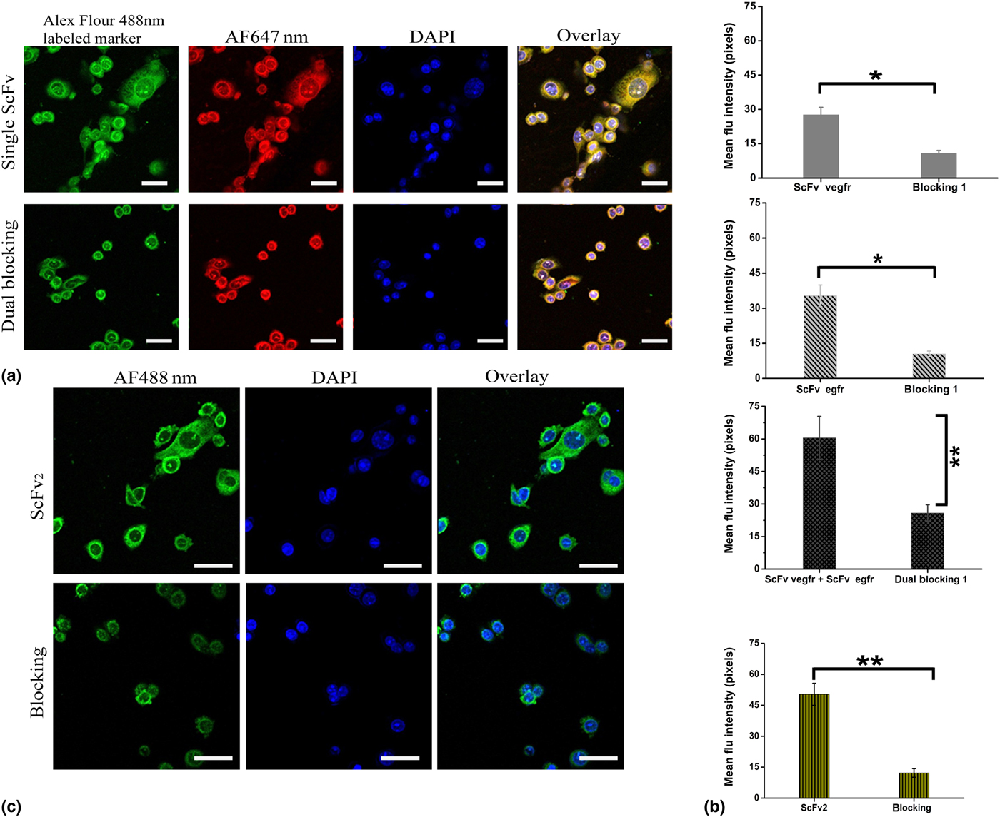
In vitro targeting assay was performed by confocal microscopy. As shown in Fig. 3(a), ScFvvegfr labeled with AF488 (green signal) was incubated with HCT116 cells, showing that the green flu-dots were focused on the cytomembrane of HCT116 cells. Similarly, ScFvefgr@AF647 (red signal) presented a slightly stronger signal intensity than ScFvvegfr@AF488. VEGFR mAb and EGFR mAb were preincubated with HCT116 cells for 2 h, resulting in successful blocking and proving that ScFvvegfr and ScFvegfr could specifically target HT116 cells. Furthermore, two ScFv fragments could co-localize on the surface of cells and the flu-intensity was stronger than that of the single ScFv fragment [Fig. 3(b)]. The ScFv2 fragments labeled with AF488 were incubated with HCT116 cells [Fig. 3(c)]. The green flu-signals were widely distributed on the surface of cells and efficiently targeted relative receptors on the HCT116 cytomembrane. Compared with the single ScFv fragments, the blocking capability of EGFR mAb and EGFR mAb to ScFv2 was stronger, indicating that bispecific ScFv2 combination enhanced targeting specificity. It might be induced via the cross-talk synergistic reaction mechanism of EGFR and VEGF(R).
Confocal imaging of ScFv2, ScFvegfr, and ScFvvegfr targeting HCT116 cells. (a) Single ScFvegfr/AF647 (red) and ScFvvegfr/AF488 (green) targeting HCT116 cells, DAPI (blue). EGFR, VEGFR mAb were preincubated with HCT116 cells for 2 h for blocking. (b) Mean flu-intensity quantitation by Image J 2X. (c) ScFv2/AF488 (green) targeting HCT116 cells, DAPI (blue). EGFR, VEGFR mAb were simultaneously preincubated with HCT cells for 2 h, for dual blocking. P* < 0.05; P** < 0.01; P*** < 0.001.

EGFR is an ideal target for tumor therapy, as it is commonly overexpressed or constitutively activated in many different cancers, which is associated with tumor malignancy. Multiple agents targeting EGFR have been developed for clinical use, which can be divided into two groups, including small molecule tyrosine kinase inhibitors (TKIs) and mAbs. Angiogenesis plays a crucial role in tumor growth and metastasis, which is mediated by multiple VEGFRs and their ligands. Inhibition of VEGF/VEGFR by mAbs or TKIs is widely used in anticancer therapy. Moreover, EGFR regulates VEGF expression via different transcription factors, STAT3, Sp1, and HIFs, and the MAPK and PI3 K signaling cascades.[Reference Larsen, Ouaret, El Ouadrani and Petitprez20, Reference Jebali and Dumaz21, Reference Abdelgawad, Bakr, Alkhoja and Mohamed24]
In vivo NIRF and MR imaging
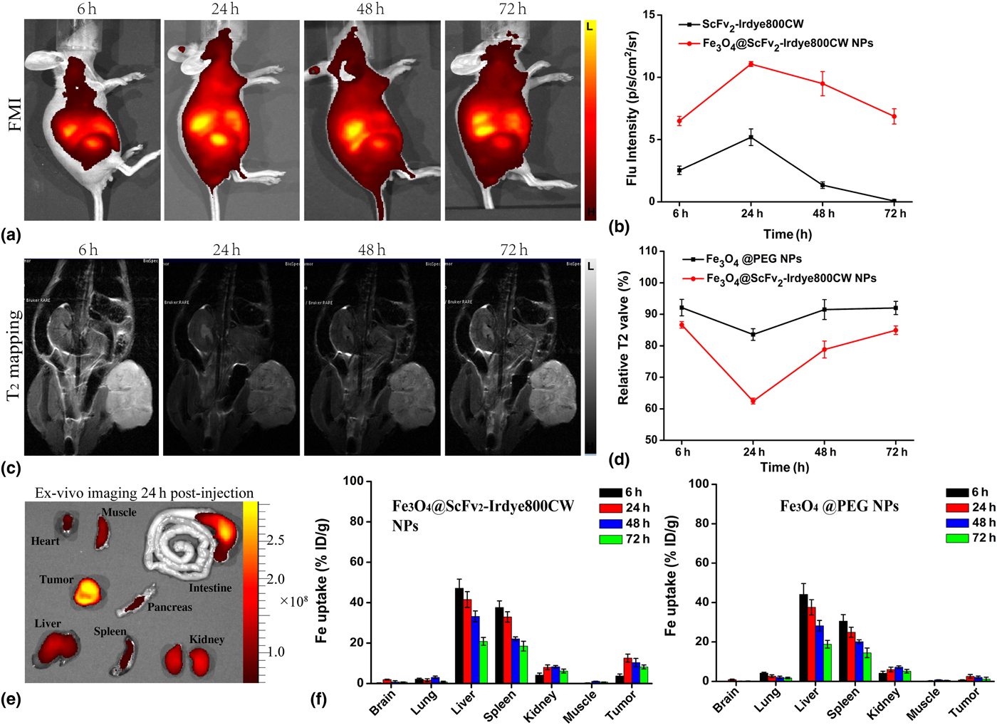
Firstly, the conjugated Dye800 enables the optical imaging of the Fe3O4-Dye800-ScFv2 NPs. The whole-body NIRF images of model mice bearing subcutaneous and orthotopic implanted tumors were acquired before and after tail-vein injection of Fe3O4-Dye800-ScFv2 NPs. The FMI and MRI T 2 mapping images of model mice are shown in Figs. 4(a) and 4(c). It was noted that the flu-intensity in tumor tissue peaked 24 h post injection, showing distinct tumor localization. The flu-signal in the liver was predominant 24 h post injection, suggesting the clearance of probes from the liver by the MPS. Furthermore, the flu-signal in the liver showed no significant decline after 72 h. The high flu-signal in tumor tissues declined after 24 h [Fig. 4(b)].
In vivo biodistribution of Fe3O4-Dye800-ScFv2 NPs. (a) In vivo NIRF images of CRC-bearing nude mice 6, 24, 48, and 72 h post injection of Fe3O4-Dye800-ScFv2 NPs. (b) The enhanced subcutaneous tumors can be detected with varying time and fluorescent intensity of tumor region. (c) In vivo MR images of CRC-bearing nude mice 6, 24, 48, and 72 h post injection of the Fe3O4-Dye800-ScFv2 NPs and Fe3O4@PEG NPs at a dosage of 10 mg Fe/kg. (d) The T 2 signal in the tumor region at different time points. (e) Ex vivo images of main organs and tumor at 24 h post-injection were shown. (f) Fe uptake of tumor and other main organs 6, 24, 48, and 72 h post injection of the Fe3O4-Dye800-ScFv2 and Fe3O4@PEG NPs at a dosage of 10 mg Fe/kg.

As seen in the T 2 mapping images, the relative T 2 value decreased by 63% 24 h post injection, indicating the lowest signal in tumor tissues and the optimal contrast among tumor and other tissues [Fig. 4(d)]. Then the absolute T 2 value began to rise. The accumulation of probes in the tumor region could last 72 h. Magnetic iron oxide decreased the T 2 -weighted tissue signal to enhance the contrast by shortening the transverse relaxation time. Similarly, the distributions of the T 2 signal in vivo were consistent with the flu-signal of the whole body. And the ex vivo fluorescent images of main organs and subcutaneous tumor 24 h post-injection via tail vein were shown in Fig. 4(e), the strong fluorescent intensity of tumor tissues meant the excellent take-up of the probe, and little phagocytosis in liver, kidney, and intestines could be known. The Fe3O4-Dye800-ScFv2 NPs were developed with good optical and T 2 -weighted imaging capability. Meanwhile, it was conjugated with the ScFv2 fragment, causing the nanoprobe to selectively accumulate in the CRC tumor tissue, showing prolonged retention.[Reference Shang, Chang, Kan, Majetich and Lee33] Herein, Fe3O4-Dye800-ScFv2 NPs could be specific T 2-targeting contrast agents for CRC tumors in model mice. It might also be a promising and potential T 2-targeting contrast agent for the clinical diagnosis of CRC.
Next, to provide further support to the specific targeting ability of the nanoprobe, the Fe3O4@PEG NPs from the control tissues, tumor tissues, and tissues from other main organs were harvested at different time points (6, 24, 48, and 72 h) after MRI experiments. The Fe contents in the tumor and other organs were measured by ICP-AES. As shown in Fig. 4(f), Fe quantitation was consistent with the imaging results shown in Fig. 5. The nanoprobe modified by the ScFv2 fragment presented a better uptake by tumor tissues, greater enhancement, and longer blood cycle time. Nevertheless, the biodistribution of the Fe3O4-Dye800-ScFv2 NPs in vivo was similar to that of the control probe, because of the MPS in the liver and spleen. It also meant that the liver and spleen were major metabolic organs for Fe3O4-Dye800-ScFv2 NPs, via hepatoenteral circulation. Only a small amount of nanoprobe was cleared by the kidneys.
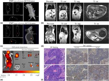
Detection of tiny-volume and large-volume orthotopic HCT116 tumors using Fe3O4-Dye800-ScFv2 NPs and confirmation of their pathology. 3D optical imaging for (a) tiny- and (c) large- HCT116 tumors, and T 2 images presented pre-contrast and after tail-vein injection of the Fe3O4-Dye800-ScFv2 NPs were shown as (b) and (d), correspondingly. The dots indicate the CRC tumor regions. (e) Fluorescent imaging of tiny-volume orthotopic HCT116 tumor at 24 h post-injection. And ex vivo images of main organs and tumor were shown. (f) Pathology results verified by HE staining and IHC staining (VEGFR2 and EGFR antibody). The scale bar above corresponds to 200 µm and the one at the bottom corresponds to 100 µm.

Finally, 3D-NIRF and T 2-weight images of tiny and large orthotopic HCT116 tumors in the model mice before injection and 24 h post-injection were obtained And in vivo and ex vivo fluorescent images of orthotopic HCT116 tumors mice were shown in Fig. 5(e). Without the T 2 contrast agent, the intraperitoneal tissues widely presented a high T 2 signal because of a large amount of liquid and abdominal fat; some mixed signals resulted from feces in the intestinal region. Twenty-four hours post-injection, the T 2 signal of both the small and large tumors decreased due to the enhancement of the negative signal. The tumor and its region could be easily observed due to the intense contrast between the decreased T 2 signal of the tumor and high T 2 signal of the intraperitoneal tissues. Furthermore, 3D-NIRF imaging was performed to verify the tumor location in Figs. 5(a) and 5(c).
Pathology
The tumor tissues were harvested after the in vivo experiments for hematoxylin and eosin staining and immunohistochemical staining [Fig. 5(f)]. The results of the pathological analysis showed the typical features of adenocarcinoma, and those of immunohistochemical staining indicated that VEGFR and EGFR were overexpressed in the HCT116 tumors. These observations indicate that the Fe3O4-Dye800-ScFv2 NPs, as specific T 2 contrast agents, may be a potential strategy for the detection of tumors overexpressing VEGFR and EGFR.
Conclusion
In summary, the Fe3O4-Dye800-ScFv2 probe was successfully developed. The characteristic results showed that the probe was biocompatible and nontoxic, with a regular morphology. The in vitro experiments demonstrated that the ScFv2 fragment conjugated onto the Fe3O4-Dye800-ScFv2 NPs could specifically recognize the VEGFR/EGFR-overexpressing cells, including CRC cells. In addition, the Fe3O4-Dye800-ScFv2 NPs also exhibited a high r 2 relaxivity and excellent MR imaging capability for human CRC xenografts in nude mice in vivo. Whole-body NIRF imaging showed that the CRC tumors could be easily visualized by the Fe3O4-Dye800-ScFv2 NPs because of their exclusive accumulation in the CRC tumor regions, similar to the case for the cells in vitro. Therefore, our Fe3O4-Dye800-ScFv2 NPs could be promising contrast agents for the MRI/NIRF dual-modal imaging of VEGFR/EGFR-overexpressing tumors, including CRC tumors.
Supplementary material
The supplementary material for this article can be found at https://doi.org/10.1557/mrc.2018.127.
Acknowledgments
This work was financially supported by the National Natural Science Foundation of China under Grant No. 81671757 and the CAMS Innovation Fund for Medical Sciences under No.2016-I2 M-1-001 and Beijing Nova Program under Grant No. Z181100006218061. All the authors in this paper thank the Beijing Gegen Biotechnology Co, Ltd for providing the ScFv fragments and the Key Laboratory of Molecular Imaging, Institute of Automation, Chinese Academy of Sciences for imaging equipment.