Introduction
Schwannomas or neurilemmomas are slow-growing, benign neoplasms derived from Schwann cells, the sheath cells that cover myelinated nerve fibres. Schwannomas are slowly growing tumours that have a predilection for the head, neck and flexor surface of the upper and lower extremities. Intraoral lesions are unusual and intraosseous schwannomas are even rarer, representing less than 1 per cent of benign primary tumours of the bones. The site most commonly involved is the mandible, particularly in the posterior segment of the body and ramus.Reference Chi, Carey and Muller1
The aim of this report was to present a clinical case of schwannoma in the mandibular angle, mimicking a keratocystic odontogenic tumour, with a complicated posterior evolution.
Case report
An asymptomatic, 60-year-old man presented to our centre for a maxillofacial examination. One month earlier, a dentist in private practice had taken a panoramic radiograph which had revealed a radiolucency in the left mandibular angle. Otherwise, the medical and dental history was unremarkable.
Clinical examination revealed no expansion of the mandible, facial swelling or dental pain on percussion. There was no trismus or paraesthesia. The overlying mucosa was intact and of normal colour.
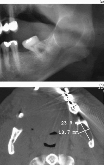
Panoramic radiography and computed tomography (CT) identified a well circumscribed, unilocular, radiolucent lesion in the left mandibular angle, along the intraosseous path of the inferior alveolar nerve (Figure 1). Our first diagnosis was odontogenic keratocyst.
(a) Panoramic radiography and (b) axial computed tomography images, revealing a well circumscribed, unilocular, radiolucent lesion located in the left mandibular angle.

Subsequently, the patient underwent total removal of the lesion under general anaesthesia. A 5 cm incision was made in the left buccal mucosa and the tumour was bluntly dissected from its cavity and removed. The course of the inferior alveolar nerve was not clearly identified. The lesion was oedematous and solid, with a greyish colour and no apparent cystic cavities.
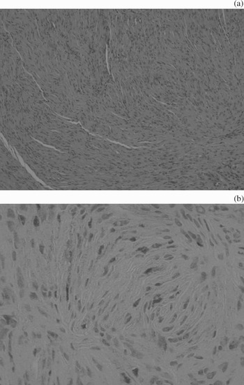
Microscopically, the tissue consisted of encapsulated, well demarcated tumour lobules composed of spindle-shaped cells with aligned long nuclei, similar to Schwann cells (Figure 2a). Some of these nuclei formed typical palisades around acellular eosinophilic areas (Verocay bodies). Antoni A tissue represented the predominant microscopic pattern, alternating with occasional Antoni B areas. Immunohistochemical staining for S-100 protein was diffusely positive. No signs of haemorrhage, necrosis, calcification or hyalinisation were observed (Figure 2a). The diagnosis was intraosseous schwannoma.
(a) Photomicrograph depicting tumour lobules composed of spindle-shaped cells with nuclear palisading (H&E; original magnification ×100). (b) Immunohistochemical staining for S-100 protein was diffusely positive (original magnification ×100).

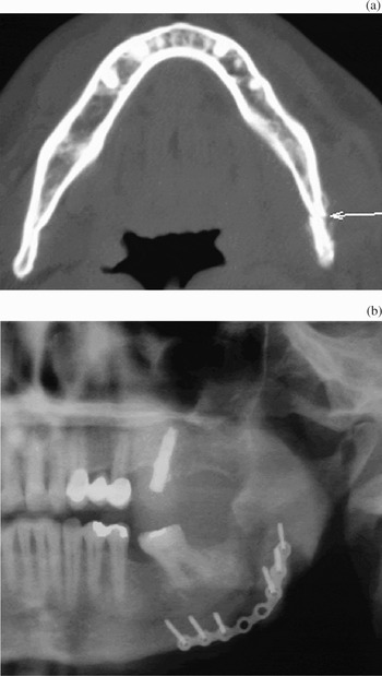
In the early post-operative course, the patient complained of paraesthesia of the inferior alveolar nerve. Forty-five days after surgery, the patient developed left hemifacial swelling and severe pain. A panoramic radiograph and CT revealed a pathological fracture of the left mandibular angle at the surgical site (Figure 3a).
(a) Axial computed tomography image showing pathological fracture at the surgical site (arrow). (b) Panoramic radiograph showing the fracture reduction and reconstruction plate.

Under general anaesthesia, the patient underwent open reduction of the fracture with a reconstruction plate (Figure 3b).
This time, the post-operative course was uneventful and healing proceeded normally. Six months later, there were no signs of recurrence.
Discussion
Neurilemmomas or schwanommas are infrequent, usually benign tumours derived from the neuroectodermal Schwann cells, which have a predilection for the flexor surfaces of extremities and the head and neck. Schwannomas rarely occur in the oral cavity. Intraosseous schwannomas are rare (less than 1 per cent), but when they do occur the mandible is the most commonly affected site. In a 2003 literature review, Chi et al. documented 43 cases of intraosseous schwannomas of the jaws, 38 of which were in the mandible.Reference Chi, Carey and Muller1 Most cases reported in the mandible had a posterior location, corresponding to the intraosseous course of the inferior alveolar nerve.
Clinically, neurilemmomas are slow-growing tumours which may be present for years before becoming symptomatic. Approximately half of all intraosseous mandibular schwannomas produce noticeable swelling.Reference Martins, Taghloubi, Bussadori, Fernandes, Palo and Martins2 Other reported symptoms include pain and paraesthesia. There is a female predilection, with a 1.6:1 female-to-male ratio.Reference Nakasato, Katoh, Ehara, Tamakawa, Hoshino and Izumizawa3 Most neurilemmomas are diagnosed in patients younger than 50 years, with a peak prevalence in the second and third decades of life. Our patient was 60 years old at the time of diagnosis.
Radiological diagnosis of schwannomas is difficult. The typical radiographic presentation of mandibular and maxillary schwannomas is that of a well defined, unilocular radiolucency with a thin, sclerotic border.Reference Shrime, Wilson, Eisig and Haddad4 Radiographically, this appearance can be suggestive of a benign process, such as an odontogenic keratocyst, periodontal cyst or ameloblastoma, but is otherwise nonspecific.
• Intraosseous schwannoma is an unusual neoplasm
• This paper describes a case of schwannoma in the mandibular angle mimicking a keratocystic odontogenic tumour, with a complicated evolution
• The authors discuss the problematic diagnosis of this tumour and its correct management
Histopathological analysis of specimens is extremely important in order to establish the final diagnosis. When seen as a gross specimen, the schwannoma tissue is solid, roundly lobulated and moderately firm. Most tumours are encapsulated. Microscopically, the tumour is classified into two basic types of tissue, Antoni A and Antoni B.Reference Sadeghi, Koenig and Clark5 Antoni A areas consist of closely packed spindle cells with their nuclei lying in rows and displaying a palisade effect and Verocay bodies. In contrast, Antoni B tissue comprises less cellular, less organised, nonorganoid areas, often with prominent, thickened blood vessels. Diffuse immunoreactivity for S-100 protein is routinely observed in schwannomas, but staining is somewhat diminished in Antoni B areas. The microscopic features of intraosseous and soft tissue schwannomas are identical.
Schwannoma treatment consists of total surgical removal of the lesion. Periodic follow-up examinations are indicated, but recurrence is extremely rare. Radiation therapy is not recommended; schwannomas are characteristically radioresistant.Reference Chi, Carey and Muller1, Reference Sadeghi, Koenig and Clark5
Conclusion
We report an unusual case of intraosseous neurilemmoma of the mandible. The clinical presentation was that of an asymptomatic tumour in the left mandibular angle, identified during a routine radiographic examination in a patient in the sixth decade of life. Further radiographic examination revealed a unilocular radiolucency, prompting an initial diagnosis of keratocystic odontogenic tumour. The lesion was completely excised, with no signs of recurrence six months later. However, the patient developed a post-operative pathological fracture at the surgical site. To our knowledge, this is the first reported case of this unusual tumour having such a complicated evolution.