Transoesophageal echocardiography is an important imaging modality in congenital cardiac disease. The technique can provide comprehensive diagnostic information on intracardiac structures with high spatial and temporal resolution. It is particularly important in the assessment of atrioventricular valves, left and right ventricular outflow tracts and of the atrial septum.Reference Sutherland and Stumper1, Reference Ayres, Miller-Hance and Fyfe2 In many centres, transoesophageal echocardiography during surgery and catheter interventions is regarded as the optimal technique to guide the procedure and to reduce the incidence of residual lesions.Reference Ayres, Miller-Hance and Fyfe2
The application of transoesophageal echocardiography has to date been limited by patient size. Current paediatric multiplane probes may be technically difficult to insert in patients weighing less than 4 kilograms, particularly if a sheath is used to minimise cross-infection. Complications may include difficulty with probe insertion, airway compression, displacement of an endotracheal tube, and damage to surrounding structures, particularly in smaller patients.Reference Stevenson3 Despite the fact that very small transoesophageal echocardiogram probe prototypes have been reported over the past 15 years,Reference Shiota, Lewandowski and Piel4–Reference Scohy, Gommers, Jan ten Harkel, Deryck, McGhie and Bogers7 the commercial availability of a very small probe would extend transoesophageal echocardiography to smaller infants, but only if probe insertion is atraumatic and image quality is adequate.
In older children and adults, there might also be a place for a very small transoesophageal echocardiogram probe to facilitate probe insertion and minimise discomfort, particularly when using conscious sedation rather than general anaesthesia.
We report the initial experience of a micro multiplane transoesophageal echocardiogram probe used at a tertiary congenital heart centre applied to the assessment of congenital cardiac defects in all age groups, in normal clinical practice. The aims of the study were to document the quality of all of the imaging modalities of the probe and to report adverse events related to probe placement or manipulation.
Materials and methods
Micro transoesophageal echocardiogram probe
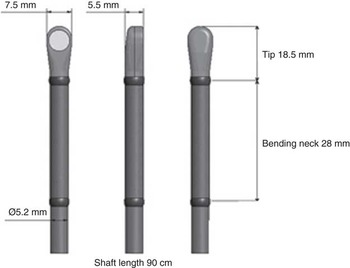
A micro transoesophageal echocardiogram S8-3t probe has recently been released for clinical use (Philips Medical Systems, Andover, Massachusetts, United States of America). It has a 32-element sector array with an extended operating frequency range of 3–8 megahertz The shaft diameter is 5.2 millimetres, and the tip dimensions are 7.5 millimetres (width) × 5.5 millimetres (height) × 18.5 millimetres (length). The total probe length is 90 centimetres (Fig 1). Similar to larger standard probes, the micro transoesophageal echocardiogram probe offers multiplane imaging from 0 to 180 degrees and manual control of anterior and posterior flexion.
Micro transoesophageal echocardiogram dimensions.

The standard paediatric transoesophageal echocardiogram (S73-t) probe by comparison has a tip length of 27 millimetres, width of 10.6 millimetres, height of 8 millimetres, and shaft of 8 millimetres. The adult three-dimensional transoesophageal echocardiogram (X7-2t) matrix array transducer has 2500 elements with a shaft length of 1 metre, and a diameter of 1 centimetre. The tip has a width of 1.7 centimetres, height of 1.35 centimetres, and length of 3.8 centimetres.
Standard two-dimensional imaging, steerable pulse wave Doppler, continuous wave Doppler, colour flow Doppler, advanced XRES, M-mode, and harmonic imaging are available. The transoesophageal echocardiogram was used to evaluate the appearance of endocardial borders, atrioventricular and semilunar valves, systemic and pulmonary venous inflow, as well as atrial and ventricular septums using standard views and positions. This included high oesophageal, mid-oesophageal, and transgastric views. Pulse wave, continuous wave, and colour flow Doppler were applied in these projections.
Study design
We prospectively studied elective and emergency transoesophageal echocardiogram imaging at our institution using the micro transoesophageal echocardiogram probe in all patients over a 3-month period – between 15 December, 2009 and 15 March, 2010. All patients underwent transoesophageal echocardiography for clinical indications according to our usual institutional practice. For the initial part of the study, the images of the micro transoesophageal echocardiogram probe were compared with images obtained using the conventional paediatric or adult-sized multiplane transoesophageal echocardiogram probes in larger patients. For patients too small for either the standard paediatric or adult probes, studies were performed using only the micro transoesophageal echocardiogram probe and no comparison was made. With further experience, we did not change probes if the micro transoesophageal echocardiogram images were diagnostic. All probes were used in conjunction with the Philips iE33 ultrasound system (Philips Medical Systems, Andover, Massachusetts, United States of America). We routinely use a disposable sheath on all transoesophageal echocardiogram probes for infection control.
Image quality
All studies were performed or directly supervised and assessed by either an experienced paediatric cardiologist or a certified echocardiographer qualified to report independently. The assessors were required to rate the imaging quality at the time of the study using a subjective scale (1 = poor, 2 = adequate, 3 = good, 4 = excellent, 0 = not assessed). The imaging modalities assessed included the quality of two-dimensional and colour flow Doppler imaging, pulse wave and continuous wave Doppler. Further information was collected on maximal frame rates with two-dimensional imaging and colour Doppler, along with maximal overall image depth achieved. There were five observers. Retrospectively, two observers analysed the image quality related to near-field and far-field structures to assess the maximal depth of diagnostic images. We defined this depth as the level at which there was a loss of image definition illustrated by the appearance of the “banana effect”, with horizontal spread of the echo signal.Reference Thijssen8
Ethical approval
The study was approved as per institutional policy. Our institutional ethics committee has approved retrospective analysis of all clinical echocardiographic data for purposes of teaching and research. The micro transoesophageal probe is CE marked and therefore not an experimental probe. Standard patient consent was obtained for all transoesophageal echocardiogram examinations. All patients or carers received a standard transoesophageal echocardiogram information sheet. The study was submitted through the appropriate institutional channels. Specific additional consent for sequential transoesophageal echocardiogram probe placement was not deemed necessary.
Results
Study group
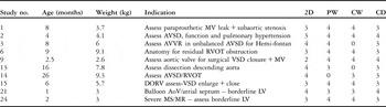
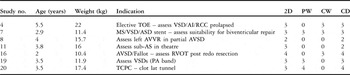
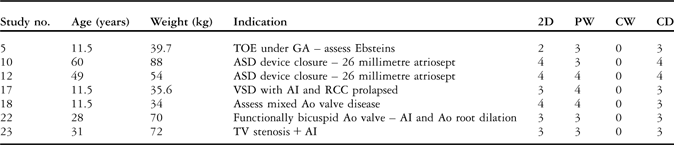
A total of 24 studies were performed on 23 patients with a median weight of 11.7 kilograms (2.6–72 kilograms) and a median age of 3 years (0.16–60 years). All patients were intubated when under general anaesthesia at the time of study as per usual institutional practice. Studies were divided into three patient groups: less than 10 kilograms (n = 10), 10–30 kilograms (n = 7), and greater than 30 kilograms (n = 7). The patient demographics and indications for imaging are listed in Tables 1–3. Imaging was performed intra-operatively (n = 10), during cardiac catheter intervention (n = 3), urgent assessment on the intensive care unit (n = 2), and as part of semi-elective imaging investigations (n = 9).
Patient and image characteristics using micro transoesophageal echocardiogram probe: Group 1, < 10 kg.

AoV = aortic valve; AVSD = atrioventricular septal defect; AVVR = atrioventricular valve regurgitation; CD = colour Doppler; CW = continuous wave Doppler; DORV = double outlet right ventricle; LV = left ventricle; MR = mitral regurgitation; MS = mitral stenosis; MV = mitral valve; PW = pulse wave Doppler; RVOT = right ventricular outflow tract; VSD = ventricular septal defect; 2D = two dimensional
Patient and image characteristics using micro transoesophageal echocardiogram probe: Group 2, 10–30 kg.

AI = aortic incompetence; ASD = atrial septal defect; AVSD = atrioventricular septal defect; AVVR = atrioventricular valve regurgitation; CD = colour Doppler; CW = continuous wave Doppler; MS = mitral stenosis; PA = pulmonary artery; PW = pulse wave Doppler; RCC = right coronary cusp; RVOT = right ventricular outflow tract; sub-AS = sub-atrial septum; TOE = transoesophageal echocardiography; TCPC = total cavopulmonary connection; VSD = ventricular septal defect; 2D = two dimensional
Patient and image characteristics using micro transoesophageal echocardiogram probe: Group 3, >30 kg.
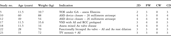
AI = aortic incompetence; Ao = ascending aorta; ASD = atrial septal defect; CD = colour Doppler; CW = continuous wave Doppler; GA = general anaesthesia; PW = pulse wave Doppler; RCC = right coronary cusp; TOE = transoesophageal echocardiography; TV = tricuspid valve; VSD = ventricular septal defect; 2D = two dimensional
Probe insertion and complications
The micro transoesophageal echocardiogram probe was passed without difficulty in all paediatric patients, without the need for direct vision. Of the patients included in the study, one neonate (weight 2.6 kilograms) had transient bradycardia on probe insertion, which resolved without the need for specific intervention. Of the four adults – aged 28–60 years, two patients had to have the probe passed under direct vision because the smaller probe coiled up in the oropharynx when passed blindly via the oral route. No adjustment of ventilation was required, and a consistent end tidal carbon dioxide trace was maintained with no elevation of measured levels noted during the transoesophageal echocardiogram examinations. We did not attempt to measure mean airway pressure changes. There were no other complications in the study group.
Cross-sectional imaging
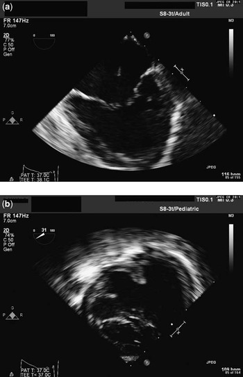
The assessment scores for two-dimensional image quality for all studies ranged from 2 to 4, with a mean of 3.3 and a median of 3. This was similar in the less than 10-kilogram group with a mean score of 3.2. The image definition obtained in those less than 10 kilograms was clearly defined and of full diagnostic value. An example of images obtained is displayed in Figure 2a and b.
(a) Zero degree “four-chamber” view of unbalanced atrioventricular septal defect. Note the horizontal “banana effect” in the far-field structures as the image quality reduces. (b) Transgastric view in the same patient demonstrating the common atrioventricular valves and central loss of coaptation resulting in atrioventricular valve regurgitation.

The median scores for two-dimensional imaging in the 10- to 30-kilogram group was 3 (mean 3). In the greater than 30-kilogram group, the median score was also 3 (mean 3.28).
Image depth
The maximal image depth achieved in our patients was 12 centimetres. In all studies, there was consistently good imaging quality up to a depth of 4–6 centimetres, after which the image degraded. This was despite moving the region of the focus range to a region of higher depth. This phenomenon was present at a similar depth for colour flow Doppler.
The imaging quality of near-field structures for all three groups was good. However, in the 10- to 30-kilogram group, there was some loss of quality in imaging. There was difficulty in maintaining adequate contact, and the probe was maintained in a slightly flexed position in order to improve this. This affected the quality of the image, with foreshortening of cardiac structures in some planes.
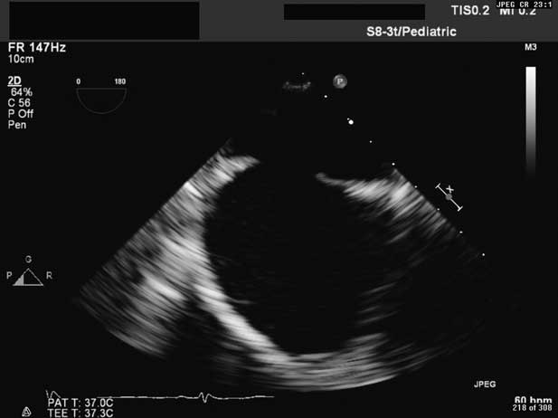
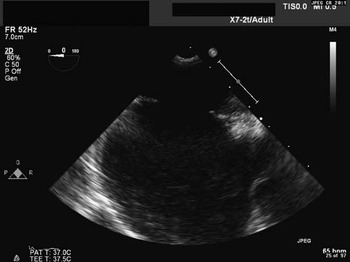
The adult-sized patients were suitable for imaging of the near-field structures alone, namely the atrial septum and its margins (Figs 3 and 4). We were able to guide percutaneous device closure of the atrial septum using an atriosept device (Fig 5). Further evaluation of other cardiac structures was not possible.
Two-dimensional image of atrial septum using micro transoesophageal echocardiogram probe in Patient 12.
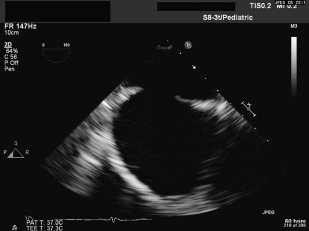
Comparative three-dimensional transoesophageal echocardiogram probe image of atrial septum in Patient 12.

Doppler and colour flow Doppler quality
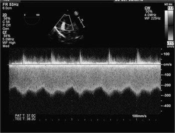
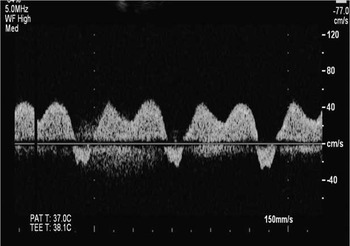
The mean assessment scores for the overall group for pulse wave is equal to 3.42 (range 3–4, median 4), continuous wave is equal to 3.5 (range 3–4, median 4), and colour flow Doppler 3.2 (range 2–4, median 3). The mean scores were 3.7 (range 3–4, median 4), 3.75 (range 3–4, median 4), and 3.3 (range 3–4, median 3), respectively, for the pulse wave, continuous wave, and colour flow Doppler in the less than 10-kilogram group. There was consistently good pulse wave Doppler and continuous wave Doppler where this was applied (Figs 5 and 6).
Continuous wave Doppler signal across the atrial septum.

Pulse wave Doppler signal in pulmonary veins.

Frame rates
The frame rates on two-dimensional imaging were significantly higher than the paediatric and adult probes, which only allowed frame rates of 52–130 hertz. Frames rates with the micro transoesophageal echocardiogram probe were generally in the region of 147 hertz. Frame rates greater than 200 hertz were obtained when utilising the zoom function. The frames rates when utilising colour flow Doppler were also higher at 28–72 hertz compared with standard transoesophageal echocardiogram probes with frame rates of 11–34 hertz. We had set the optimal machine settings for each probe. It should be noted that the refresh frequency of monitors is often below the actual frame rates of the acquired image, limiting the visible frequency.
Comparative data
A comparison with a suitable alternative larger probe was made in 12 studies in patients with age range from 3.5 years to 60 years, and median weight was 34.8 kilograms (11.9–72 kilograms). The standard paediatric transoesophageal echocardiogram probe (S7-3t) was used as the alternative probe in five studies, and the adult three-dimensional (X7-2t) probe was used in seven studies.
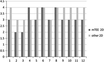
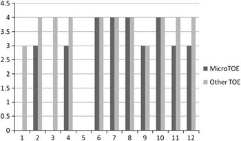
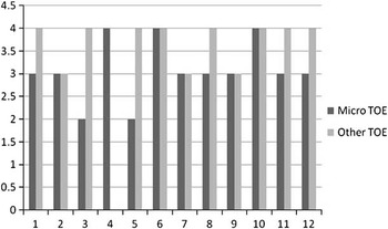
In these 12 patients, the mean two-dimensional image quality score was 3.08 (median 3) for micro transoesophageal echocardiogram studies and 3.67 (median 4) for other probes (p = 0.02). The comparative scores for two-dimensional, pulse wave and colour Doppler imaging between the micro transoesophageal echocardiogram and alternative probes are illustrated in Figures 7–9.
Comparison of two-dimensional image quality using micro transoesophageal echocardiogram versus “other transoesophageal echocardiogram probes” (paediatric/adult three-dimensional transoesophageal echocardiogram probe).

Comparison of pulse wave image quality using micro transoesophageal echocardiogram versus “other transoesophageal echocardiogram probes” (paediatric/adult three-dimensional transoesophageal echocardiogram probe).

Comparison of colour Doppler image quality using micro transoesophageal echocardiogram versus “other transoesophageal echocardiogram probes” (paediatric/adult three-dimensional transoesophageal echocardiogram probe).

Additional information
In the less than 10-kilogram group, the micro transoesophageal echocardiogram study provided additional information in eight patients and resulted in a change of management in six out of ten (60%). Patients one and six had poor transthoracic acoustic windows. Patient one (3.7 kilogram) had severe chronic lung disease with emphysematous lung changes. This patient had suspected paravalvular mitral valve prosthesis leak, which was confirmed on transoesophageal echocardiogram. Patient six had cardiac surgery 12 hours before the transoesophageal echocardiogram study, which confirmed residual right ventricular outflow tract obstruction requiring re-operation. Intra-operative imaging identified an iatrogenic perforation of the left coronary cusp in Patient 9, requiring additional intra-operative repair. Dissection of the aorta was excluded in Patient 13, who had a raised endothelial flap following cardiac catherisation 6 weeks before surgery. Patient 24 with a borderline left heart was shown to have an inadequate mitral valve for a biventricular repair and went on to have a Norwood 1 operation. We were able to guide catheter intervention in Patient 21, who underwent static ballooning of the atrial septum and balloon aortic valvuloplasty in the same procedure.
Of the 10 patients in the 10- to 30-kilogram group, two patients had findings that affected management. Patient 16 had residual right ventricular outflow tract obstruction requiring additional intra-operative relief of the subpulmonary obstruction. Patient 20 had a clot in the lateral tunnel immediately after a total cavopulmonary anastomosis. There were no new findings from the transoesophageal echocardiogram in the greater than 30-kilogram patient group. No additional information from the micro transoesophageal echocardiogram was obtained or missed when compared with the adult transoesophageal echocardiogram probe.
Manipulation of the probe
A consistent comment from operators was that a degree of flexion of the probe was required in order to maintain contact for adequate imaging quality. The probe was felt to be easy to manipulate, and the transgastric views in the less than 10-kilogram group were particularly easy to obtain.
Discussion
We utilised the micro transoesophageal echocardiogram probe in a variety of clinical settings and across a wide spectrum of patient ages and weights, all with congenital cardiac disease. Two-dimensional and colour flow Doppler imaging were consistently achieved at high frame rates compared with standard transoesophageal echocardiogram probes, which may be desirable in smaller and younger patients with fast heart rates. The micro transoesophageal echocardiogram probe provided good imaging quality up to a depth of around 4–6 centimetres, and the best image quality was observed in patients less than 10 kilograms and near-field structures in adult patients. A fairly consistent comment was that there was a need to flex the probe to maintain good contact. Pulse wave Doppler and continuous wave Doppler quality was consistently rated well and matched the quality of standard probes.
Our results in adult-sized patients undergoing closure of atrial septal defects (n = 2) demonstrated very good near-field imaging of the atrial septum. The application of the micro transoesophageal echocardiogram probe for this purpose is yet to be described in the literature. Combined with conscious sedation and pernasal insertion, this less-invasive micro transoesophageal echocardiogram imaging may then allow more catheter interventions in a larger patient population unsuitable for general anaesthesia. No pernasal insertion of the micro transoesophageal echocardiogram probe was attempted in the study, as all patients were consented for transoesophageal echocardiogram.
The older child (10–30 kilograms) had the poorest results when compared with standard transoesophageal echocardiogram imaging, and the technique of flexing the micro probe alone as in the other patient groups was not always sufficient to improve image quality. Our results are consistent with the observations of Scohy et al.Reference Scohy, Gommers, Jan ten Harkel, Deryck, McGhie and Bogers7, Reference Scohy, Gommers, Schepp, McGhie, de Jong and Bogers9 The reason for this observation is difficult to explain. The observation might relate to the shorter bending neck of 40 millimetres to the micro multiplane probe when compared to standard paediatric and adult probes, making it difficult to reach the left ventricular apex to acquire transgastric views in bigger patients.Reference Scohy, Gommers, Schepp, McGhie, de Jong and Bogers9 However, it does not explain the loss of image quality on standard oesophageal views as well.
It is widely accepted that intra-operative transoesophageal echocardiogram in paediatric patients offers clear clinical benefits for the identification of residual lesions and reduced re-operation rates.Reference Scohy, Gommers, Jan ten Harkel, Deryck, McGhie and Bogers7, Reference Bettex, Schmidlin and Bernath10–Reference Reynolds, Spevack and Shah12 The high rate of change in management in our patient group relates to the targeted use of transoesophageal echocardiography in our patients. We focussed on the lower weight group where conventional imaging would have previously involved using a standard paediatric probe with an internal shaft diameter of 7.4 millimetres. The use of these standard paediatric probes has been previously described to include neonates weighing 2.2 kilograms.Reference Scott, Blackburn, Wharton, Wilson, Dickinson and Gibbs13 Passing such a probe can be difficult in small neonates and carries a relatively high risk of airway compression, oesophageal trauma, and risk to adjacent structures.Reference Stevenson3, Reference Greene, Alexander and Knauf14, Reference Muhiudeen, Miller-Hance and Silverman15 The use of the micro transoesophageal echocardiogram probe was consistently atraumatic in our experience, with only one transient bradycardia related to its insertion in a single infant. This is consistent with findings by Zyblewski et alReference Zyblewski, Shirali and Forbus16 who recently assessed the safety profile of this probe when used intra-operatively in infants less than 5 kilograms. They also noted that the imaging findings were diagnostic in this weight group, but there was no assessment of image quality.
The micro transoesophageal echocardiogram approach in small infants may supersede the use of intracardiac echocardiogram probes by the transoesophageal route, which are single-plane, non-renewable, and carry a higher cost.Reference Drinker, Camitta and Herlong17, Reference Cannesson, Hénaine and Metton18 Epicardial echocardiography remains an option, regardless of weight, but this technique may be limited by the training of surgeons to acquire images, limited acoustic windows, and disturbance of the flow of the procedure.
The limitations of our study include the small number of studies, the subjective grading of the images, and the use for multiple operators to assess image quality. However, our study does represent a broad-based assessment relating to “real” clinical cases and includes the first description of any comparative assessment of image quality for the micro multiplane transoesophageal echocardiogram probe.
Conclusion
The micro transoesophageal echocardiogram probe provides imaging of diagnostic quality in our assigned group of less than 10 kilograms. In larger patients, we were able to obtain good imaging of the near-field structures, such as the atrial septum, where they may be a role for micro transoesophageal echocardiogram-guided interventions, even in the conscious adult. We were unable to demonstrate a clear benefit from the micro transoesophageal echocardiogram probe in the intermediate-sized child. In these patients, the standard paediatric probe continues to provide better imaging.
The micro transoesophageal echocardiogram probe is a useful adjunct to transoesophageal echocardiogram, but should supplement and not replace the standard transoesophageal echocardiogram probes used in a congenital cardiac unit. It is particularly useful in the neonatal population.