INTRODUCTION
The eastern shore of the Adriatic Sea is a region of Dinaric karst, characterized by porous rock and numerous related geo-morphological phenomena, such as anchialine caves. From a bio-geographical perspective, the fauna of the Dinaric coast and its associated islands generally exhibits a paralittoral distribution (Sket, Reference Sket1994). There are about 30 anchialine caves along the Croatian coastline. These caves extend up to 100 m from the sea, are small in size and occur in low tidal areas.
Many new copepods from marine cave ecosystems in recent years have been found in deep-sea hyperbenthos and in anchialine caves (Huys, Reference Huys1988; Boxshall & Iliffe, Reference Boxshall and Iliffe1990; Boxshall & Jaume, Reference Boxshall and Jaume2000; Jaume et al., Reference Jaume, Cartes and Boxshall2000, Reference Jaume, Boxshall and Gracia2008). Owing to the efforts of the Bio-Speleological Society in Zagreb, preliminary investigations into calanoid copepods in anchialine caves have been undertaken along the Croatian coast and on nearby islands (Kršinić, Reference Kršinić2005a, Reference Kršinićb).
The genus Stephos comprise small copepods, measuring up to 1 mm in total length, living in hyperbenthos and in anchialine caves. A total of 30 species have been described (Carola & Razouls, Reference Carola and Razouls1996). In the Mediterranean Sea, there are five known species in its western part: Stephos gyrans (Giesbrecht, Reference Giesbrecht1892); Stephos margalefi Riera et al., Reference Riera, Vives and Gili1991; Stephos marsalensis Costanzo et al., Reference Costanzo, Campolmi and Zagami2000; Stephos cryptospinosus Zagami et al., Reference Zagami, Campolmi and Costanzo2000 and Stephos vivesi Jaume et al., Reference Jaume, Boxshall and Gracia2008. A new genus and species of the family Stephidae, Speleohvarella gamulini, has been described by Kršinić (Reference Kršinić2005a) from marine cave ecosystems. The latest investigations into anchialine caves along the eastern coast of the Adriatic Sea have revealed two new copepod species of the genus Stephos. One of these species, Stephos boettgerschnackae Kršinić (Reference Kršinić2012), was found in an anchialine cave in the northern part of the eastern Adriatic shoreline, in Urinj Cave, not far from the city of Rijeka. The other species were solely collected in anchialine Lenga Pit on the island of Mljet, in the southern part of the Adriatic Sea, which shall be analysed in the present paper.
MATERIALS AND METHODS
Material was collected during sampling expeditions carried out in Lenga Pit on Mljet Island, near the city of Dubrovnik in the South Adriatic, on 4 October 2004, 21 June, 4 September and November 2006, and 28 May 2007. Lenga Pit is about 100 m from the shoreline, measuring approximately 25 m in length and 9 m in depth. There is a small, 9 m deep lake at the end of the cave. The cave has been generally described by Miculinić et al. (Reference Miculinić, Bilandžija, Cvitanović, Jalžić, Jalžić, Lukić, Pavlek, Durbešić and Benović2011), and topographically by Cukrov et al. (Reference Cukrov, Bilandžija, Cukrov and Jalžić2011). The Lenga Pit has very stable conditions. The salinity in the water column below depths of 3 m is between 21.00 and 39.00, while the temperature is approximately 15.5°C. Divers from the Croatian Natural History Museum (Zagreb) and the Croatian Bio-Speleological Society (Zagreb) made collections with a hand-held 20 cm diameter Nansen net, 125 µm mesh, sampling between depths of 2 and 8.5 m.
All samples were preserved in a 2.5% formaldehyde–seawater solution neutralized with CaCO3. Specimens were dissected on slides in lactophenol. Drawings were made with the aid of a camera lucida on an Olympus BX51 differential interference contrast microscope. Specimens were measured using an ocular micrometer. The descriptive terminology follows Huys & Boxshall (Reference Huys and Boxshall1991).
RESULTS
SYSTEMATICS
Order CALANOIDA G.O. Sars, 1903
Family STEPHIDAE G.O. Sars, 1902
Genus Stephos T. Scott, 1892
Stephos grieveae sp. nov.
(Figures 1–6)
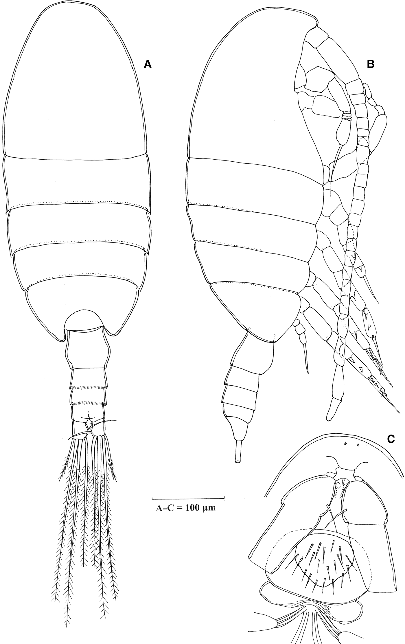
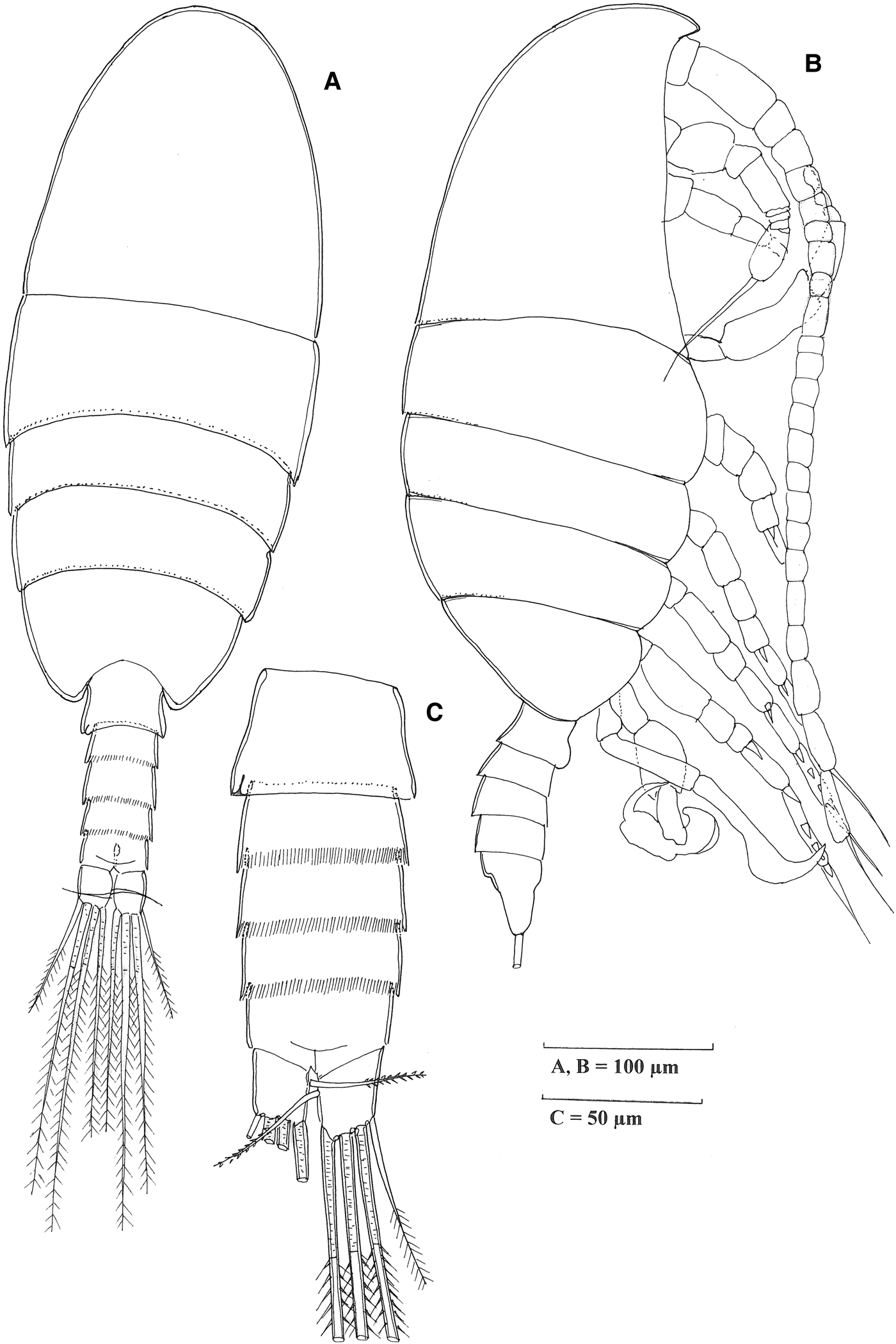
Stephos grieveae sp. nov., adult female. A, habitus, dorsal view; B, habitus, lateral view; C, rostral area ventral view.

TYPE MATERIAL
Holotype: adult male, no. HPM-BSZ, 1774, 600 µm in length, Lenga Pit, Mljet Island, South Adriatic, Croatia (42°47′42.2″N 17.5°23′22.3″E), collected on 28 May 2007.
Paratypes: no. HPM-BSZ, 1775, seven adult females and eight males, collected from same location as holotype. Types are deposited at the Croatian Natural History Museum, Zagreb.
Additional material: about 20 specimens, females and males, from the samples collected on 4 October 2004, 21 June 4 September and November 2006, collected from Lenga Pit, deposited at the Institute of Oceanography and Fisheries, Split.
ETYMOLOGY
This species is named in honour of Dr Janet M. Bradford-Grieve, a prominent New Zealand copepod taxonomist.
DESCRIPTION
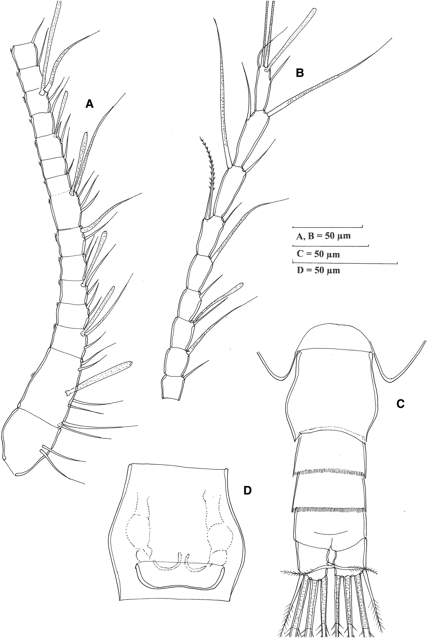
Female (Figure 1A, B), total length (excluding caudal setae): 600–670 µm (620 ± 23.5 µm, N = 9). Body oval in dorsal aspect. Nauplius eye absent. Cephalosome and first pedigerous somite completely separated, pedigerous somites 4 and 5 fused. Rostral area, with two sensillae medially, rostrum represented by rounded knob (Figure 1C). Last pedigerous somite slightly asymmetrical. Proportional lengths of prosome somites measured mid-dorsally; 48.5:15.5:9.8:10.7:15.5 = 100. Ratio of prosome and urosome including caudal rami as 2.9:1. Prosome 2.3 times as long as wide. Urosome of 4 free somites (Figure 2C). Proportional lengths of urosomites and caudal rami 37.4:18.6:17:13.5:13.5 = 100. Genital double-somite (Figure 2D) about as long as wide, symmetrical, with proximolateral margins produced in dorsal aspect. External genital area symmetrical in ventral view, paired egg-laying duct, paired gonopores situated medially near posterior edge, covered by single operculum. Caudal rami symmetrical, 1.25 times longer than wide, ramus with four terminal setae corresponding to setae III–VI, seta VII small, pinnate, setae I and II absent (Figure 2C).
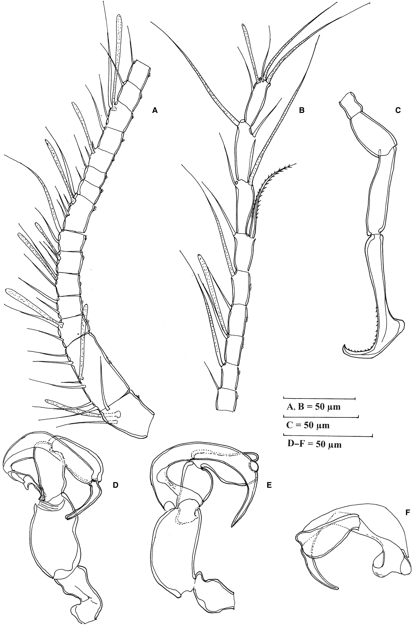
Stephos grieveae sp. nov., adult female. A, B, antennule (A, segments 1–15; B, segments 16-24); C, urosome and caudal rami, dorsal view; D, genital double-somite, ventral.
Antennules (Figure 2A, B), symmetrical, 24-segmented, reaching almost to end of third urosomite. Armature and fusion pattern of segments as follows: segment 1 (ancestral I) 2; segment 2 (II–IV), 6 setae + ae; segment 3 (V) 2 + ae; segment 4 (VI) 2 setae; segment 5 (VII) 2 setae + ae; segments 6 and 7 (VIII, IX) 2 setae each; segment 8 (ancestral X–XI) 4 setea + ae; segments 9–10 (XII, XIII) 1 seta each; segment 11 (XIV) 2 + ae; segment 12 (XV) 1 seta; segment 13, (XVI) 2 setae + ae; segments 14–17 (XVII – XX) 1 seta each; segment 18 (XXI) 1 seta + ae; segments 19 and 20 (XXII, XXIII) 1 seta each; segment 21 (XXIV) 1 + 1 seta; segment 22 (XXV) 1 + 1 seta; segment 23 (XXVI) 1 + 1 seta; segment 24 (XXVII –XXVIII), 4 + ae. Segment 2 with patch of spinules at posterior mid-length; segments 3–20 bearing row of small posterior spinules, comb-like, along distal border.
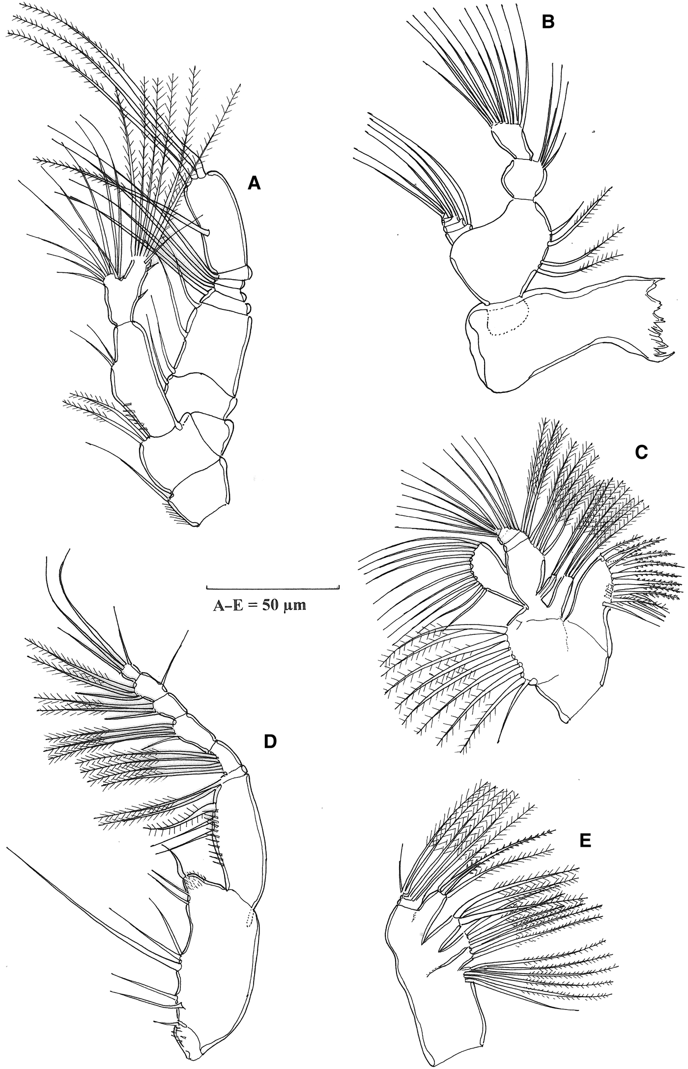
Antenna (Figure 3A), biramous, with exopod longer than endopod. Coxa with one and basis with two short setae distally; exopod 7-segmented; 1.3 times as long as endopod; armature as follows: 1, 3, 1, 1, 1, 1, 4. Endopod 2-segmented, proximal segment bearing two small setae subdistally. Proximal segment armed with row of setules laterally, distal segment with nine unequal setae on lateral margin and seven setae distally, with the two outer border spinules.
Stephos grieveae sp. nov., adult female; A, antenna; B, mandible; C, maxillule; D, maxilla; E, maxilliped.
Mandible (Figure 3B), gnathobase cutting edge with isolated unicuspid tooth, eight heterogeneous teeth and one dorsal spinulose seta. Mandibular palp biramous, basis length and width similar with four setae on inner margin. Endopod 2-segmented, first segment with four unequal setae and terminal segment truncate bearing 11 setae. Exopod 5-segmented, setal formula 1, 1, 1, 1, 2.
Maxillule (Figure 3C), praecoxal arthrite bearing nine stout marginal spines and four elements on posterior surface. Coxal epipodite armed with nine, and coxal endite with four, setae. Proximal basal endite with three setae, distal basal endite with five, basal exite unarmed. Exopod bearing 11 setae. Endopod articulated to basis, indistinctly 3-segmented, with setal formula 3, 4, 7.
Maxilla (Figure 3E), praecoxal endites with six and three setae, respectively. Coxal endites each with three setae. Allobasis with proximal endite powerfully developed with three setae, one stouter than rest. Endopod 3-segmented, with setal formula 1, 1, 4.
Maxilliped (Figure 3D), praecoxal endite with one seta and row of spinules; syncoxal endites, setal formula 2, 3, 3 on medial margin, distal seta thorn-like; distal margin with row of spinules. Basis with longitudinal row of spinules and with three setae, free endopod 6-segmented, armature formula 2, 4, 4, 3, 3 + 1, 4.
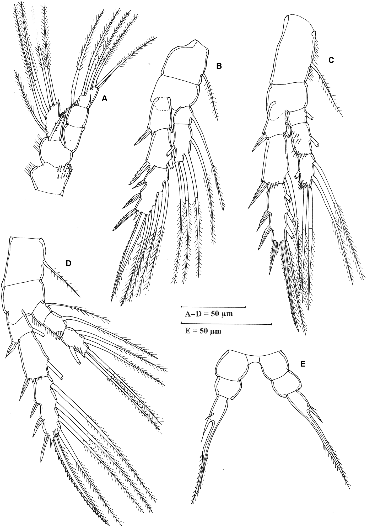
Swimming legs 1–4 biramous, exopods 3-segmented (Figure 4), armature formula as follows:
Stephos grieveae sp. nov., adult female; A, leg 1; B, leg 2; C, leg 3; D, leg 4; E, leg 5.
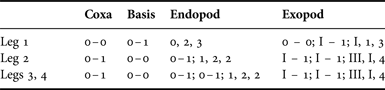
Endopods of leg 1, 1-segmented, of leg 2, 2-segmented, of legs 3 and 4, 3-segmented. Leg 1 endopod with very fine spinules on outer lobe, exopod naked. Endopod of legs 3 to 4 with one transverse row of spinules distally on segments 2 and 3. Coxa of leg 3 is longer than wide; legs 3 and 4 exopod segments distally with spinules. Fifth legs (Figure 4E) symmetrical, uniramous, 3-segmented, proximal segment separated from intercoxal sclerite. Third segment very long, armed with setules distally along both margin; distal segment armed with single small spine on anterior surface and spiniform process laterally.
Male (Figure 5A, B), total length: 550–620 µm (585 ± 23.6 µm, N = 15). Prosome 2.3 times as long as wide. Habitus similar to those of adult female. Posterior pedigerous somite symmetrical. Rostral area as in female. Prosome: urosome ratio = 2.85:1. Urosome of 5 somites (Figure 5C). Proportional lengths of urosomites: 30:16.5:20:18.5:15 = 100. Genital somite slightly asymmetrical viewed dorsally, wider than long, genital aperture located on left side (Figure 5C). Caudal rami as in female.
Stephos grieveae sp. nov., adult male; A, habitus, dorsal view; B, habitus, lateral view; C, urosome dorsal view.
Antennule (Figure 6A, B) similar to those of female, 24-segmented with similar disposition of setae and aesthetascs, although there is an additional aesthetasc on ancestral segment XVIII. Armature of antenna, mouthparts and swimming legs 1 to 4 as in female (Figures 3 & 4).
Stephos grieveae sp. nov., adult male; A, B, antennule (A, segments 1–15; B, segments 16–24); C, right leg 5; D, E, F, left leg 5 (F, segments 4 and 5).
Fifth legs uniramous and asymmetrically (Figure 6C, D). Right leg 4-segmented and very long (Figure 6C); segment 2 (basis) longer than coxa, segment 4 very elongate, distal part is more robust than proximal, hook-like distally with thickened knee-shaped projection and inner margin with small denticles. Left leg 5-segmented (Figure 6D, F), segments unarmed, segment 2 (basis) elongate and robust, segments 3 and 4 shorter and of similar length, segment 5 crescent shaped, longer than segment 2, articulated to segment 4 at approximately one quarter of length of segment, shorter part in form of heavy, tapering processes, while longer part forms a club-shaped curved extension (Figure 6F).
DISCUSSION
The morphological characteristics of the family Stephidae, and a key to the determination of the genera were provided by Boxshall & Halsey (Reference Boxshall and Halsey2004). Bradford-Grieve (Reference Bradford-Grieve1999) categorized the species of the genus Stephos into four groups based on the morphological of the male fifth legs. According to this classification, nine species belong to group IV, where segment 4 of the male left fifth leg is narrow: Stephos angulatus, Stephos pacificus, Stephos tsuyazakiensis, Stephos morii, Stephos rustadi, Stephos pentacanthos, Stephos vivesi, Stephos marsalensis and Stephos geoijinensis. The present Stephos species with male fifth leg, typical of species group IV, have been discovered in the Adriatic Sea in the Eastern Mediterranean.
There are six known Stephos species from the Mediterranean Sea. Stephos gyrans (Giesbrecht, Reference Giesbrecht1892) was found only in the Naples aquarium, while Stephos cryptospinosus (Zagami et al., Reference Zagami, Campolmi and Costanzo2000) and Stephos marsalensis (Costanzo et al., Reference Costanzo, Campolmi and Zagami2000) have only been reported from coastal waters, while Stephos margalefi (Riera et al., Reference Riera, Vives and Gili1991) and Stephos vivesi (Jaume et al., Reference Jaume, Boxshall and Gracia2008) have been recorded exclusively from caves, as is the case with Stephos boettgerschnackae (Kršinić, Reference Kršinić2012), which was recorded from Urinj Cave in the Adriatic Sea.
The new species is characterized by significant differences from all the species found in the Mediterranean Sea as well as representatives of the group IV, with the exception of Stephos rustadi. Stephos rustadi and this new species have narrow segment 4 of the male left fifth leg, and simple segment 5, without appendages. Stephos rustadi was described from material collected in Lysefjorden out of Bergen at depths of 63–80 m (Strömgren, Reference Strömgren1969). The author possessed a large number of individuals; therefore, I accept the description with figures at face value. The new species, collected from an anchialine cave in the Adriatic Sea, is on average ~25% smaller in body length, but has slightly longer antennules than Stephos rustadi. The antennal exopod of the new species is 7-segmented, whereas, in the majority of Stephos spp, such as Stephos rustadi, the exopod is 6-segmented. The caudal rami at Stephos rustadi have two strong and one small seta, which is unique for the genus Stephos. Regardless of the flawed data for appendages, it is hard to believe that the setae were lost during microscopic processing. Female fifth legs of the new species have the first segment (coxa) separated from intercoxal sclerite, the long terminal segment is armed with spinules distally along both margins and a single small spine is located on the anterior surface. A spiniform process is situated laterally, while in Stephos rustadi the proximal segment is fused to the intercoxal sclerite, the outer edge of terminal segment has setules and is without a small spine proximally. Males of the new species are distinguished from Stephos rustadi by characteristics of the fifth legs. The distal segment of the left leg is crescent shaped, and is connected to segment 4 at approximately one quarter of its length. The shorter part tapers into a heavy process, while the longer part forms a club-shaped, curved extension, whereas Stephos rustadi carries the short extension tapers and the longer extension is bifid. In addition, the female genital double somite of Stephos rustadi appears to be slightly asymmetrical, with a rounded swelling on the left (in dorsal view) and on the left a protrusion with a narrower base on the right; the female fifth leg is without an additional setule on segment 3; on maxilliped terminal and penultimate segments of the endopod have very long outer-border setae; the male fifth leg right segment 2 is wide and about 1/2.3 time length of segment 3; the concave border of the right terminal hook-like segment of the left fifth leg is naked; exopod segment 3 has terminal bifurcation with long branches.
Stephos rustadi was found in water with a salinity of about 34, a temperature of 7°C and a very low oxygen concentration of 1.57 mg O2l−1, between 3 and 12% saturation (Strömberg, Reference Strömgren1969). Previously described species from the Adriatic Sea, Stephos boettgerschnackae, and new species have very different geographical distributions. Stephos boettgerschnackae is widely distributed along the eastern coast of the Adriatic, and is present in almost all anchialine habitats from Rovinj in the northern Adriatic Sea to the island of Hvar in the central Adriatic. The bulk of the population was found in Urinj Cave, near Rijeka, North Adriatic, with many individuals present. For basic hydrographic and chemical data in Urinj Cave, see Cukrov et al. (Reference Cukrov, Jalžić, Omanović and Cukrov2006). However, the new species occurs only in two caves located in the southern Adriatic, on the island of Mljet: Lenga Pit and Zaglavica Cave (Kršinić, unpublished data). Due to the location of the island of Mljet in the Adriatic Sea, both caves are influenced by the water of the southern Adriatic. In the Lenga Pit, the salinity varied between 1.5 at the surface and 39 at the bottom, and the temperature ranged between 14.2° and 16.6°C. The oxygen concentration was at its lowest at a depth of 4 m with 0.14 mg O2l−1, while significantly higher values were found near the bottom, during January 2009 (Žic et al., Reference Žic, Truesdale, Cuculić and Cukrov2011). A majority of the population of this new species was found in the deeper parts of the cave, which had significantly higher values of oxygen and salinity. Increased concentrations of dissolved organic carbon and trace metals such as Zn and Cu were found in the water and sediment (Cuculić et al., Reference Cuculić, Cukrov, Kwokal and Mlakar2011) in Lenga Pit. Also, bacterial abundance was higher in the cave than in the surrounding sea water (Krstulović et al., Reference Krstulović, Šolić, Šantić, Maršić-Lučić, Ordulj and Šestanović2013). In addition to Stephos grieveae, calanoid copepods such as Stygocyclopia balearica, Speleohvarella gamulini and Badijella jalzici were found in the Lenga Pit. Therefore, this cava is a very interesting habitat for multidisciplinary research into succession, survival and the role of copepods in extreme conditions, and, for this purpose, it should be fully protected.
ACKNOWLEDGEMENTS
I would like to thank Branko Jalžić from the Croatian Natural History Museum (Zagreb) and members of the Croatian Bio-Speleological Society in Zagreb for collecting the material on which this work was based. I would also like to thank the anonymous reviewers and Dr Janet Bradford-Grieve, Emeritus Researcher, for their suggestions on the improvement of this paper.