Marfan syndrome is an autosomal dominant disorder of the connective tissue, whose cardinal features affect eyes, musculoskeletal, and cardiovascular system.Reference Weyman and Scherrer-Crosbie1 In the large majority of cases, Marfan syndrome is caused by mutations of FBN1 gene, which codes for fibrillin-1. However, FBN1 gene mutation cannot be considered its unique genetic cause, since several other genes are potentially involved (e.g., TGFBR1, TGFBR2, COL1A1).Reference Attias, Stheneur and Roy2,Reference Gentilini, Oliveri and Fazia3 Due to the variable expression of this genetic syndrome, diagnosis is based on major and minor criteria (2010 revised Ghent nosology).Reference Loeys, Dietz and Braverman4 Although family history plays a role in the diagnostic workup, Marfan syndrome presents as sporadic in a significant number of patients.Reference Gray, Bridges and West5 The most frequent cardiovascular abnormalities are mitral valve prolapse and dilation of the sinuses of Valsalva,Reference Figueiredo, Martins, Lima and Alvares6,Reference Murdoch, Walker, Halpern, Kuzma and McKusick7 associated to an increased risk of aortic and mitral regurgitation, endocarditis, aortic dissection, and heart failure.
The natural history of bicuspid aortic valve, the most common congenital cardiac abnormality,Reference Williams8,Reference Ward9 is characterised by haemodynamic valvular impairment (present at birth or, more commonly, acquired by ageing) and by increased prevalence of aortic abnormalities, ranging from reduced aortic elasticity to aortic dilatation, aneurysm, and dissection.Reference Nistri, Sorbo, Marin, Palisi, Scognamiglio and Thiene10–Reference Sticchi, De Cario and Magi13 Despite prevalence and natural history of bicuspid aortic valve and other cardiovascular manifestations are well known in adults with Marfan syndrome, little is known about children and young patients.Reference Mueller, Stark and Steiner14,Reference van Karnebeek, Naeff, Mulder, Hennekam and Offringa15
The aim of this study was to describe a well-characterised, by a comprehensive clinical and genetic investigation, cohort of consecutive children and young patients with Marfan syndrome, looking at the impact of family history and presence of bicuspid aortic valve on disease severity.
Methods
Study population
We retrospectively reviewed clinical finding in children consecutively diagnosed with Marfan syndrome at the Inherited and Rare Cardiovascular Disease Clinic of the University of Campania “Luigi Vanvitelli” between 2009 and 2108. The diagnosis of Marfan syndrome was based on the revised Ghent criteria.Reference Loeys, Dietz and Braverman4
Study protocol
All patients underwent a comprehensive clinical–instrumental–genetic evaluation, including family and personal history, physical examination, standard 12-lead electrocardiogram, echocardiography, blood tests, 24-hour ambulatory electrocardiogram monitoring, exercise stress test, and, if required, cardiac MRI and/or genetic analysis. Clinical and instrumental evaluation was repeated every 6–12 months. The clinical genetics evaluation, including generation of a revised Ghent systems scoreReference Loeys, Dietz and Braverman4 as well as genetic testing, was performed by a board-certified clinical geneticist.
Echocardiography
Left ventricular wall thickness, left ventricular chamber dimensions, systolic function, and diastolic function were measured as absolute values by M-mode, two-dimensional, and Doppler echocardiography. Mitral and tricuspid valves were evaluated in particularly the parasternal long-axis and apical four-chamber views. Particular attention was posed on the evaluation of echocardiographic features of myxomatous degeneration of the cardiac valves, including thickening of the valve leaflets, prolapse of the mitral, tricuspid, and aortic valve leaflets, or billowing during systole of the mitral or tricuspid leaflets, or billowing during diastole of the aortic leaflets.Reference Foster16 Mitral or tricuspid prolapse was defined as the displacement, by more than 2 mm above the high points of the mitral or tricuspid annulus, of the abnormal echocardiographic billowing of one or more mitral or tricuspid leaflets, as recorded in either the parasternal or apical long-axis views.Reference Foster16,Reference Barlow17
The aortic root diameters were measured at four levels (aortic annulus, sinus of Valsalva, sinotubular junction, and ascending aorta) with the use of a parasternal long-axis view.
According to the current recommendations,Reference Lang, Badano and Mor-Avi18 aortic annulus was measured at midsystole from inner edge to inner edge, while other aortic root measurements were made at end-diastole, in a perpendicular plane to that of the long axis of the aorta using the L–L convention. A z score was also calculated for each aortic root measurement.
Genetic analysis
Genetic analysis was performed after obtaining informed written consent, according to the procedure established by the local ethics committee. Sequencing methods varied according to different year and sequencing method available. In patients evaluated between 2009 and 2012, molecular genetic testing was guided by the clinical phenotype and performed using direct Sanger sequencing. Next-generation sequencing was available from 2012, and since that year, all patients underwent next-generation sequencing panel.
Statistical analysis
Normally distributed continuous data are presented as mean ± sd and were compared by t-test. Categorical variables were expressed as number (percentage) and analysed by the chi-square test or Fisher exact test when appropriate. p-Values <0.05 (two-tailed) were considered significant.
Results
Clinical characteristics
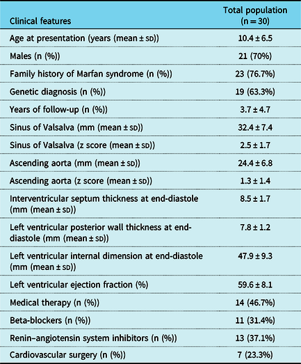
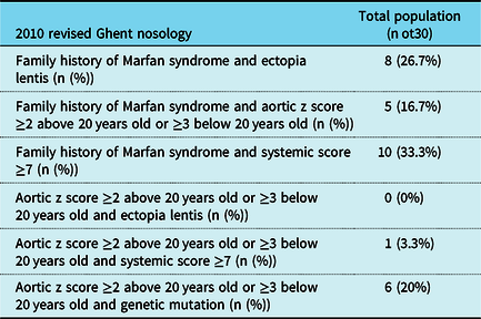
Clinical characteristics of the examined cohort are shown in Table 1. The age at clinical diagnosis was 10.4 ± 6.5 (median 9, age range 0–25), 70% were males. Family history was present in 23 patients (76%), and a genetic testing was performed in 19 (63%) (7 patients underwent Sanger sequencing and 12 next-generation sequencing panel), showing a disease-causing mutation of FBN1 gene in 18 of them (95%; Marfan syndrome) and of TGFBR2 in 1 (5%; Loyes–Dietz syndrome). The subgroup of patients who have not performed genetic testing had a positive family history of Marfan syndrome. Distribution of Ghentʼs diagnostic criteria in the examined cohort is reported in Table 2.
Clinical characteristics of the examined cohort

2010 revised Ghent nosology in our population

In our cohort, the large part of patients was diagnosed given the presence of family history of Marfan syndrome and another one diagnostic criteria: 33% of patients showed family history and a systemic score ≥7 points, 26% family history and ectopia lentis, and 17% family history and aortic dilation. In the latter subgroup, two patients with diagnosis of bicuspid aortic valve were identified.
In patients without family history, diagnosis was based on aortic dilation (z score ≥2 above 20 years old or ≥3 below 20 years old) and another one diagnostic criteria: genetic mutation in 20% of patients, while systemic score ≥7 points in 3%. In the single patients with Loyes–Dietz syndrome, the first clinical diagnosis was Marfan syndrome, based on the presence of aortic dilation and systemic score ≥7; however, the next-generation sequencing panel showed a TGFBR2 mutation, suggesting a final diagnosis of Loyes–Dietz syndrome.
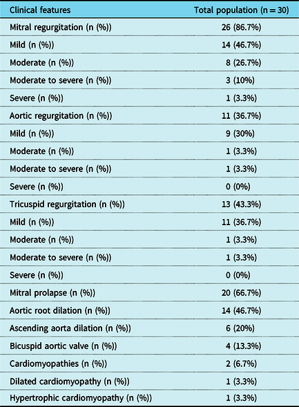
In our cohort, all patients showed cardiovascular abnormalities (Table 3). The most common, present in 26 patients (86.7), was mitral regurgitation (54% mild, 31% moderate, 11% moderate to severe, 4% severe), followed by mitral prolapse, present in 20 (67%). All patients with moderate, moderate to severe, and severe regurgitation showed mitral prolapse. Other cardiovascular abnormalities were aortic root dilation, present in 14 patients (47%); ascending aorta dilation, in 6 patients (20%); aortic regurgitation, in 11 patients (37%) (82% mild, 9% moderate, 9% moderate to severe); tricuspid regurgitation, in 13 patients (43%) (84% mild, 8% moderate, 8% severe); bicuspid aortic valve, in 4 patients (13%), and cardiomyopathies in 2 patients (6.6%): 1 patient (3%) with significant left ventricular hypertrophy (16 mm), suggesting non-obstructive hypertrophic cardiomyopathy and 1 (3%) with dilated left ventricle and midly reduced cardiac function (ejection fraction 45%), suggesting dilated cardiomyopathy.
Cardiovascular abnormalities in total population

Prevalence and clinical impact of bicuspid aortic valve and family history
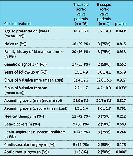
Bicuspid aortic valve was identified in four patients (13% of the total cohort) with Marfan syndrome. Compared to patients with tricuspid aortic valve, those with bicuspid aortic valve were younger (3.2 ± 4.3 versus 10.7 ± 6.8 years old, p-value = 0.043) and showed greater z score of aortic sinus diameters (4.2 ± 0.9 versus 2.2 ± 1.6 z score, p-value = 0.033) (Table 4). Moreover, patients without family history of Marfan syndrome (n = 7, 24% of the total cohort) showed higher prevalence of aortic sinus dilation (87 versus 32%, p-value 0.009) and greater aortic sinus diameters (4.2 ± 2.1 versus 1.9 ± 1.1 z score, p-value 0.002).
Clinical characteristics of the examined cohort according to the type of aortic valve

* p-values <0.05 were considered statistically significant.
Follow-up
During a mean follow-up of 3.7 (±4.7) years (median 3 years, range 0–16 years), four patients developed progressive left ventricle dilation and worsening of left ventricular function, due to mitral and/or aortic valve regurgitation, and an 11-year-old male patient, showing severe aortic sinus dilation (54 mm, z score 6.75), experienced sudden death due to aortic dissection.
Moreover, during follow-up, seven patients (23%) underwent cardiac surgery: an 8-year-old male patient, showing mitral prolapse and severe regurgitation, received the resection of a papillary fibroelastoma of mitral valve chordae, mitral valve repair, and annuloplasty; an 8-year-old male patient, showing severe mitral regurgitation, underwent mechanical replacement of the mitral valve; two patients, an 8-year-old female and a 10-year-old male, showing mitral valve prolapse and moderate to severe regurgitation, received mitral valvuloplasty; a 4-year-old male patient, showing mitral valve prolapse and moderate to severe regurgitation, received mitral valvuloplasty and, 3 years later, due to the development of severe aortic root and ascending aorta dilation, underwent complete aortic root replacement using Bentall procedure; two male patients, a 3-year old, carrying a mutation in TGFBR2 gene, and an 11-year old, showing severe aortic root dilation (24 mm, z score 6.28, and 27 mm, z score 5.31, respectively), underwent valve-sparing aortic root replacement using David procedure and complete aortic root replacement using Bentall procedure, respectively.
Both patients who underwent aortic root replacement using Bentall procedure showed bicuspid aortic valve. Compared to patients with tricuspid aortic valve, those with bicuspid aortic valve underwent more frequently aortic root replacement during follow-up (50 versus 4%, p-value = 0.004).
Moreover, patients with sporadic Marfan syndrome (without family history) showed higher rate of aortic surgery (37 versus 0%, p-value = 0.002) and of cardiovascular surgery (including mitral valve or aortic surgery) during follow-up (50 versus 14%, p-value = 0.037).
Discussion
Marfan syndrome is a genetic disease in which connective tissue becomes weak secondary, in the large part of cases, to mutation in the gene coding for fibrillin-1 (FBN1), resulting in aortic dilation, aortic dissection, aneurysm formation, aortic regurgitation, and mitral valve prolapsed.Reference Weyman and Scherrer-Crosbie1–Reference Figueiredo, Martins, Lima and Alvares6 Despite prevalence and natural history of the cardiovascular abnormalities is well known in adults, little is known in children and young patients.Reference Mueller, Stark and Steiner14,Reference van Karnebeek, Naeff, Mulder, Hennekam and Offringa15
In our cohort of children and young adults with Marfan syndrome, all patients showed cardiovascular abnormalities, of which the most frequent were mitral regurgitation, mitral valve prolapsed, and aortic root dilation (in particular involving sinus of Valsalva), present in 40, 67, and 47%, respectively. The results were found to be similar to previous studies including children with Marfan syndrome.Reference Mueller, Stark and Steiner14,Reference van Karnebeek, Naeff, Mulder, Hennekam and Offringa15
An interesting finding of our study was the severity of aortic disease in patients with sporadic Marfan syndrome (without family history of disease). In particular, these patients, compared to patients with family history of Marfan syndrome, showed higher prevalence of aortic sinus dilation, greater aortic sinus diameters, and higher rate of cardiovascular surgery during follow-up, especially aortic surgery. This finding is consistent with a previous study including childrenReference van Karnebeek, Naeff, Mulder, Hennekam and Offringa15; however, the reason of this association remains unclear.
In our study, we showed a relatively high prevalence of bicuspid aortic valve, present in 13% of total cohort. Bicuspid aortic valve is the most common congenital cardiac abnormality, affecting 1–2% of the general population.Reference Williams8,Reference Ward9 This condition is associated with several disorders, including aortic stenosis and/or regurgitation, and aortic root dilation, usually associated with bicuspid aortic valve and may lead to aortic aneurysm, dissection, and rupture.Reference Nistri, Sorbo, Marin, Palisi, Scognamiglio and Thiene10–Reference Sticchi, De Cario and Magi13 A recent study found bicuspid aortic valve in 3.3% of patients with Marfan syndrome and reported that patients with bicuspid aortic valve had larger aortic dimension.Reference Nistri, Porciani, Attanasio, Abbate, Gensini and Pepe19 In our cohort, patients with bicuspid aortic valve were younger, showed greater aortic diameters, and underwent more frequently aortic root replacement during follow-up compared to patients with tricuspid aortic valve.
These results show that paediatric onset of Marfan syndrome can be associated with severe phenotype and prognosis, especially in patients without family history of disease and in those with bicuspid aortic valve. Therefore, considering that these characteristics may worse the natural history of aorthopathy in children, in their presence may be required a close follow-up and, if necessary, a preventive surgery at an early age.
Limitations
This study is limited by the low number of patients studied (including the subgroup with Marfan syndrome and bicuspid aortic valve), and the fact that genetic analysis has been performed in only 63% of patients with clinical diagnosis of Marfan syndrome. Larger studies are needed to better characterise the clinical course of aortopathy in infants and children with Marfan syndrome and to better understand the possible clinical implication. We also reported two patients with cardiomyopathy. Primary (or secondary) cardiomyopathies have been well described in Marfan syndrome. However, we are not able to exclude, without a dedicated genetic panel on sarcomeric and/or overlapping disease, the coexistence of two diseases in the patient affected by non-obstructive hypertrophic cardiomyopathy.
Conclusions
In our cohort of children and young adult patients with Marfan syndrome, the absence of family history and the presence of bicuspid aortic valve were associated to severe aortic dilation and worse prognosis.
Acknowledgements
The authors acknowledge Prof. Antonio Novelli for his precious contribution.
Financial Support
This research received no specific grant from any funding agency, commercial or not-for-profit sectors.
Conflicts of Interest
None.
Ethical Standards
The authors assert that all procedures contributing to this work comply with the ethical standards of the relevant national guidelines on human experimentation and with the Helsinki Declaration of 1975, as revised in 2008, and has been approved by the institutional committees.