INTRODUCTION
The study of chemosynthetic ecosystems in the deep sea represents a challenging issue due to the difficulty of sampling, which involves the use of modern technologies such as manned submersibles. Vent animals were useful as flux indicators of hydrothermal activity for geologists prospecting for vents, who appreciated the need for an explanation of the large biomass of animals and the means by which species could be maintained at vents in the face of local extinctions (Desbruyères et al., Reference Desbruyères, Segonzac and Bright2006). Since the first studies of chemosynthetic ecosystems in the 1970s, many charismatic vent organisms have been discovered in these chemosynthetic environments. Thus, dozens of new species of clams, mussels, limpets, crabs or cephalopods, among others, have been identified and later classified, increasing our knowledge of these extreme environments.
Based on the samples obtained in one of these studies, a new Octopoda genus and species Vulcanoctopus hydrothermalis González & Guerra, 1998 was erected. This classification was made based on two male specimens. These individuals were caught during the French cruise HOT 96, two metres from the main black smoker of the Genesis site, which is located at 12°48.43′N–103°56.41′W, at 2600 m depth on the East Pacific Rise (EPR) (González et al., Reference González, Guerra, Pascual and Briand1998). The possibility of raising a new sub-family (Vulcanoctopodinae) within the Octopodidae was then discussed for the first time. The parasites found in these two individuals also allowed us to describe a new genus and species of copepod (López-Gonzalez et al., Reference López-González, Bresciani, Huys, González, Guerra and Pascual2000).
Among the cephalopods, only V. hydrothermalis has been recognized as endemic of hydrothermal vents (Desbruyères et al., Reference Desbruyères, Segonzac and Bright2006). It inhabits an isolated extreme environment very close to the base of the chimneys and is also observed on the pillow lava at several metres from the active areas. This benthic species has characters that represent adaptations either to the deep-sea or to a hydrothermal vent habitat (González et al., Reference González, Guerra, Pascual and Briand1998).
Based on twenty-five specimens filmed between 2600 and 2650 m depth in the EPR, the most important behaviour patterns were described for the first time (Rocha et al., Reference Rocha, González, Segonzac and Guerra2002). Afterwards, the capture of seventeen new males allowed us to describe the main morphological variations of this species (González et al., Reference González, Guerra, Rocha and Briand2002). The analysis of the stomach contents showed that V. hydrothermalis preys mainly on Crustacea: Decapoda (Rocha et al., Reference Rocha, González, Segonzac and Guerra2002; Voight, Reference Voight2005).
An intriguing aspect of the samples studied was the absence of females, which suggested the existence of a spatial segregation by sex (González et al., Reference González, Guerra, Rocha and Briand2002).
The objective of the French cruise BIOSPEEDO (Biologie-Sud Pacifique Est-Etude de la Dorsale Océanique) using the submersible ‘Nautile’, onboard the RV ‘L'Atalante’, was to study the biodiversity and geodiversity of the hydrothermal vents along the EPR between 7°24′S and 21°33′S. During this cruise, the first female specimen of V. hydrothermalis was collected at the hydrothermal vent site Gromit in April 2004 (Jollivet et al., Reference Jollivet, Lallier and Barnay2004).
The aim of this paper is to describe, for the first time, the female of V. hydrothermalis, to complete its diagnosis and description.
MATERIALS AND METHODS
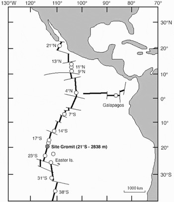
A female Vulcanoctopus hydrothermalis of 34.9 mm mantle length (ML) was caught during the dive PL 1578 (Figure 1), by the robotized arm grab of the manned submersible ‘Nautile’, on the site Gromit (EPR, 21°33 66′S, 114°17 98′W) at 2832 m depth in April 2004 (Jollivet et al., Reference Jollivet, Lallier and Barnay2004).
Map showing the locality where the Vulcanoctopus hydrothermalis female has been collected (solid circle).

The specimen was collected near a high temperature hydrothermal vent, on a mussel bed of Bathymodiolus thermophilus and Calyptogena magnifica, crawling among stalked barnacles (Neolepas cf. rapanui), bythograeid crabs Bythograea spp., several fish species (Causse et al., Reference Causse, Biscoito and Briand2005), and chiridotid holothurian Chiridota sp. The animal was caught in very good condition. It was fixed in formaldehyde (4% in seawater) and preserved in 70% ethanol.
Definition of counts, measurements and indices followed Roper & Voss (Reference Roper and Voss1983), Clarke (Reference Clarke1986) and González et al. (Reference González, Guerra, Pascual and Briand1998). Maturity was assessed according to Guerra (Reference Guerra1975).
The female was dissected and the digestive tract, reproductive tract, circulatory and excretory systems, and eyes were removed for detailed description. The reproductive tract was sectioned for analysing the stage of maturity, number and size of oocytes and also to perform histological analysis on sections stained with haematoxylin and eosin. All oocytes in the ovary were measured, counted and photographed using the Image Analysis System Eclipsenet®. The specimen is stored at the Marine Ecology and Biodiversity Research Group (ECOBIOMAR, Instituto de Investigaciones Marinas, CSIC) cephalopod collection with the reference number VH06030.
RESULTS
Female external and internal description
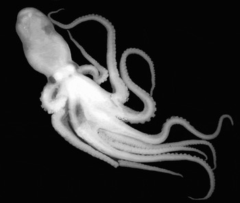
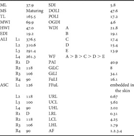
The external morphology of the female is very similar to that of the male (Figure 2). However, no parasitic cysts were found in the skin as seen previously in several males of EPR-13°N (López et al., Reference López-González, Bresciani, Huys, González, Guerra and Pascual2000). Measurement, counts and indices after preservation are summarized in Table 1.
Photograph of the unique female Vulcanoctopus hydrothermalis collected to date.

Measurements (mm), counts and indices of the Vulcanoctopus hydrothermalis female follow Roper & Voss (Reference Roper and Voss1983), Clarke (Reference Clarke1986) and González et al. (Reference González, Guerra, Pascual and Briand1998) except: damaged (D); distal oviduct length index (DOLI)— length of the distal part of the oviduct as a percentage of mantle length (ML); oviducal gland diameter index (OGDI)—diameter of the oviducal gland as a percentage of ML; proximal oviduct length index (POLI)—length of the proximal part of the oviduct as a percentage of ML.

The mantle cavity and digestive tract of the female have similar characteristics to those of males (González et al., Reference González, Guerra, Pascual and Briand1998). However, the dark swelling observed and described for males was not present in the female specimen. The digestive tract of the female contained some food remains, which were impossible to identify. The circulatory, excretory and nervous systems are also similar to those of males.
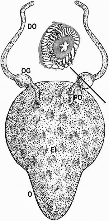
The reproductive tract of the female comprises a pear shaped ovary and two oviducts (Figure 3). No external seminal receptacles were found. The posterior edge of the ovary fits into the posterior part of the mantle. In this specimen, the proximal part of the oviducts is short (proximal oviduct length index = 17.2) and narrow, indicating it is at a maturing stage, with spermatophores attached to the external part of the ovary. The oviducal glands are almost spherical and of uniform white colour, their maximum diameter measuring 1.6 mm (oviducal gland diameter index = 4.6%). The distal part of the oviducts is relatively long (distal oviduct length index = 46.7%).
The reproductive system of the Vulcanoctopus hydrothermalis female and the histological structure of the oviducal glands showing the absence of spermatophores reservoir. DO, distal oviduct; EI, external view by transparency of the egg insertion; O, ovary; OG, oviducal gland; and PO, proximal oviduct.

The histological structure of the oviducal glands is composed of an outer endothelial layer and an underlying connective tissue sheet, which appears profusely innervated and vascularized. In the inner part are the proximal and distal glands, both multilobulated and rounded by smooth muscular tissue covered by connective tissue and outer endothelium. The above glands are also innervated and vascularized, and they contain the lumen of the oviduct which is 0.4–0.5 mm diameter. This lumen is surrounded by a thick wall composed of a smooth muscular coat formed by two layers of fibres lying in a particular orientation (the outer circular and the inner longitudinal). The wall of the lumen is covered by an endothelilal layer, which at regular intervals form expansions of this tissue. Each oviducal gland is structurally divided into two concentric glands around the oviduct and separated by muscular and connective tissue. Seminal receptacles (spermathecae) were absent (Figure 3).
The ovary wall is very slender and translucent. It is adhered to the internal side of the mantle. The ovary wall is 65 µm thick (mean size) and it is formed of three layers: the outer squamous endothelial tissue, an intermediate lax connective tissue and an inner squamous cell layer of endothelial tissue.
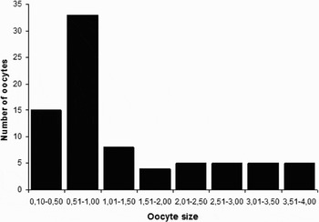
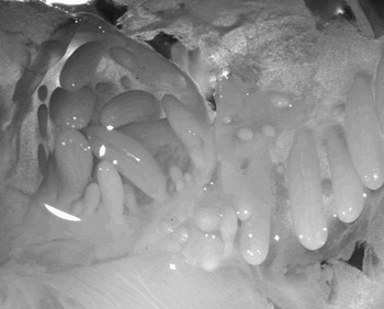
Eighty finger-like oocytes of different sizes, ranging from 0.15 to 4.0 mm maximum length and between 0.5 and 0.6 mm of maximum width, were counted in the ovary. Figure 4 shows the oocytes size distribution. The oocytes were fixed by their bases to the inner side of the ovary wall and lack peduncles (Figure 5).
Size (maximum length in mm)–egg distribution within the ovary of Vulcanoctopus hydrothermalis.

The oocytes of Vulcanoctopus hydrothermalis lack peduncles and are fixed by their bases to the inner side of the ovary wall.

Amended diagnosis of the species including female characters Vulcanoctopus hydrothermalis González & Guerra, 1998: body translucent, with a muscular consistency; mantle pear-shaped and posteriorly mitre-like. The arms are 1.5–4.3 times ML and with two rows of suckers; no enlarged suckers; arm formula typically 1.2.4.3 or 1.2.3.4. The maximum depth of the largest sector of the web is about 22% of the longest arm. Right arm III hectocotylized (hectocotylized arm 1.5–2.1 times ML) in males. Ligula short (8–10% of hectocotylized arm), lance-shaped and without transverse ridges. Calamus represents 30 to 50% of the ligula length in fully mature specimens. Presence of a large white body (which covers the optic nerves and the optic lobe), a crop, and a multilobulate digestive gland (8–12 lobes); ink sac absent; gills with 7–8 lamellae per demibranch. Spermatophore length 70–125% of ML. Oviducal glands without spermathecae. Finger-like oocytes (80) ranging from 0.15 to 4 mm maximum length, lacking peduncles.
DISCUSSION
One of the most remarkable characters of the V. hydrothermalis female is the lack of spermathecae. The absence of these reservoirs in the oviducal glands has been previously reported in four Eledone species (E. cirrhosa, E. moschata, E. massyae and E. gaucha), which suggest that this is a consistent characteristic of this uniserial arm sucker genus (Pérez et al., Reference Pérez, Haimovici and Cousin1990). The inner structure of the oviducal glands of V. hydrothermalis is identical to that described in Pteroctopus tetracirrhus (Morales, Reference Morales1973), which indicated that this species also lacks spermathecae. Therefore, this is not unique to the subfamily Eledoninae, as it occurs also in some members of the Octopodinae such as P. tetracirrhus, and could be a common character among the Octopoda.
The spermatophores of the Octopodinae and Bathypolypodinae enter the oviducts and the spermatozoa are stored in the spermathecae of the oviducal glands after the spermatophoric reaction event (Mann et al., Reference Mann, Martin and Thiersch1970; Froesch & Marthy, Reference Froesch and Marthy1975). Fertilization of the oocytes occurs in the oviducal glands just before spawning. However, this is not the case of the four eledonid species mentioned above or V. hydrothermalis, which lack spermatechae. In these eledonid octopuses, the spermatophores penetrate in the oviducts and oviducal glands reaching the ovarian cavity, in which the spermatophoric reaction takes place. Bladders of spermatangia are found in the ovary of E. cirrhosa and E. moschata mated females (Boyle, Reference Boyle and Boyle1983; Mangold, Reference Mangold and Boyle1983). However, in E. massyae and E. gaucha the spermatangia are attached to the apical filaments of the oocytes (Pérez et al., Reference Pérez, Haimovici and Cousin1990). The oocytes of V. hydrothermalis lack apical filaments and no spermatangia were observed inside the ovary (Figure 5). Therefore, the fertilization mechanism remains unknown in this species.
Whatever the fertilization mechanism, the existence of an Octopodinae such as P. tetracirrhus lacking spermathecae and with two rows of suckers in the arms, such as V. hydrothermalis, point out the possibility of the colonization of hydrothermal vents by a group of octopods with biserial suckers on the arms. Phylogenetic relationships of V. hydrothermalis are still unknown. However, considering structural, biogeographical and ecological reasons, some species of the genera Benthoctopus and Bathypolypus would be the most suitable octopod ancestor of V. hydrothermalis. Perhaps the deep-sea Benthoctopus, which has smooth skin, moderate size ligula and lack of laminae would be the best candidate. Nevertheless, molecular analyses are necessary to test this hypothesis.
ACKNOWLEDGEMENTS
We acknowledge Didier Jollivet (Chief Scientist, Roscoff, France) and the team of the N/O ‘L'Atalante’ and the submersible ‘Nautile’, for collecting the specimen studied here. We thank also Violaine Martin and Patrick Briand (IFREMER, Brest) for Figure 1 and Figure 2, respectively, Mrs Fiona Read for the English corrections, and the referees for their valuable comments and suggestions. The cruise was supported by the French programme DORSALE and the ECCHIS-GDR (CNRS & IFREMER).