Introduction
Radiotherapy (RT) is associated with a range of skin toxicities including radiation dermatitis, lichenoid eruptions and radiation induced neoplasms. It has rarely been associated with cases of autoimmune blistering skin diseases including bullous pemphigoid (BP), pemphigus vulgaris and epidermolysis bullosa acquisitis.Reference Cuperus, Albregts and Toonstra1–Reference Aguado, Marquina, Pretel, Ruiz-Carrillo and Espana3
This case raises two hypotheses regarding a possible radiation-induced BP that was triggered within 2 months after RT for breast cancer or a radiation recall dermatitis (RRD) induced by adjuvant Trastuzumab.
Case Report
A 64-year-old woman with a previous history of hypertension and type 2 diabetes had underwent radical mastectomy and axillar lymph node dissection for an infiltrative ductal carcinoma (IDC). The histopathology revealed a 25 mm, grade III IDC, HR+, Her2+++, 1N+/15N.
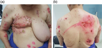
Adjuvant treatment comprised of six courses of chemotherapy (three anthracycline and three docetaxol), Hormone therapy (Anastrozole), Trastuzumab and 3D conformal RT to a total dose of 50 Gy/25 fractions. Target volumes were chest wall, internal mammary nodes region (IMN) and supraclavicular fossa. Two months after the completion of RT and after eight cycles of trastuzumab, an erythematous and pruritic vesicular eruption developed initially in the irradiated area and extended later on over other skin areas. (Figures 1a and 1b)
(a and b) Vesiculobullous eruption developed initially in the irradiated site and then extended to the chest.

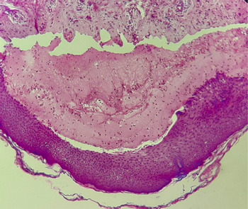
Skin biopsy showed BP, with subepidermal blisters and an inflammatory infiltrate with abundant eosinophils (Figure 2).
Histopathological examination of skin biopsy [Hematoxylin and eosin stain (20x)]: subepidermal blister and an inflammatory infiltrate with abundant eosinophils.

The patient was treated with oral corticosteroids and lesions evolved favourably.
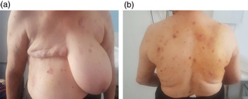
At her 2-year follow-up appointment, BP lesions were well-controlled, although she continued to develop occasional blisters, which were successfully treated with oral corticosteroids (Figures 3a and 3b).
(a and b) Bullous pemphigoid lesions after 2 years of follow-up.

Discussion
BP is the most common autoimmune subepidermal blistering disease of the skin and mucous membranes. It is characterised by autoantibodies against structural proteins of the dermal–epidermal jonction and responsible for localised or generalised bullous lesions.Reference Schmidt and Zillikens4
RT is one of several physical agents including ultraviolet light, thermal burns and skin grafts that have been reported to induce BP in normal skin.Reference Danescu, Chiorean, Macovei, Sitaru and Baican5 Radiation-induced bullous pemphigoid (RIBP) affects mainly women with breast carcinomas,Reference Campa, Mansouri, Wilcox and Griffin6–Reference Cabrera-Rodriguez, Munoz-Garcia, Quiros Rivero, Ropero Carmona and Rios Kavadoy8 but it has been reported also with other cancers (vulva, cervix, lung and esophagus).Reference Nguyen, Kwan and Ahmed9
Mul et al., reported a large series of BP associated with radiotherapy. BP occurred usually more than 1 year after radiotherapy, only 5/27 patients developed BP within weeks. No dose threshold was found, BP was observed after 20 Gy (three cases), but median dose was 50 Gy.Reference Mul, van Geest and Pijls-Johannesma10
The mechanisms of RIBP remain unclear; radiation tissue damage could induce BP antigens. Another explanation is that patients may already have circulating autoantibodies before RT was performed.
Radiation also influences the levels of matrix metallopeptidase 9 (MMP-9) and the expression of endothelial growth factor (VEGF). It is known that both molecules are involved in the pathophysiology of the blistering.Reference Nguyen, Kwan and Ahmed9
The diagnosis is based on histopathological examination which revealed subepidermal blisters, dermal papillae and the epidermis is preserved.Reference Schmidt and Zillikens4
Therapy for RIBP does not differ from that of ‘de novo’ BP, treatment consists mainly of the administration of local corticosteroids, oral corticosteroids and immunosuppressive treatments depending on the extension of the disease with favourable outcome within weeks.Reference Schmidt and Zillikens4,Reference Joly, Roujeau and Benichou11
In our case, Trastuzumab was administered concomitantly with Docetaxel and BP appeared after the eighth cycle, but we cannot rule out that it may have caused BP or there have been any synergistic effect between RT and Trastuzumab. In the literature, only PB due to programmed cell death protein-1 (PD-1) and programmed death ligand-1 (PD-L1) inhibitors has been described.Reference Lopez, Khanna, Antonov, Audrey-Bayan and Geskin12 As far as we know, no reported study has established any link between induced BP and Trastuzumab. However, few cases of RRD induced by adjuvant Trastuzumab have been previously reported.Reference Chung, Stuart and Keyes13,Reference Moon, Koo, Suh, Yoon, Bae and Lee14 Indeed, RRD is defined as the development of skin reactions in a previously irradiated area after exposure to a recall-triggering drug. Anti-cancer agents are the most commonly involved drugs. The diagnosis of RRD should be considered in patient developing a skin reaction in a previously irradiated area that was normal prior to the introduction of a new drug. RRD can also appear after several exposures to triggering drugs.Reference Camidge and Price15 Dermatological reactions may include maculopapular eruptions, erythema, vesicle formation, hemorrhage and necrosis. The time interval between completion of radiation therapy and the recall reaction ranged from days to years. There are no specific therapies for RRD, corticosteroids and non-steroidal anti-inflammatory agents could be used or lesions may resolve spontaneously if the reaction is not severe.Reference Azria, Magné and Zouhair16
Conclusion
We suggest that BP could be a side effect of RT, but also a RRD induced by Trastuzumab or may be its development during the course of breast cancer is entirely coincidental. How can we determine the definite etiology? The answer to this question remains difficult but we should keep in mind that BP is a relatively benign skin reaction that could develop during the course of several cancers. Therefore, oncologists should be aware of this entity because early diagnosis allows adequate management.
Financial Support
None.
Declaration of Competing Interest
The authors declare that there is no conflict of interest.