Introduction: S-layers as a universal microbial strategy
The prokaryotic cell surface serves as the primary interface through which microorganisms interact with their physical, chemical, and biological environment. It is now well recognized that one of the most frequently observed prokaryotic cell envelope surface structures are monomolecular arrays of protein or glycoprotein subunits referred to as surface (S)-layers (Sleytr and Pum, Reference Sleytr and Pum2025). These porous crystalline lattices are nearly ubiquitous among Archaea, where they frequently represent the exclusive wall structure, and are widely distributed across diverse bacterial taxa (Sleytr and Sára, Reference Sleytr and Sára1997; Messner et al., Reference Messner, König and ClausHand Varma2010; Albers and Meyer, Reference Albers and Meyer2011; Johnston et al., Reference Johnston2024; Isbilir et al., Reference Isbilir2025). Despite their apparent structural simplicity, S-layers represent a significant metabolic investment, accounting for up to ~10% of total cellular protein synthesis (Sleytr et al., Reference Sleytr1993). A closed S-layer on a rod-shaped cell contains ~5 × 10⁵ protein subunits, requiring the continuous synthesis, secretion, and lattice incorporation of hundreds of copies per second during rapid growth (Sleytr et al., Reference Sleytr1999). This immense biosynthetic flux highlights their fundamental physiological importance as self-assembling surface barriers with high morphogenetic potential. They can therefore be regarded as the simplest biological boundary layer systems (Sleytr, Reference Sleytr1975; Sleytr et al., Reference Sleytr1988).
Depending on the organism, S-layer lattices exhibit oblique (p1, p2), square (p4), or hexagonal (p3, p6) symmetry, with hexagonal arrangements predominating in Archaea. Unit cell spacings range from ~5 to 30 nm, generating a mathematically precise nanoscale mosaic of pores with diameters of ~2–10 nm (Sleytr et al., Reference Sleytr1999; Pum et al., Reference Pum2013; Pum and Sleytr, Reference Pum and Sleytr2014; Schuster and Sleytr, Reference Schuster and Sleytr2020; Schuster and Sleytr, Reference Schuster and Sleytr2021; Isbilir et al., Reference Isbilir2025; Sleytr and Pum, Reference Sleytr and Pum2025). Despite wide variation in sequence, symmetry, and biological context, S-layers share striking structural commonalities at the level of surface chemistry and topology. S-layers have been attributed roles as protective coats, molecular sieves, scaffolds for extracellular enzymes and virulence factors, and, in Archaea, as determinants and stabilizers of cell shape and to enhance nutrient acquisition (Messner et al., Reference Messner, Pum, Sára, Stetter and Sleytr1986a; Pum et al., Reference Pum, Messner and Sleytr1991; Engelhardt, Reference Engelhardt2007; Li et al., Reference Li2018; von Kügelgen et al., Reference Von Kügelgen2024; Sleytr and Pum, Reference Sleytr and Pum2025; Sleytr and Schuster, Reference Sleytr and Schuster2026). While these functions are context-dependent, they do not by themselves explain the remarkable universality and long-term evolutionary persistence of S-layers.
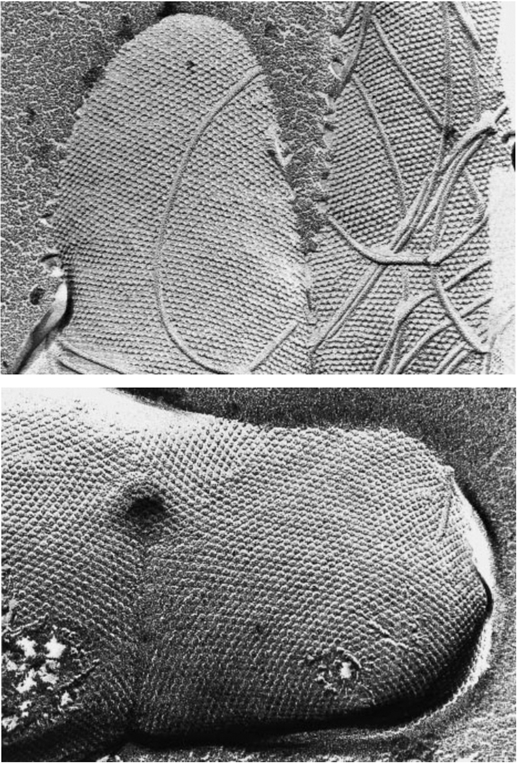
Historically, freeze-etching (FE) electron microscopy of intact cells provided the first compelling evidence that S-layers completely cover the cell surface as a coherent layer and may function as antifouling surfaces (Figure 1). Due to the high cooling rates (approximately 10.000 °C/s) associated with FE preparation, the resulting images are considered to be representative of native in vivo surface characteristics (Sleytr and RobardsReference Sleytr and Robards1977; Robards and Sleytr, Reference Robards and Sleytr1985). From early FE studies of Bacillus, Clostridium, Acinetobacter, Spirillum spp., and selected archaeal extremophiles, S-layer lattices consistently appeared clean and sharply resolved. Remarkably, this was observed even when bacterial or archaeal cells were isolated from sediment-rich soils or macromolecule-dense environments such as decaying organic matter, complex growth media, blood-serum and hot springs (Sleytr, Reference Sleytr1970, Reference Sleytr1978; Sleytr et al., Reference Sleytr, Adam and Klaushofer1968, Reference Sleytr1969a, Reference Sleytr, Adam and Klaushofer1969b, Reference Sleytr1999; Hollaus and Sleytr, Reference Hollaus and Sleytr1972; Beveridge and Murray, Reference Beveridge and Murray1976; Messner et al., Reference Messner, Pum, Sára, Stetter and Sleytr1986a; Pum et al., Reference Pum, Messner and Sleytr1991; Sára and Sleytr, Reference Sára and Sleytr2000). This antifouling effect also notably extended to the bacterial and archaeal flagella. Even when collapsed onto the cell surface in FE-preparations, the helical structure of flagellar filaments and hook regions remained sharply resolved and free of adsorbed debris (Sleytr and Glauert, Reference Sleytr and Glauert1973; Sleytr, Reference Sleytr1978; Sleytr and Pum, Reference Sleytr and Pum2025; see also Figure 1). This observation suggests that maintaining a pristine cell surface is a fundamental biological strategy in chemical and physically complex environments. Mechanistic interpretation of this phenomenon became possible only with the advent of atomic-resolution structural biology, including cryo-electron microscopy (cryo-EM), cryo-electron tomography (cryo-ET), X-ray crystallography, and protein-structure prediction approaches (Figure 2) (Pavkov et al., Reference Pavkov2008; Pavkov-Keller et al., Reference Pavkov-Keller, Howorka and Keller2011; Baranova et al., Reference Baranova2012; Bharat et al., Reference Bharat2015, Bharat et al., Reference Bharat2021; Sogues et al., Reference Sogues2023, Reference Sogues2025; Buhlheller et al., Reference Buhlheller2024, Reference Buhlheller, Sagmeister, Grininger, Gubensäk, Sleytr, Usón and Pavkov-Keller2025; Gambelli et al., Reference Gambelli2024; von Kügelgen et al., Reference Von Kügelgen2024; Isbilir et al., Reference Isbilir2025). Independent experimental evidence for the antifouling properties of S-layers emerged from efforts to engineer isoporous ultrafiltration membranes (UFMs) based on S-layer protein lattices. In the mid-1980s, controlled deposition of S-layers on porous solid supports enabled the fabrication of UFMs with uniform pore size and geometry at macroscopic scale (Sára and Sleytr, Reference Sára and Sleytr1987a, Reference Sára and Sleytr1987b). These membranes exhibited pronounced resistance to non-specific adsorption and clogging, attributed to the molecular smoothness of the crystalline lattice, its precise isoporosity, and the zwitterionic character of the surface. Subsequent studies established S-layer-based UFMs as robust, low-fouling interfaces for biotechnological and biomedical applications (Sára and Sleytr, Reference Sára and Sleytr1987a, Reference Sára and Sleytr1987b; Schuster and Sleytr, Reference Schuster and Sleytr2021).
Why Dynamic Lattice Fluctuations of S-Layers Are Expected In Vivo
-
• Dynamic S-layers are a physical necessity, not a special case: Under physiological conditions, any two-dimensional protein lattice must respond to thermal energy, geometric constraints, and growth. S-layer dynamics therefore emerge as a generic physical consequence of operating a crystalline system on a living, curved, and expanding surface.
-
• Architecture defines mechanical behavior: S-layers are not rigid crystals, but modular assemblies of protein subunits linked by flexible hinges. This reduces the effective stiffness of the lattice and places it in the regime of elastically compliant, thermally active two-dimensional materials. As a result, subunits undergo continuous, low-amplitude fluctuations around their equilibrium positions while long-range order is preserved.
-
• Curvature enforces defect-mediated dynamics: The embedding of a two-dimensional lattice on a curved cell surface introduces unavoidable geometric frustration. Non-zero Gaussian curvature cannot be accommodated without topological defects, making dislocations and disclinations intrinsic, energetically required features. Their presence – and crucially their mobility – generates persistent, localized rearrangements within the lattice.
-
• Growth prevents a static energy minimum: In vivo , S-layers are continuously expanded and reorganized. The incorporation of new subunits at defect sites, followed by lateral redistribution toward lower free-energy configurations, necessitates repeated bond breaking and reformation. Consequently, the lattice exists in a dynamic steady state, sampling an ensemble of closely related configurations rather than occupying a single minimum.
-
• Fluctuations are structured, not disordered: These dynamics do not imply loss of crystallinity. Instead, they represent thermally driven, defect-coupled micro-motions within an ordered lattice. Experimental imaging captures static snapshots of this behavior as nanoscale heterogeneity but does not resolve its temporal dimension.
-
• Coupling to the underlying envelope enables mobility: Dynamic behavior requires a balance between stability and reversibility at the interface with the cell envelope. Whether mediated by SCWPs (Gram-positive bacteria), LPS (Gram-negative bacteria), or fluid archaeal membranes, anchoring interactions must allow transient detachment and lateral redistribution while maintaining continuous surface coverage.
-
• Interfacial consequences: dynamically regulated boundaries: The combination of thermal fluctuations and defect mobility produces a continuously reorganizing interface. This limits the formation of persistent structural gaps and constrains the configurational freedom of approaching macromolecules, contributing to reduced surface residence times.
-
• Hydration as a coupled dynamic field (proof-of-principle): Model systems demonstrate that crystalline S-layer lattices can modulate the structure and mobility of adjacent water layers at nanometer scales. Although obtained under simplified conditions, these results support the concept that lattice dynamics and hydration structure are coupled, potentially preventing the formation of long-lived, kinetically trapped hydration domains.
-
• Take-home message: Dynamic lattice fluctuations are an inevitable outcome of combining thermal motion, elastic architecture, curvature frustration, and growth. S-layers should therefore be understood not as static protective shells but as intrinsically dynamic, physically active interfaces whose functionality – including antifouling – emerges from this steady-state dynamism.
Biophysical Impact of S-Layer Glycosylation.
Building directly on the hydration-dominated interfacial properties of the protein S-layer lattice, glycosylation extends the crystalline lattice into a hydrated glycan phase that governs the cell–environment interface.
This glycan canopy introduces the following synergistic biophysical effects:
-
• Surface property modulation
Glycans alter surface charge, wettability, and the zeta-potential of the S-layer, thereby tuning interactions with environmental macromolecules and the underlying cell envelope.
-
• Hydration-amplified antifouling barrier
A dense network of hydroxyl groups and an increased solvent accessible surface stabilize large hydration shells. Displacement of this structured water imposes a high free energy penalty on nonspecific adsorption. Charged glycan residues further enhance this effect through electrostatic repulsion.
-
• Two functional modes
-
1. Hydrated mode: Glycans behave as entropic polymer brushes governed by steric and hydration forces, maintaining a highly slippery interface.
-
2. Dehydrated mode: Loss of water induces hydrogen-bond-mediated collapse into a cohesive and mechanically compliant shield that protects the cell envelope under osmotic or desiccation stress.
-
• Divalent ion interaction
Charged glycans can serve as reservoirs or diffusion barriers for divalent cations such as Ca2+ and Mg2+, supporting structural integrity of both the S-layer lattice and the lipid membrane in Archaea.
Structural Evidence for S-layer Antifouling.
Atomic-resolution studies confirm that antifouling is a direct consequence of the 2D-crystalline architecture:
-
• Surface hydration: Outward-facing surfaces and pore walls are dominated by polar/charged residues, maximizing the desolvation penalty to oppose non-specific adsorption.
-
• Modular plasticity: Rigid domains connected by flexible hinges enable curvature adaptation via dislocations and disclinations. Atomic models confirm foundational geometric predictions (Messner et al., Reference Messner, Pum, Sára, Stetter and Sleytr1986a; Pum et al., Reference Pum, Messner and Sleytr1991; Sleytr and Pum, Reference Sleytr and Pum2025) that these defects are essential for maintaining a continuous barrier during growth.
-
• Selective nanochannels: Solvent-filled pores act as hydration-gated filters, where transport is governed by size exclusion and charge-mediated hydration shells.
-
• Universal glycan canopies: In many Bacteria and most Archaea , S-layer proteins scaffold dense, flexible glycan chains. This glycosylation extends the hydration-stabilized interface into a dynamic, entropic shield.
Key insight: Antifouling is symmetry-independent; it emerges from the periodic density of hydration-optimizing residues and the lattice’s modular flexibility.
Transmission electron micrographs of freeze-etched preparations of whole cells of Thermoanerobacter thermohydrosulfuricus L111–69 (top) and Methanocorpusculum sinense DSM 4274 (bottom), both exhibiting hexagonal (p6) S-layer lattice symmetry. The cells are partially embedded in ice and display their distinct S-layer lattice structure. The M. sinense cell is captured at an advanced state of cell division (bottom). Numerous wedge disclinations can be seen in the hexagonal lattices. Flagella, collapsed onto the cell surface during cell centrifugation, are visible in T. thermohydrosulfuricus (top). Although the organisms were cultured in complex media, their S-layer lattices are not masked by adhering macromolecules, illustrating the excellent antifouling properties of these surface structures. (Adapted from Sleytr et al., Reference Sleytr1999. Copyright © 1999 Wiley-VCH. Reproduced with permission.)

Figure 1. Long description
A two-panel vertical T E M display.
* The top panel shows a cell of Thermoanerobacter thermohydrosulfuricus L 1 1 1 69. The cell surface is covered in a highly ordered, repeating hexagonal p 6 lattice. Several long, thin, cord-like flagella are draped across the lattice surface, appearing as curved lines. The lattice shows distinct wedge disclinations where the regular pattern of the S-layer is interrupted or shifts orientation.
* The bottom panel shows a cell of Methanocorpusculum sinense D S M 4 2 7 4. This cell also exhibits a prominent p 6 hexagonal lattice symmetry across its rounded surface. The texture appears as a dense field of uniform, microscopic bumps. A few circular depressions or irregularities in the lattice represent wedge disclinations. The cell is partially submerged in a smooth, darker background representing the ice matrix from the freeze-etching process.
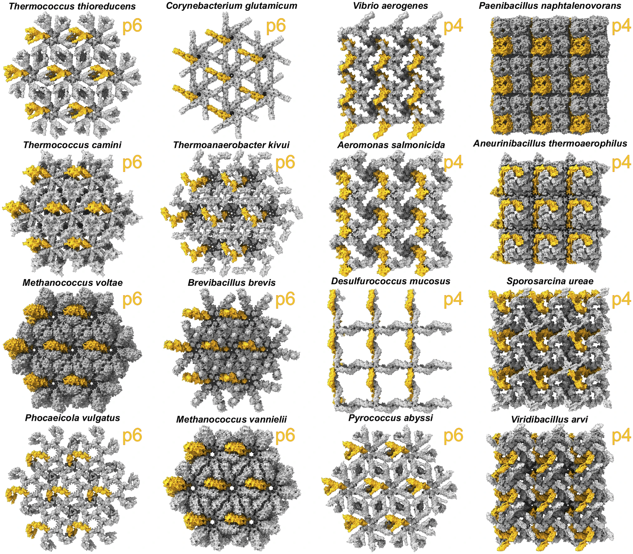
Representative SymProFold calculated assemblies of different S-layers are shown in surface representation from the top side (outside of the cell, facing the environment). One monomer of the assembly unit is shown in orange. (Buhlheller et al., Reference Buhlheller2024; CC BY 4.0 license international (http://creativecommons.org/licenses/by/4.0/)).

Figure 2. Long description
The 16 panels are arranged in a 4 by 4 grid. Each panel displays a gray molecular surface representation of an S-layer assembly with one monomer highlighted in orange. A yellow label in the top-right of each panel indicates the symmetry type, either p 4 or p 6.
Row 1 from left to right
* Thermococcus thioreducens: p 6 symmetry with a dense hexagonal lattice.
* Corynebacterium glutamicum: p 6 symmetry with a thin, spindly hexagonal lattice.
* Vibrio aerogenes: p 4 symmetry with a diagonal woven appearance.
* Paenibacillus naphtalenovorans: p 4 symmetry with a dense, block-like square lattice.
Row 2 from left to right
* Thermococcus camini: p 6 symmetry with a thick, interlocking hexagonal structure.
* Thermoanaerobacter kivui: p 6 symmetry with a complex, radiating hexagonal pattern.
* Aeromonas salmonicida: p 4 symmetry with an open, cross-hatched square lattice.
* Aneurinibacillus thermoaerophilus: p 4 symmetry with a dense grid of rounded subunits.
Row 3 from left to right
* Methanococcus voltae: p 6 symmetry with a very dense, almost solid hexagonal surface.
* Brevibacillus brevis: p 6 symmetry with a star-shaped hexagonal lattice.
* Desulfurococcus mucosus: p 4 symmetry with a very thin, open square grid.
* Sporosarcina ureae: p 4 symmetry with a dense, overlapping square lattice.
Row 4 from left to right
* Phocaeicola vulgatus: p 6 symmetry with a delicate, floral hexagonal pattern.
* Methanococcus vannielii: p 6 symmetry with a dense hexagonal arrangement of large subunits.
* Pyrococcus abyssi: p 6 symmetry with an open hexagonal lattice of triangular subunits.
* Viridibacillus arvi: p 4 symmetry with a dense, textured square lattice.
Historically, these recurring observations were repeatedly noted throughout S-layer literature but were never elevated to a unifying functional principle. The consistent appearance of remarkably clean S-layer surfaces in ultrastructural FE studies (Figure 1) (Sleytr and Glauert, Reference Sleytr and Glauert1973; Sleytr, Reference Sleytr1978; Sleytr et al., Reference Sleytr1988; Pum et al., Reference Pum, Messner and Sleytr1991; Sleytr et al., Reference Sleytr1999) and the unexpectedly low fouling behavior reported for early S-layer-based UFMs (Sleytr and Sára, Reference Sleytr and Sára1986; Sára and Sleytr, Reference Sára and Sleytr1987a, Reference Sára and Sleytr1987b) were typically described as striking properties rather than interpreted as manifestations of a fundamental biological strategy. Even later comprehensive reviews emphasized the exceptional resistance of S-layer lattices to unspecific adsorption without placing it into a broader mechanistic context (Sleytr et al., Reference Sleytr2014; Sleytr and Pum, Reference Sleytr and Pum2025; Sleytr and Schuster, Reference Sleytr and Schuster2026). As a consequence, the idea that resistance to non-specific adsorption might represent a central evolutionary function of S-layers remained implicit for decades.
In this review, we explicitly revisit these long-standing observations and integrate them into a coherent mechanistic and evolutionary framework. By combining ultrastructural evidence with atomic-resolution structural data and biophysical principles, we argue that S-layers generate a globally non-adhesive surface while enabling spatially restricted, specific interactions at the prokaryotic interface. We propose that this inherent antifouling capacity represents a pivotal evolutionary trait, granting microorganisms a decisive survival advantage by preventing the non-specific adsorption of macromolecules and debris. This advantage becomes particularly evident when considering that prokaryotic life operates exclusively within a low Reynolds number regime (see Section “Life at low Reynolds numbers,” Box 4).
Physics at the Interface: Life at Low Reynolds Numbers.
Microbial life operates in a hydrodynamic regime where viscous forces dominate inertia (see also https://en.wikipedia.org/wiki/Reynolds_number). At the scale of a single cell (~1 um), the Reynolds number (Re) typically ranges from 10−5 to 10−3. In this regime, fluid motion is laminar and kinematically reversible, a ‘sticky’ world where the ‘scallop theorem’ dictates that reciprocal motion yields no net progress and surfaces are encased in stagnant boundary layers (Purcell, Reference Purcell1977).
-
1. The diffusion constraint
At the micrometer scale, the cell–fluid interface constitutes a fundamental transport bottleneck. Because convection is negligible at the cell surface, nutrient uptake and metabolite export depend entirely on diffusion. The flux (J) of molecules to the surface is governed by Fick’s first law:

where D is the diffusion coefficient and
$ \frac{\mathrm{d}C}{\mathrm{d}x} $
 is the concentration gradient. In the absence of convective renewal, these gradients are established over very short distances and are highly sensitive to the physicochemical properties of the outermost surface. Any ‘fouling’, the non-specific adsorption of environmental macromolecules, increases the effective diffusive path length and reduces the gradient, potentially starving the cell.
-
2. S-layers as optimized physicochemical filters
S-layers (glyco)protein lattices form a structural solution to these physical constraints by maintaining a ‘clean’ interface:
-
• Antifouling properties: The molecularly smooth, highly ordered, and strongly hydrated lattice suppresses non-specific adsorption. This prevents the accumulation of debris that would otherwise thicken the stagnant boundary layer.
-
• Size-selective permeability: Regular nanopores (2–10 nm) ensure unhindered diffusion of small solutes while maintaining a defined molecular weight cutoff, excluding larger molecules that contribute to fouling.
-
• Interface definition: By masking the heterogeneities of underlying envelope components (e.g., peptidoglycan or LPS), S-layers impose a spatially uniform boundary condition, minimizing local ‘stickiness’ and micro-scale turbulence.
-
3. Functional implication
In a regime where transport is set at the interface and cannot be restored by flow, maintaining a permeable surface is a physical requirement for sustained metabolism. S-layers represent a minimal, robust evolution to life under diffusion-limited conditions. Their occurrence across diverse prokaryotic lineages reflects a universal strategy to preserve an open diffusion interface in a hydrodynamic environment where surface fouling would otherwise be prohibitive.
Lattice dynamics
S-layers have long been described as rigid, crystalline surface coats; however, accumulating evidence suggests that their defining property lies not in static order but in regulated dynamical behavior. Early digital image analyses of electron micrographs of negatively stained preparations revealed systematic lattice distortions incompatible with a brittle crystalline architecture (Crowther and Sleytr, Reference Crowther and Sleytr1977). Freeze-etching studies of vitrified intact cells further showed that S-layers maintain long-range order while conforming to highly curved geometries, including hemispherical cell poles, despite substantial mechanical strain during growth and division (Sleytr and Glauert, Reference Sleytr and Glauert1975; Sleytr, Reference Sleytr1978; Sleytr et al., Reference Sleytr1999; Pum et al., Reference Pum2021). These observations are consistent with a model in which S-layers behave as curvature-adaptive crystalline lattices governed by the interplay of elasticity, topology, and thermally driven dynamics.
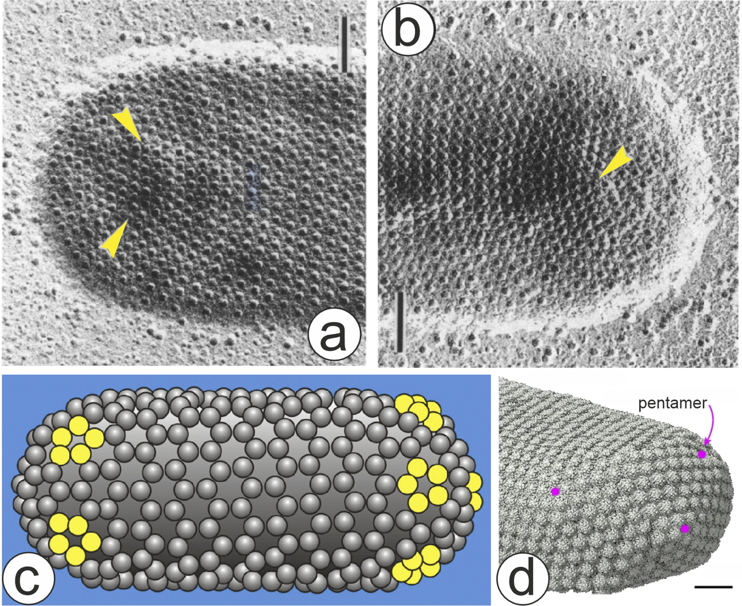
Within the framework of two-dimensional crystals constrained to curved manifolds, a fundamental geometrical constraint is that long-range order cannot be maintained on surfaces of non-zero Gaussian curvature without the introduction of topological defects. Consistent with this requirement, S-layers accommodate complex cellular geometries through dislocations and disclinations while preserving lattice coherence (Figure 3) (Harris and Scriven, Reference Harris and Scriven1970; Messner et al., Reference Messner, Pum and Sleytr1986b; Gruber and Sleytr, Reference Gruber and Sleytr1988; Pum et al., Reference Pum, Messner and Sleytr1991). This curvature tolerance is further supported by recrystallization experiments, in which isolated S-layer subunits self-assemble into ordered lattices on substrates with small radii of curvature, including carbon nanotubes, liposomes, emulsomes, and magnetic beads (Küpcü et al., Reference Küpcü1995; Ücisik et al., Reference Ücisik2013; Breitwieser et al., Reference Breitwieser2016, Reference Breitwieser2019; Sleytr and Pum, Reference Sleytr and Pum2025), as well as by the spontaneous formation of highly curved cylindrical assemblies in vitro (Sleytr and Plohberger, Reference Sleytr, Plohberger, Baumeister and Vogell1980; Messner et al., Reference Messner, Pum and Sleytr1986b; Pum et al., Reference Pum2021). In cylindrical regions of rod-shaped cells, lattices often retain a defined orientation relative to the longitudinal axis, indicating persistent curvature-induced elastic strain (Sleytr and Glauert, Reference Sleytr and Glauert1975; Gruber and Sleytr, Reference Gruber and Sleytr1988; Sleytr and Pum, Reference Sleytr and Pum2025).
Electron micrograph of freeze-dried and shadowed preparations of envelopes of Thermoproteus tenax labelled with polycationic ferritin. The marker binds to negatively charged domains on the extracellular surface of the S-layer in a regular fashion. Depending on the orientation of the cell during adsorption, two (a) or one (b) local wedge disclinations (‘pentamers’) can be seen in the hexagonal lattice (arrows). Scale bar, 500 nm. (c) Schematic drawing of the minimum number of pentagons required at the hemispherical caps as revealed in (a, b). (Modified after Messner et al., Reference Messner, Pum, Sára, Stetter and Sleytr1986a, with permission.) (d) Molecular structure of the Nitrosopumilus maritimus S-layer in intact cells. Map of the subtomogram positions in the cellular S-layer showing the presence of pentameric defects (pink) at the edge of the cell. Scale bar, 50 nm. (von Kügelgen et al., Reference Von Kügelgen2024; CC BY license (http://creativecommons.org/licenses/by/4.0/)).

Figure 3. Long description
A four-panel figure labeled a through d.
Panel a: An electron micrograph of a Thermoproteus tenax cell envelope. The surface is covered in a regular hexagonal lattice of small dark dots. Two yellow arrowheads point to specific regions where the hexagonal pattern is interrupted by wedge disclinations. A vertical scale bar representing 500 nanometers is in the top right.
Panel b: A similar electron micrograph showing a different orientation of a cell. A single yellow arrowhead points to a pentameric defect in the lattice. A vertical scale bar is in the bottom left.
Panel c: A schematic drawing of a rod-shaped cell composed of gray spheres. At the hemispherical caps on both ends, specific clusters of spheres are colored yellow to represent the minimum number of pentagons required to close the hexagonal lattice at the poles.
Panel d: A three-dimensional molecular map of the Nitrosopumilus maritimus S-layer. The surface is a dense, textured gray lattice. Three pink dots mark the positions of pentameric defects at the curved edge of the cell. A pink arrow labeled pentamer points to one of these dots. A horizontal scale bar representing 50 nanometers is in the bottom right.
S-layer reassembly on curved synthetic supports provides a controllable platform to interrogate how surface geometry modulates nucleation pathways, growth kinetics, and elastic accommodation. Assembly proceeds via an induction period followed by cooperative lattice expansion, with rates set by protein concentration, substrate chemistry, pH, ionic strength, and the availability of divalent cations such as Ca2+ (Pum et al., Reference Pum2021; Sleytr and Pum, Reference Sleytr and Pum2025).
Curvature reshapes the local free-energy landscape for protein subunit addition and step propagation, biases orientation selection, and regulates the creation, mobility, and annihilation of defects during growth, thereby coupling elastic fields to kinetic processes (Tscheliessnig et al., Reference Tscheliessnig2021). These effects manifest as curvature-dependent nucleation densities, domain coarsening rates, and orientational correlation lengths. Substrate compliance further tunes the accommodation pathway. On deformable interfaces, strain can be partially dissipated by substrate remodeling, whereas on rigid supports it is relaxed primarily through defect transport and lattice reorganization. Time-dependent structural recovery after perturbation reflects the reversibility of inter-subunit bonds and the dynamic rearrangement of defect populations within the lattice.
In hexagonal lattices, curvature can be accommodated through pentameric and heptameric coordination units, as observed in Methanocorpusculum sinense (Figure 1) (Pum et al., Reference Pum, Messner and Sleytr1991), Thermoproteus tenax (Figure 3) and Thermoproteus neutrophilus (Messner et al., Reference Messner, Pum, Sára, Stetter and Sleytr1986a), and more recently by using electron cryotomography and subtomogram averaging from Nitrosopumilus maritimus cells (Figure 3) (von Kügelgen et al., Reference Von Kügelgen2024). Comparable defect structures in square lattices support the generality of defect-mediated adaptation (Sleytr and Glauert, Reference Sleytr and Glauert1975). These observations are consistent with a description of S-layers as curvature-frustrated two-dimensional crystals, in which defects act as intrinsic degrees of freedom that relax elastic stress and minimize free-energy costs.
At the molecular level, S-layer lattices are composed of relatively rigid protein subunits interconnected by flexible hinges (Horejs et al., Reference Horejs2011a, Reference Horejs2011b; Tscheliessnig et al., Reference Tscheliessnig2021; Foo et al., Reference Foo2025). This architecture reduces the effective elastic modulus and enables thermally driven nanoscale fluctuations around equilibrium positions. Such fluctuations facilitate repeated breaking and reformation of inter-subunit contacts required for lattice expansion, cell division, and the formation of ordered arrays at newly generated cell poles (Sleytr et al., Reference Sleytr1999; Pum et al., Reference Pum2021; Sleytr and Pum, Reference Sleytr and Pum2025). Long-range order is thus maintained in the presence of continuous, defect-coupled micro-motions, placing S-layers in the regime of elastically compliant, thermally active crystalline materials.
Dynamic behavior is further modulated by coupling to the underlying cell envelope. In Gram-positive bacteria, S-layer proteins bind to secondary cell wall polymers (SCWPs) (Sára, Reference Sára2001; Ferner-Ortner et al., Reference Ferner-Ortner2007; Messner et al., Reference Messner2009), whereas in Gram-negative organisms anchoring occurs via lipopolysaccharides (LPS) of the outer membrane (Nomellini et al., Reference Nomellini1997). In most Archaea, the plasma membrane provides a laterally fluid support (Albers and Meyer, Reference Albers and Meyer2011; Sleytr and Pum, Reference Sleytr and Pum2025). Despite these differences, anchoring can be described in terms of thermally driven on–off binding equilibria that permit continuous lattice remodeling. Recrystallization on planar and vesicular lipid membranes, including tetraether systems, provides experimental support for this dynamic coupling (Györvary et al., Reference Györvary1999; Hirn et al., Reference Hirn1999; Schuster et al., Reference Schuster2003; Schuster and Sleytr, Reference Schuster and Sleytr2014; Ücisik et al., Reference Ücisik2015a). In membrane-associated systems, the lattice may therefore behave as a quasi-two-dimensional crystalline sheet on a viscoelastic substrate, combining lateral mobility with orientational order.
Cell growth and division impose additional constraints that drive the system away from equilibrium. Newly synthesized subunits are preferentially incorporated at defects, particularly gliding dislocations, which act as growth seams (Harris and Scriven, Reference Harris and Scriven1970; Sleytr and Glauert, Reference Sleytr and Glauert1975; Gruber and Sleytr, Reference Gruber and Sleytr1988; Sleytr and Pum, Reference Sleytr and Pum2025). These sites enable insertion without loss of global order, followed by relaxation processes that redistribute strain and reduce free energy. The lattice is therefore better described as a dynamic steady state than as a single energy minimum, continuously sampling a set of closely related configurations.
The consequences of this defect-mediated dynamics extend to interfacial properties. Continuous lattice reorganization maintains topological closure and limits the persistence of nanoscale gaps that could act as nucleation sites for nonspecific adsorption. At the same time, thermal fluctuations generate a temporally varying interfacial energy landscape. For approaching macromolecules, this dynamic environment can restrict accessible conformational states, leading to an unfavorable entropic contribution (ΔS < 0) upon confinement and, under appropriate conditions, to effective repulsive interactions.
In addition, model and computational studies indicate that crystalline S-layer lattices can modulate the structure and dynamics of adjacent interfacial water over nanometer length scales, with local variations linked to lattice rigidity and charge distribution (Tscheliessnig et al., Reference Tscheliessnig2021). Although obtained under simplified in vitro conditions, these findings support a coupling between lattice organization and hydration dynamics that may reduce the formation of kinetically trapped adsorption states.
Taken together, these observations support a physical picture in which S-layers function as mechanically active, non-equilibrium interfaces. Their properties emerge from the interplay of curvature-induced frustration, defect dynamics, elasticity, thermal fluctuations, and interfacial coupling. In this framework, S-layer lattices in vivo can be understood as curvature-frustrated, defect-mediated two-dimensional crystals operating out of equilibrium, whose intrinsic dynamics contribute directly to their biological function.
Antifouling mechanisms of S-layer protein surfaces
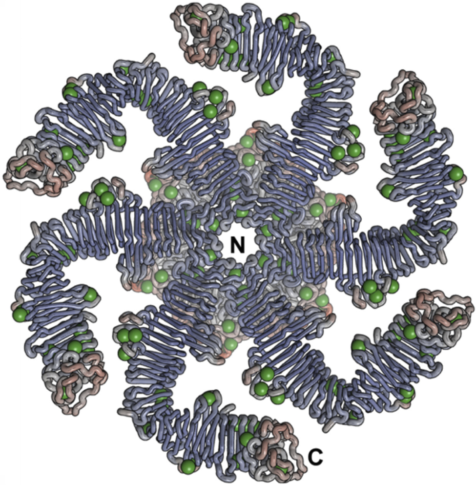
Atomic-resolution structural analyses reveal that the outward-facing surfaces of S-layer protein lattices constitute some of the most strongly hydrated biological interfaces known. Hydrophobic residues are largely sequestered within protomer cores or buried at lattice junctions, whereas the exposed surface is dominated by serine, threonine, asparagine, acidic residues, and other polar or charged side chains. These residues form dense, spatially organized hydrogen-bonding networks that stabilize tightly bound hydration shells. These shells are frequently reinforced by divalent cations, most notably Ca2+ (Figure 4). The latter not only increases the residence time and structural ordering of interfacial water but are also essential for lattice integrity by screening repulsive electrostatic interactions between adjacent protomers and enabling precise self-assembly (Bharat et al., Reference Bharat2017; Herdman et al., Reference Herdman2022). In this context, it is worth noting that Fe2+ has been shown to act as a self-assembly inducer under anaerobic conditions, which are thought to resemble the early stages of biological evolution (Iturri et al., Reference Iturri2018).
Example of ion-binding on the S-layer protein of Caulobacter crescentus as illustrated by the RsaA hexamer protein structure with bound Ca2+ ions depicted as green spheres. (Herdman et al., Reference Herdman2022; CC BY license (http://creativecommons.org/licenses/by/4.0/)).

Figure 4. Long description
At the center of the hexameric structure is a small circular pore labeled with a capital N. Surrounding this central pore is a dense core of intertwined protein chains in shades of light blue and pale pink. Moving outward from this core, six distinct arms radiate in a pinwheel fashion. Each arm is composed of tightly packed, parallel beta-sheet strands that form a curved, elongated shape. Numerous small green spheres, representing calcium ions, are embedded along the inner and outer edges of these arms, particularly concentrated at the junctions where the arms meet the central core and at the distal ends. The outermost tip of the bottom-right arm is labeled with a capital C. The overall symmetry is six-fold, with each arm displaying a similar pattern of protein folding and ion binding sites.
Any macromolecule approaching the S-layer protein lattice must first disrupt and partially displace this structured hydration layer, incurring a substantial desolvation penalty that renders nonspecific adsorption thermodynamically unfavorable (Nakasako, Reference Nakasako2004; Chen et al., Reference Chen2010; Del Grosso et al., Reference Del Grosso2020). High-resolution three-dimensional frequency-modulation atomic force microscopy has directly visualized ordered, non-bulk-like water layers on protein surfaces, demonstrating that hydration shells at biomolecular interfaces are spatially structured rather than diffuse (Kimura et al., Reference Kimura2010; Ido et al., Reference Ido2022). Consistently, molecular-dynamics simulations and ultrafast spectroscopic measurements show that water molecules in protein hydration shells exhibit markedly retarded dynamics, with dominant residence and relaxation times ranging from a few to several tens of picoseconds, while more strongly bound subpopulations can reach hundreds of picoseconds depending on local surface chemistry and topology (Qin et al., Reference Qin2016; Laage et al., Reference Laage2017).
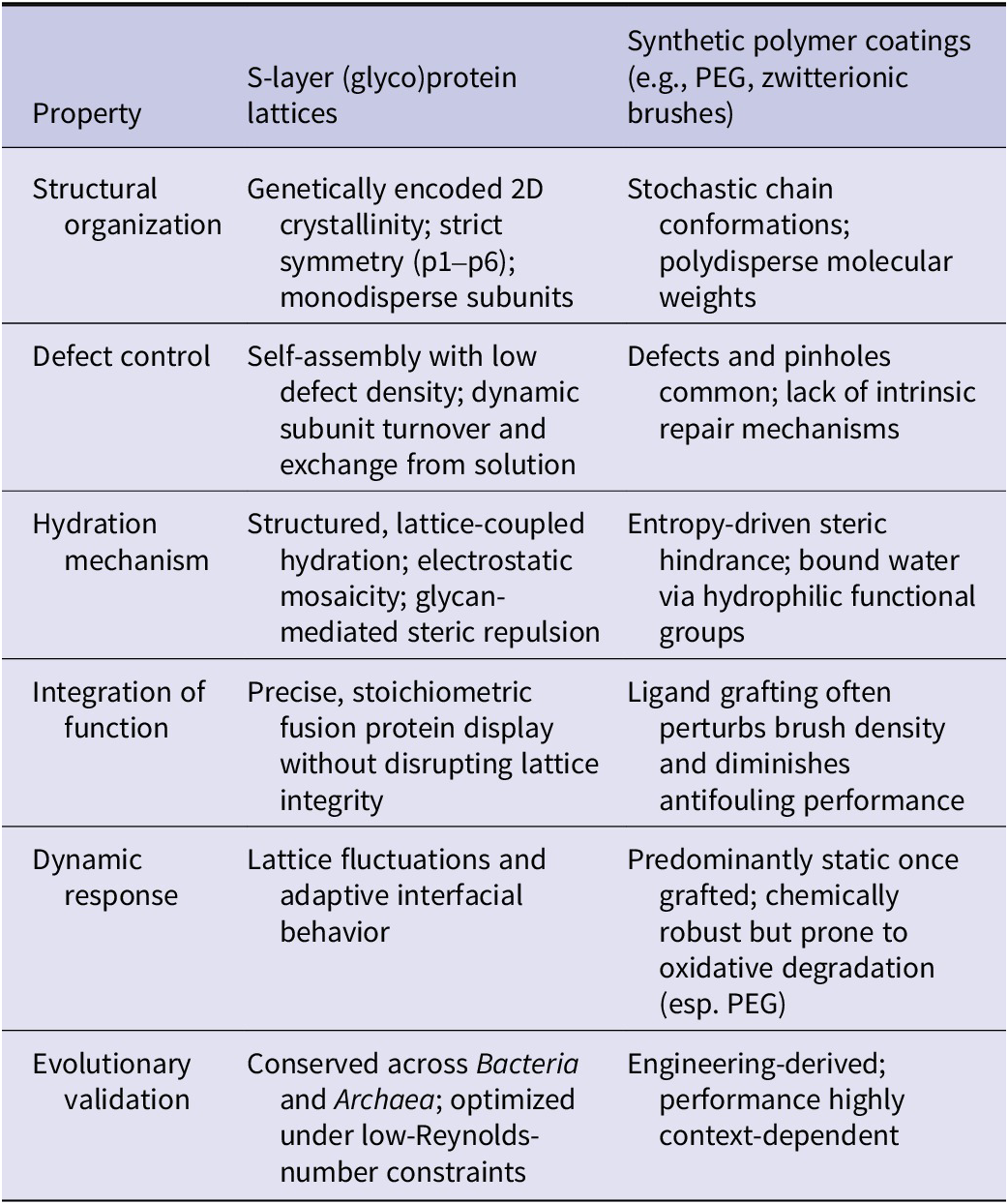
This hydration-mediated antifouling barrier is reflected in the unusually high solvent-accessible surface area (SASA) of the outward-facing S-layer lattice. In protein-only S-layers, SASA is maximized for polar and charged residues, while hydrophobic exposure is minimized. Because of the energetic cost of dehydration scales with the stability of bound interfacial water, hydrophilic SASA can be regarded as a thermodynamic proxy for antifouling strength. Atomistic simulations explicitly show that regions of high polar SASA coincide with slowly exchanging, tightly bound hydration water, thereby directly linking surface chemistry to the free-energy barrier opposing adsorption (Laage et al., Reference Laage2017). In this respect, S-layers function as biological analogues of synthetic zwitterionic or highly hydrophilic polymer coatings, in which tightly bound hydration layers suppress protein and cell adhesion through the same fundamental thermodynamic principle (Shao and Jiang, Reference Shao and Jiang2015; Moayedi et al., Reference Moayedi2024). Experimental model studies further indicate that crystalline S-layer lattices can measurably influence the organization and dynamic behavior of interfacial water layers at the nanometer scale (Tscheliessnig et al., Reference Tscheliessnig2021). While these observations were obtained under simplified in vitro conditions and do not imply classical hydrodynamic turbulence, they support the concept that periodically structured protein lattices can actively modulate hydration-layer dynamics. Such modulation is expected to influence the kinetic landscape of macromolecular adsorption by preventing the formation of long-lived, highly structured hydration domains that may otherwise facilitate nonspecific surface residence. In this context, hydration-mediated effects should be regarded as contributing factors that modulate adsorption kinetics rather than as single dominant antifouling mechanisms (Ball, Reference Ball2008; Israelachvili and Wennerström, Reference Israelachvili and Wennerström1996). Thus, the antifouling properties of S-layer surfaces most likely emerge from the combined action of surface topology, electrostatic heterogeneity, hydration structuring, and lattice dynamics.
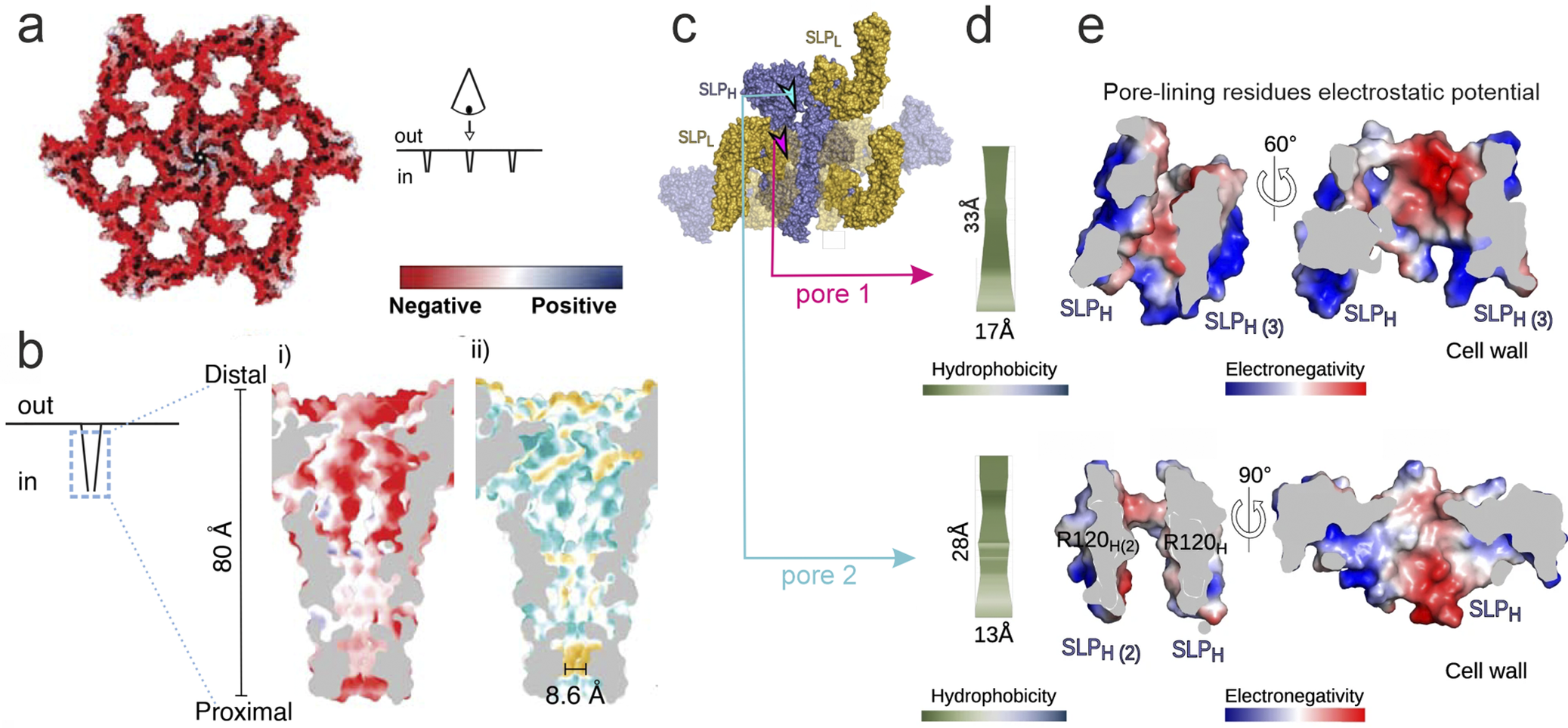
An additional, closely coupled parameter governing antifouling behavior is the net surface charge of the S-layer and its relation to the isoelectric point (pI) of the lattice-forming protein. Many bacterial S-layer proteins exhibit pI values close to neutrality, resulting in weak net surface charges at physiological pH and minimizing long-range electrostatic attraction to proteins or cells. However, several phylogenetically and ecologically distinct organisms, notably numerous Lactobacillus species, Corynebacterium glutamicum, and many Archaea, possess S-layer proteins with low pI values and consequently a pronounced negative net surface charge under their native environmental conditions (Figure 5a) (Buck et al., Reference Buck2005; Hynönen and Palva, Reference Hynönen and Palva2013; Gerbino et al., Reference Gerbino2015; Kong et al., Reference Kong2022; Sagmeister et al., Reference Sagmeister2024; Sogues et al., Reference Sogues2025). Differences in S-layer surface net charges, correlating with the optimal environmental pH, were documented early on in T. tenax (Figure 3) and T. neutrophilus (Messner et al., Reference Messner, Pum, Sára, Stetter and Sleytr1986a). Importantly, this feature does not compromise antifouling performance but instead reflects adaptation to acidic or otherwise extreme habitats.
(a, b) The S-layer of Corynebacterium glutamicum is negatively charged. (a) Extracellular view shows the S-layer colored according to its charge distribution (see indication bar). (b) Cross-section of the large pore with the lumen colored according to charge distribution (i) positive and negative charges as indicated by the bar and hydrophobicity (ii) from hydrophobic regions in yellow to hydrophilic regions in blue. The narrowest constriction of the pore is indicated. (Sogues et al., Reference Sogues2025; Creative Commons Attribution License 4.0 (CC BY)). (c–e) Pores in S-layer from Clostridioides difficile. (c) Surface representation of wild-type S-layer protein (SLP) SLPH/SLPL-complex showing pores in the 3D crystal lattice. Positions of pores marked with arrowheads. (d) Calculated hydrophobicity characteristics of the residues lining in pores 1 and 2 ranging from hydrophilic (green) to hydrophobic (blue), as per the hydrophobicity gradient key. (e) Poisson–Boltzmann electrostatic potential calculated for residues lining in pores 1 and 2 in SLPH/SLPL-complex represented as a charge distribution (positive in blue and negative in red, as per electronegativity gradient key). Arginine residues at the bottleneck of pore 2 are indicated. SLPH: SLP with high molecular weight; SLPL: SLP with low molecular weight. (Lanzoni-Mangutchi et al., Reference Lanzoni-Mangutchi2022; Creative Commons Attribution License 4.0 (CC BY; http://creativecommons.org/licenses/by/4.0/)).

Figure 5. Long description
Panel a shows a top-down extracellular view of a hexagonal S-layer lattice colored red for negative charge. A schematic to the right shows the ‘out’ to ‘in’ orientation. Panel b provides a cross-section of a pore with a vertical scale of 80 Angstroms from distal to proximal. Sub-panel i shows a red-dominant negative charge distribution. Sub-panel ii shows hydrophobicity with yellow hydrophobic regions and blue hydrophilic regions, noting a narrow constriction of 8.6 Angstroms. Panel c displays a surface representation of the S L P H and S L P L complex with arrowheads marking pore 1 in pink and pore 2 in light blue. Panel d shows the calculated hydrophobicity gradients for both pores. Pore 1 has a length of 33 Angstroms and width of 17 Angstroms. Pore 2 has a length of 28 Angstroms and width of 13 Angstroms. Panel e illustrates the Poisson-Boltzmann electrostatic potential for the residues lining pores 1 and 2. Pore 1 is shown at 0 and 60 degree rotations. Pore 2 is shown at 0 and 90 degree rotations, specifically labeling Arginine residues R 1 2 0 H and R 1 2 0 H sub 2 at the bottleneck. Color keys indicate negative/electronegativity in red and positive in blue.
In low-pH environments, such as fermented food matrices, the gastrointestinal tract, or acidothermal niches occupied by Archaea, a surplus of acidic residues within the S-layer contributes to proton buffering, cation binding, and lattice stabilization, while still supporting strong surface hydration. Under these conditions, electrostatic repulsion of approaching macromolecules is complemented by enhanced hydration forces, and antifouling efficacy emerges from the combined effects of charge density, water structuring, and desolvation energetics rather than from charge neutrality alone (Avall-Jääskeläinen and Palva, Reference Avall-Jääskeläinen and Palva2005; Albers and Meyer, Reference Albers and Meyer2011; Hynönen and Palva, Reference Hynönen and Palva2013). Crucially, even in S-layers with an overall negative net charge, antifouling efficacy is enhanced by pronounced electrostatic mosaicity, a high-frequency nanoscale patchwork of positively and negatively charged domains that limits long-range Coulombic interactions while preserving strong local hydration. This spatial decoupling of net charge from local charge distribution weakens stable adsorption geometries and increases the free-energy penalty for dehydration at the interface. These principles extend into the lattice pores, where structural and biochemical analyses reveal that pore walls are enriched in polar and charged residues (Figure 5b,e). The resulting hydrated nanochannels permit rapid diffusion of small solutes while effectively excluding larger or adhesive macromolecules, thereby combining permeability with antifouling selectivity (Sára and Sleytr, Reference Sára and Sleytr1987a, Reference Sára and Sleytr1987b; Sleytr and Sára, Reference Sleytr and Sára1997; Schuster and Sleytr, Reference Schuster and Sleytr2021; Tscheliessnig et al., Reference Tscheliessnig2021).
In glycosylated S-layers, outward-projecting glycans further amplify these hydration-driven effects by forming polymer-like, highly hydrated brushes that sterically and entropically repel potential foulants (Figure 6a,b). Notably, glycosylation frequently co-occurs with low-pI, negatively charged S-layer proteins in acid-adapted Bacteria and Archaea, suggesting a synergistic optimization of charge regulation and hydration under extreme environmental conditions. The specific contributions of glycan chemistry, dynamics, and environmental responsiveness to antifouling behavior are addressed in detail in Section “Biophysical properties of S-layer glycosylation.”
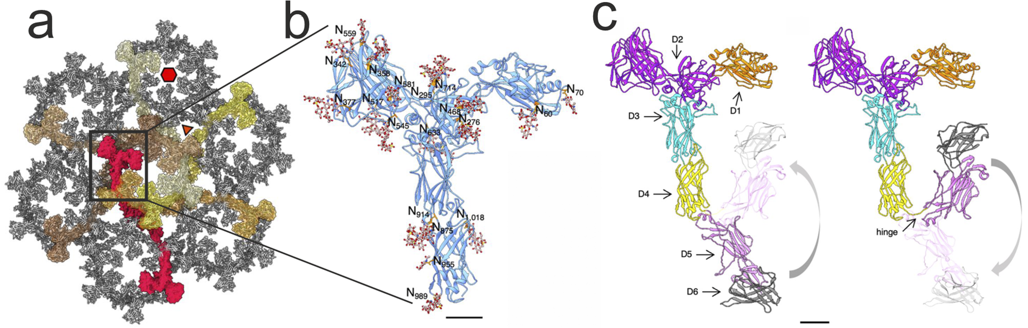
(a) Extracellular view of the S-layer of Sulfolobus acidocaldarius . The assembled SlaAis shown in ribbon representation with SlaA dimers forming a hexagonal pore highlighted in shades of red and yellow. Each dimer spans two adjacent hexagonal pores. (b) Zoom of the boxed region: Example of S-layer protein glycosylation: N-glycosylation of S. acidocaldarius SlaA. Atomic model of SlaA in ribbon representation. N-terminal SlaA30–1069 as solved by cryoEM is in cornflower blue. 19 Asn-bound N-glycans were modelled into the cryoEM map of SlaA30–1069 (glycans rusty brown sticks, Asn in orange). In the glycans, O atoms are shown in red, N in blue, and S in yellow. (c) SlaA atomic models highlighting the six domains. D5 and D6 were predicted using Alphafold. A flexible hinge exists between D4 and D5. D5 and D6 are thus free to move relative to D1–D4 from a stretched to a flapped conformation and back. Scale bars, 20 Å. (Gambelli et al., Reference Gambelli2024; CC BY 4.0 license (http://creativecommons.org/licenses/by/4.0/)).

Figure 6. Long description
Panel a shows a top-down extracellular view of the S-layer lattice. It is a porous hexagonal assembly of SlaA proteins rendered in gray. A single hexagonal pore is highlighted with SlaA dimers in shades of red and yellow. A black box indicates a region of interest for magnification. A red hexagon and a white triangle mark specific symmetry points. A scale bar indicates 20 Angstroms.
Panel b is a zoomed-in view of the boxed region showing the atomic model of SlaA 30 to 1069 in cornflower blue ribbon representation. Rusty brown stick models represent 19 N-glycans attached to orange Asn residues. Labels indicate specific glycosylation sites including N 70, N 89, N 276, N 295, N 342, N 358, N 377, N 468, N 545, N 559, N 581, N 617, N 633, N 712, N 875, N 914, N 955, N 989, and N 1,018. A scale bar indicates 20 Angstroms.
Panel c displays two states of the SlaA atomic model to illustrate conformational flexibility. The protein is divided into six colored domains: D 1 in orange, D 2 in purple, D 3 in cyan, D 4 in yellow, D 5 in pink, and D 6 in dark gray. Arrows point to each domain. A hinge is labeled between D 4 and D 5. Curved gray arrows indicate the movement of the D 5 and D 6 domains from a stretched vertical position to a folded flapped conformation. A scale bar indicates 20 Angstroms.
Biophysical properties of S-layer glycosylation
Extending the interfacial hydration and antifouling principles established for the protein S-layer lattice in the previous chapter, many S-layers further suppress nonspecific adsorption through covalent glycosylation. In this context, glycosylation is not an isolated modification but a direct continuation of the same design principle. It extends the hydration-optimized crystalline protein lattice into a highly hydrated glycan phase that dominates the cell–environment interface (Schuster and Sleytr, Reference Schuster and Sleytr2015; Schäffer and Messner, Reference Schäffer and Messner2017).
S-layer glycoproteins were first identified by chemical analyses in the mid-1970s (Sleytr, Reference Sleytr1975; Mescher and Strominger, Reference Mescher and Strominger1976; Sleytr and Thorne, Reference Sleytr and Thorne1976). Subsequent biochemical and structural studies established glycosylation as a widespread and evolutionarily conserved feature of S-layer proteins (Messner et al., Reference Messner, König and ClausHand Varma2010; Ristl et al., Reference Ristl2011; Schäffer and Messner, Reference Schäffer and Messner2017), particularly in Archaea (Albers and Meyer, Reference Albers and Meyer2011; Calo et al., Reference Calo, Guan and Eichler2011; Guan et al., Reference Guan2016; Zaretsky et al., Reference Zaretsky2019), where glycoprotein S-layers frequently constitute the sole cell wall component and integrate mechanical stability, selective permeability, and surface protection (Eichler, Reference Eichler2003, Reference Eichler2013; Sleytr et al., Reference Sleytr2007, Reference Sleytr2014; Messner et al., Reference Messner, König and ClausHand Varma2010; Albers and Meyer, Reference Albers and Meyer2011; Jarrell et al., Reference Jarrell2014; Kandiba and Eichler, Reference Kandiba and Eichler2014; Eichler and Koomey, Reference Eichler and Koomey2017).
Carbohydrate chains are covalently attached to defined amino acids and project into the extracellular space, where they form a dense and highly hydrated glycan canopy that overlays and further extends the protein lattice into the surrounding medium (Figure 6b). In certain Gram-positive bacteria, these glycans extend up to ~40 nm from the lattice (Christian et al., Reference Christian1988; Sára et al., Reference Sára1989). At this length scale, the glycans behave similarly to polymer brushes, where steric effects and hydration forces dominate antifouling interactions. These carbohydrate chains may also carry charged groups, as observed in extremophiles such as Sulfolobus acidocaldarius , which further strengthens hydration and introduces electrostatic repulsion of fouling macromolecules (Guan et al., Reference Guan2016; Gambelli et al., Reference Gambelli2024). Freeze-etch electron microscopy reveals the dynamic nature of these carbohydrate chains. The persistence of lattice features in replicas indicates that the glycans collapse during sublimation, confirming that their structure depends on hydration rather than rigidity (Figure 1).
The biophysical effect of glycosylation arises from the same principles described for the protein lattice, namely hydration thermodynamics and the structuring of interfacial water, but is amplified by steric effects typical of polymers and, where present, by electrostatic contributions. The dense network of hydroxyl groups stabilizes extensive shells of structured and bound water, while the increase in solvent-accessible surface area expands the interfacial volume that can retain water (Schuster and Sleytr, Reference Schuster and Sleytr2015). Because the free energy penalty for nonspecific adsorption scales with both the stability and the volume of this interfacial water, glycosylation, including the contribution of charged glycans, substantially strengthens the antifouling barrier beyond that of the protein lattice alone. Consistent with this view, studies integrating cryo-EM tomography and small-angle X-ray scattering with continuum modeling have shown that recrystallized S-layer lattices can modulate the flow and electrostatics of adjacent water within the 10–50 nm regime, indicating that ordered S-layer architectures influence interfacial hydration dynamics (Tscheliessnig et al., Reference Tscheliessnig2021). Although these investigations did not isolate glycosylation effects and relied on simplified, model reconstructions, they support the broader concept that periodically organized outer cell surfaces actively shape the structure and dynamics of near-surface water (Tscheliessnig et al., Reference Tscheliessnig2021). At the same time, quantitative surface acoustic/optical measurements on native glycosylated S-layer protein SgsE from Geobacillus stearothermophilus NRS 2004/3a versus its non-glycosylated recombinant counterpart demonstrate substantially enhanced hydration and viscoelasticity for the glycosylated lattice, approximately twice the bound/coupled water localized preferentially to the extending glycans, implicating glycosylation as a key modulator of interfacial hydration at S-layer surfaces (Schuster and Sleytr, Reference Schuster and Sleytr2015). Nevertheless, direct experimental evidence that explicitly links glycosylation-dependent hydration to nanoscale water dynamics at intact S-layer–cell interfaces remains limited, and further studies are needed to clarify this connection (Schuster and Sleytr, Reference Schuster and Sleytr2015). Therefore, the proposed contribution of glycan-mediated hydration stabilization to interfacial water dynamics should be regarded as a physically grounded extension of established hydration-shell concepts rather than a mechanism demonstrated in situ at native S-layer surfaces.
In extremophiles such as S. acidocaldarius, S-layer glycans remain stable at high temperature and low pH, conditions under which non-glycosylated proteins show protein folding rearrangements and protein unfolding (Rodrigues-Oliveira et al., Reference Rodrigues-Oliveira2019; van Wolferen et al., Reference van Wolferen2022; Gambelli et al., Reference Gambelli2024). Glycosylation therefore represents a robust mechanism for maintaining the hydration-governed interfacial properties of the S-layer under extreme environmental stress.
This architecture implies two functional modes. In the hydrated state, the glycan canopy behaves as an entropic polymer brush governed by hydration and steric repulsion, maintaining a highly slippery interface. Upon dehydration, the loss of water enables hydrogen bonding within and between glycan chains, which causes collapse into a cohesive and mechanically compliant layer. This collapse does not represent failure but a protective configuration that mitigates mechanical and osmotic stress on the underlying membrane, consistent with known properties of glycans in extremophiles (Crowe et al., Reference Crowe1984; Eichler, Reference Eichler2003).
Future experimental validation could directly probe this mechanism. Atomic force microscopy and single-molecule force spectroscopy could quantify changes in entropic elasticity, adhesion, and mechanical compliance as a function of hydration state, whereas quartz crystal microbalance with dissipation monitoring could characterize changes in viscoelasticity and resolve differences in the locked-in-place hydration layer at the surfaces of glycosylated and non-glycosylated recrystallized S-layer proteins (Schuster and Sleytr, Reference Schuster and Sleytr2015). Together, these complementary techniques would link molecular hydration dynamics to macroscopic antifouling performance.
Geometric and atomic-resolution case studies
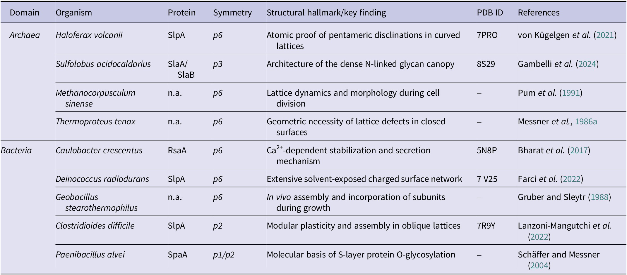
Examples of S-layer (glyco)proteins determined at atomic and near-atomic resolution, as well as foundational geometric studies relating to the data presented in Sections “Lattice dynamics”–“Biophysical properties of S-layer glycosylation,” are listed in Table 1. These structures do not introduce additional functional concepts; rather, they provide molecular confirmation that S-layer lattices embody curvature-adaptive, hydration-stabilized, and glycan-extended interfaces.
Key structural and foundational geometric studies of prokaryotic S-layers

Table 1. Long description
The table consists of seven columns: Domain, Organism, Protein, Symmetry, Structural hallmark/key finding, P D B I D, and References.
Under the Archaea domain:
* Haloferax volcanii uses protein SlpA with p 6 symmetry, showing atomic proof of pentameric disclinations in curved lattices (P D B I D 7 P R O).
* Sulfolobus acidocaldarius uses SlaA/SlaB with p 3 symmetry, detailing the architecture of the dense N-linked glycan canopy (P D B I D 8 S 2 9).
* Methanocorpusculum sinense with p 6 symmetry, focusing on lattice dynamics during cell division.
* Thermoproteus tenax with p 6 symmetry, focusing on the geometric necessity of lattice defects in closed surfaces.
Under the Bacteria domain:
* Caulobacter crescentus uses RsaA with p 6 symmetry, showing Calcium 2 plus-dependent stabilization (P D B I D 5 N 8 P).
* Deinococcus radiodurans uses SlpA with p 6 symmetry, showing an extensive solvent-exposed charged surface network (P D B I D 7 V 2 5).
* Geobacillus stearothermophilus with p 6 symmetry, focusing on in vivo assembly during growth.
* Clostridioides difficile uses SlpA with p 2 symmetry, showing modular plasticity in oblique lattices (P D B I D 7 R 9 Y).
* Paenibacillus alvei uses SpaA with p 1/p 2 symmetry, focusing on the molecular basis of S-layer protein O-glycosylation.
Note: n.a.: Protein identity not specified in the original geometric study; PDB ID: Protein Data Bank identifier for the primary structural model. Symmetry notations refer to plane group classifications.
Across Archaea and Bacteria, S-layer proteins assemble into two-dimensional crystalline lattices, most commonly hexagonal (p6), but also p3, square (p4), or oblique (p2, p1) arrangements (Messner et al., Reference Messner, König and ClausHand Varma2010; Albers and Meyer, Reference Albers and Meyer2011; Fagan and Fairweather, Reference Fagan and Fairweather2014; Rodrigues-Oliveira et al., Reference Rodrigues-Oliveira2017; Isbilir et al., Reference Isbilir2025; Sivabalasarma et al., Reference Sivabalasarma2025; Sleytr and Pum, Reference Sleytr and Pum2025). Despite extensive sequence divergence, these lattice geometries remain conserved, indicating that only a narrow range of architectures satisfies the physical constraints imposed on a continuous protein interface at the cell boundary (Pum et al., Reference Pum2021). Representative predicted and experimentally determined architectures are shown in Figure 2.
Early studies on M. sinense (see also Figure 1) and T. tenax (Figure 3a–c) had already identified the necessity of pentameric and heptameric defects to maintain a closed, continuous lattice during growth and division (see Section “Lattice dynamics”) (Messner et al., Reference Messner, Pum, Sára, Stetter and Sleytr1986a; Pum et al., Reference Pum, Messner and Sleytr1991; Sleytr and Pum, Reference Sleytr and Pum2025). In T. tenax , generally only the theoretically required number of pentamers was detected at the hemispherical cell poles, consistent with rapid establishment of a low-free-energy configuration during cell division (Messner et al., Reference Messner, Pum, Sára, Stetter and Sleytr1986a; Sleytr and Pum, Reference Sleytr and Pum2025). Decades later, mapping of S-layer repeating units on N. maritimus by subtomogram averaging revealed a hexagonal array in which hexamers coat the cell with near-continuous coverage, while pentameric defects are enriched at regions of high curvature (Figure 3d) (von Kügelgen et al., Reference Von Kügelgen2024).
The S-layers of Haloferax volcanii and S. acidocaldarius provide a particularly illustrative example of curvature adaptation at atomic resolution. Their lattices consist of immunoglobulin-like domains arranged hexagonally, with pentameric units closing the lattice over curved membrane regions (Gambelli et al., Reference Gambelli2024). Beyond static architecture, atomic structures also clarify the basis of dynamic lattice behavior. Flexible linker regions connecting quasi-rigid domains are resolved, providing molecular evidence for hinge-like architectures that enable defect-mediated plasticity while preserving long-range crystalline order (Figure 6c) (Gambelli et al., Reference Gambelli2024).
A comparable modular organization is evident in bacterial S-layers. In Caulobacter crescentus, the 2.7 Å structure of the S-layer protein RsaA reveals hexameric packing of rigid domains connected by flexible elements (Bharat et al., Reference Bharat2017). Docking into cryo-electron tomography maps yields a pseudo-atomic representation of the in vivo lattice, revealing periodic surface corrugation and well-defined nanopores. Domain-level structures from Deinococcus radiodurans (Farci et al., Reference Farci2021, Reference Farci2022; Farci and Piano, Reference Farci and Piano2024), Clostridioides difficile (Lanzoni-Mangutchi et al., Reference Lanzoni-Mangutchi2022), and Bacillus anthracis and C. glutamicum (Sogues et al., Reference Sogues2023, Reference Sogues2025) display the same design principle, i.e., rigid oligomeric building blocks connected by hinge-like elements and stabilized by extensive but non-locking interfaces.
At atomic resolution, the solvent-exposed surfaces of S-layer lattices exhibit a consistent chemical organization. Polar and charged residues dominate outward-facing surfaces and pore walls, whereas hydrophobic residues are largely buried within protomer cores and inter-subunit interfaces. For instance, in the S-layer of C. glutamicum , PS2, the pore lumen is predominantly negatively charged but becomes more neutral toward the cell (Figure 5b) (Sogues et al., Reference Sogues2025). Accordingly, the PS2 S-layer represents a semi-permeable layer with possible roles in selectivity, barrier function, and structural support (Sogues et al., Reference Sogues2025). In C. difficile , the major S-layer protein, SlpA, is posttranslationally cleaved into two S-layer proteins (SLPs): the high molecular weight and low molecular weight, referred to as SLPH and SLPL, respectively. These subunits form a complex that is incorporated in the S-layer and shapes two different pores (Figure 5c) (Lanzoni-Mangutchi et al., Reference Lanzoni-Mangutchi2022). The pore linings are hydrophilic, suggesting preferential diffusion of small, hydrated ions (Figure 5d). Pores exhibit mixed charge distributions with both positive and negative regions (Figure 5e) (Lanzoni-Mangutchi et al., Reference Lanzoni-Mangutchi2022). This residue distribution, observed across phylogenetically distant organisms, provides the structural basis for stabilization of tightly bound interfacial water. This hydration layer imposes a substantial desolvation penalty on approaching macromolecules, supporting the antifouling mechanism described in Section “Antifouling mechanisms of S-layer protein surfaces.” This chemical organization is independent of lattice symmetry and is also observed in oblique lattices such as C. difficile (Lanzoni-Mangutchi et al., Reference Lanzoni-Mangutchi2022).
In both Gram-negative and Gram-positive systems, pore walls are lined with polar residues and are frequently stabilized by bound divalent cations such as Ca2+ at protomer interfaces (Figure 4) (Herdman et al., Reference Herdman2022). These pores form hydrated nanopores that enable rapid diffusion of small solutes while excluding larger macromolecules. Functional studies on S-layer ultrafiltration membranes demonstrate sharp molecular weight cut-offs, with retention efficiencies approaching 100% for proteins above ~66 kDa (Sleytr and Sára, Reference Sleytr and Sára1986, Sleytr et al., Reference Sleytr1988; Sára and Sleytr, Reference Sára and Sleytr1987b; Schuster and Sleytr, Reference Schuster and Sleytr2021; Pfeifer et al., Reference Pfeifer2022). Permeability and antifouling selectivity arise from hydration-stabilized nanopores embedded within a periodic protein lattice.
Archaeal S-layers provide structural confirmation of the glycan canopy model introduced in Section “Biophysical properties of S-layer glycosylation.” In S. acidocaldarius , cryo-EM reconstructions show a porous lattice formed by the SlaA component decorated with dense N-linked glycans on solvent-exposed loops (Figure 6b) (Gambelli et al., Reference Gambelli2024). Glycoproteomic analyses demonstrate a high density of glycosylation sites in these regions (Peyfoon et al., Reference Peyfoon2010). The glycans are only partially resolved due to conformational flexibility, consistent with a hydrated, polymer-like state. Similar glycan decorations occur in bacterial systems, including Paenibacillus alvei (Schäffer and Messner, Reference Schäffer and Messner2004), where they extend the hydration-optimized interface into a dynamic entropic layer. The protein lattice thereby defines anchoring points for a flexible glycan canopy that enhances hydration and steric repulsion.
Taken together, these case studies reveal a conserved structural blueprint across Archaea, Gram-negative, and Gram-positive bacteria. This architecture comprises oligomeric protein modules connected by hinge-like elements, periodic nanopores lined with polar residues, and regularly patterned charge distributions, with dense glycan decoration particularly in archaeal systems. These motifs are consistent with the physical and thermodynamic principles outlined in preceding chapters. The atomic models summarized here show that S-layer lattices are not merely geometric surface arrays but molecularly encoded, curvature-adaptive, and hydration-stabilized biological interfaces. They thus provide a molecular blueprint for antifouling surfaces.
Specific interactions on antifouling S-layer lattices: How clean surfaces enable precise biology
S-layer lattices combine two surface properties that at first glance appear contradictory, namely a pronounced resistance to nonspecific adsorption and the capacity for highly specific biological interactions. This apparent paradox can be resolved by considering the crystalline organization of S-layers. These self-assembled lattices form highly ordered, porous, nanometer-scaled arrays that present repetitive surface motifs in a defined geometric arrangement while maintaining a strongly hydrated, molecularly regular background.
In diffusion-limited, low Reynolds number environments, random adsorption of macromolecules would rapidly mask surface features and interfere with selective interactions (Xie et al., Reference Xie2019; see Section “Life at low Reynolds numbers”). The antifouling character of S-layer lattices therefore plays a critical enabling role by preserving the accessibility of discrete functional domains embedded in the lattice. High-resolution structural analyses from diverse bacterial and archaeal systems show that while the lattice surface is largely smooth and hydration-dominated, specific domains protrude from this background and are positioned to mediate defined interactions.
In C. difficile , SlpA forms the outermost component of the cell envelope and is posttranslationally cleaved into SLPH and SLPL that assemble into a heterodimeric lattice (Waligora et al., Reference Waligora2001; Bradshaw et al., Reference Bradshaw2018; Lanzoni-Mangutchi et al., Reference Lanzoni-Mangutchi2022; Wang et al., Reference Wang2024a). SLPL is surface exposed and mediates binding to extracellular matrix proteins such as collagen, laminin, and fibronectin, contributing to epithelial adherence and immune interactions (Péchiné et al., Reference Péchiné2018; Waligora et al., Reference Waligora2001). Recent work further demonstrates that SlpA functions as a general receptor for multiple bacteriophages and that sequence variation among SlpA isoforms determines phage host range (Royer et al., Reference Royer2023, Reference Royer2025). Infection models show that deletion of SlpA profoundly affects colonization, motility, biofilm formation, and toxin production (Wang et al., Reference Wang2024a). These findings illustrate how defined receptor motifs are displayed within a lattice that otherwise resists nonspecific adsorption in the complex intestinal environment.
In the probiotic bacterium Lactobacillus acidophilus, the S-layer protein SlpA binds the C-type lectin receptor DC-SIGN on dendritic cells via its N-terminal domain, modulating cytokine responses and promoting mucosal tolerance (Konstantinov et al., Reference Konstantinov2008). Additional studies demonstrate interactions with mucins, heparan sulfate, and cell wall polymers. These observations show how precise host recognition can occur on a surface that remains largely non-adhesive to unrelated macromolecules.
S-layer lattices also serve as anchoring platforms for extracellular enzymes. In G. stearothermophilus , the S-layer protein binds a high-molecular-weight exo-amylase, preventing enzyme loss to the surrounding medium and localizing starch degradation directly at the cell surface (Egelseer et al., Reference Egelseer1995). In Clostridium thermocellum, S-layer–associated proteins such as OlpB and SdbA anchor the cellulosome complex to the envelope, enabling orchestrated cellulose degradation at the immediate cell interface (Lemaire et al., Reference Lemaire1995; Leibovitz and Béguin, Reference Leibovitz and Béguin1996). In both cases, the lattice provides a spatially defined, non-fouling scaffold that ensures retention and functionality of large extracellular protein assemblies.
Although archaeal S-layers are often described primarily as protective, antifouling envelopes, several well-documented examples demonstrate defined functional associations with protein partners. In Sulfolobus solfataricus, large sugar-binding protein complexes (‘bindosome’) co-purify with the S-layer glycoprotein, and disruption of the assembly system alters S-layer morphology and impairs growth on sugars, indicating a functional association between substrate-binding modules and the lattice architecture (Zolghadr et al., Reference Zolghadr2011). In S. acidocaldarius , the archaellum motor protein FlaF specifically binds to S-layer glycoproteins and serves as an anchoring point for the rotating motility apparatus (Banerjee et al., Reference Banerjee2015). Mechanistic models further suggest that ArlF/ArlG complexes interact with the S-layer to provide stator support for archaellum rotation (Nuno de Sousa Machado et al., Reference Nuno de Sousa Machado2022). These examples demonstrate that archaeal S-layers, like bacterial ones, provide a non-fouling crystalline platform that supports precise protein–protein interactions required for nutrient uptake and motility.
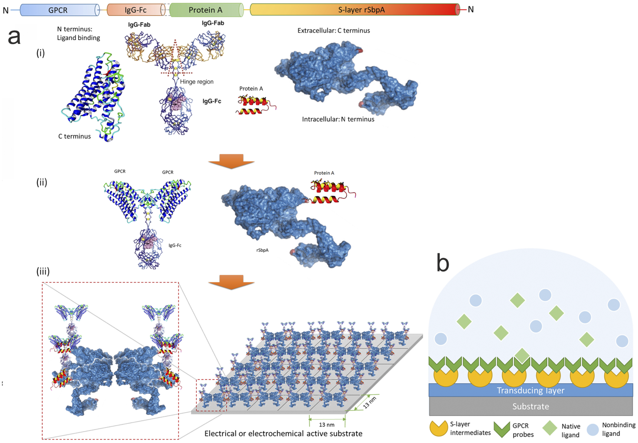
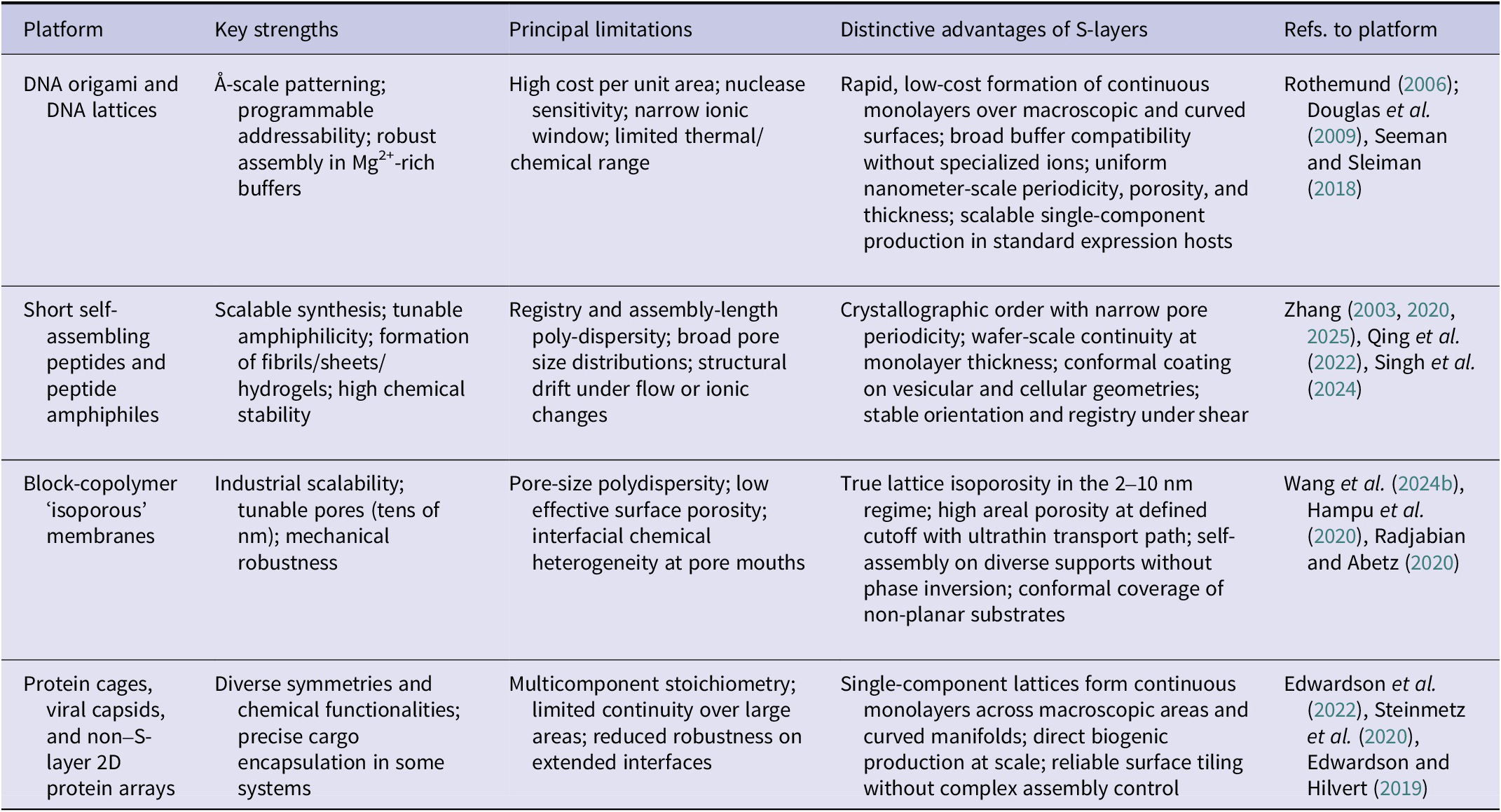
A particularly strong argument for the coexistence of antifouling properties and highly specific functionality comes from engineered S-layer fusion proteins. A wide variety of peptides and proteins ranging from 9 to 299 amino acids – including enzymes, antibody-binding domains, antigens, ligands, and metal-binding peptides – have been genetically fused to S-layer proteins without loss of function (Ilk et al., Reference Ilk2011). Notably, very short peptides such as streptavidin-binding tags (9 aa), silver- and cobalt-binding peptides (12 aa), and viral epitope mimotopes (20 aa) remain fully accessible and functional even in complex environments, including human serum. If significant nonspecific fouling of the S-layer surface occurred, such small functional peptides would be rapidly masked. Their preserved activity therefore provides experimental proof that S-layer lattices maintain a clean, accessible surface while presenting highly specific functional motifs. This duality is technologically exploited in biosensing and nanobiotechnology and reflects a remarkable degree of structural and functional plasticity (Sleytr et al., Reference Sleytr and Rosoff2001, Reference Sleytr and Ciferri2005). This concept will be revisited in Section “Translating biophysics: Application potential and future use” in the context of signal-to-noise optimization.
Across Bacteria and Archaea, S-layers thus form antifouling crystalline platforms that spatially regulate where and how interactions occur. Rather than preventing interactions, they suppress nonspecific adsorption while exposing selected recognition motifs with nanometer precision. Antifouling and specificity therefore emerge as complementary properties. The evolutionary conservation of S-layer lattices suggests that maintaining a clean, structured surface confers decisive advantages despite the trade-off of exposing receptor sites to bacteriophage exploitation.
Life at low Reynolds numbers
Prokaryotic life occurs in a hydrodynamic regime fundamentally different from that experienced by macroscopic organisms. At cell dimensions of roughly one micrometer, Reynolds numbers typically range from 10−5 to 10−3, meaning that viscous forces overwhelmingly dominate inertia and fluid motion is strictly reversible (Purcell, Reference Purcell1977; Lauga and Powers, Reference Lauga and Powers2009). Convective transport is effectively negligible, and fluid displacement stops immediately when force generation ceases. As a result, mass transport to and from the cell surface is governed almost entirely by diffusion, embedding cells in quasi-stationary boundary layers whose thickness depends on cell size, surface topology, and interfacial chemistry (Berg and Purcell, Reference Berg and Purcell1977; Dusenbery, Reference Dusenbery1997, Reference Dusenbery2009).
Even small amounts of adsorbed macromolecules, colloids, or debris increase the effective hydrodynamic radius, thicken the diffusion boundary layer, and reduce the flux of nutrients and metabolites. Fouled surfaces generate stagnant microdomains where proteins, ions, and reactive species accumulate, amplifying nonspecific interactions and environmental stress. These constraints are particularly pronounced in biofilms, (see Section “S-layers and biofilms: Reconciling antifouling with regulated surface attachment”) where diffusion is slowed and convection is virtually absent (Flemming and Wingender, Reference Flemming and Wingender2010). Under such conditions, surfaces that remain clean, hydrated, and dynamically accessible maintain nutrient flux and competitive fitness.
Within this physical framework, S-layer (glyco)protein lattices emerge as highly optimized biological interfaces. In Bacteria, S-layer pores are typically 2–8 nm in diameter and layers range from 5–15 nm in thickness. Archaeal S-layers exhibit greater structural diversity, (Albers and Meyer, Reference Albers and Meyer2011; Rodrigues-Oliveira et al., Reference Rodrigues-Oliveira2017; Bharat et al., Reference Bharat2021; Gambelli et al., Reference Gambelli2024; Schuster, Reference Schuster2024) with pore diameters of approximately 2–10 nm and thicknesses spanning 15–40 nm, strongly influenced by glycosylation and hydration. In both domains, S-layers precisely define the cell–environment boundary at the length scales that govern diffusion and interfacial physics. Their nanoscale planarity and periodicity minimize surface roughness and suppress stagnant microdomains, while pores allow rapid diffusion of small solutes and effectively exclude larger macromolecules.
A crucial and experimentally well-documented aspect is that S-layers are not static structural elements but dynamically regulated surface modules (see Section “Lattice dynamics”). Environmental isolates from Bacillaceae and Clostridiaceae have shown to retain their native S-layers only when cultivated under slow-growth, complex-medium conditions resembling natural habitats. Under optimized laboratory growth conditions in continuous culture, these organisms frequently synthesize lower-molecular-weight S-layer proteins or lose the S-layer entirely (Messner et al., Reference Messner, Hollaus and Sleytr1984; Sleytr and Beveridge, Reference Sleytr and Beveridge1999). In G. stearothermophilus PV72, environmentally induced genomic rearrangements replace the high-molecular-weight S-layer protein SbsA (~130 kDa, p6 lattice) with the smaller SbsB (~97 kDa, p2 lattice), thereby altering lattice symmetry, pore geometry, and surface chemistry in a single regulatory step (Sára et al., Reference Sára1996). In several bacteria, different S-layer genotypes within a population can switch between active and silent expression states via chromosomal inversions that relocate promoter regions, a mechanism described as phase variation of S-layer expression (Boot and Pouwels, Reference Boot and Pouwels1996).
Many S-layer proteins, particularly in Archaea, are glycosylated and subject to post-translational modification, which influences lattice stability, hydration properties, and interfacial chemistry (Eichler and Adams, Reference Eichler and Adams2005; Eichler, Reference Eichler2003, Reference Eichler2013; Albers and Meyer, Reference Albers and Meyer2011; Kandiba and Eichler, Reference Kandiba and Eichler2014). While the environmental regulation of these modifications is not fully characterized across taxa, their occurrence demonstrates that S-layer physicochemistry is not solely determined at the gene sequence level but can be modified after translation. This plasticity reflects a fundamental trade-off between biosynthetic cost and functional surface optimization. Thick, highly glycosylated lattices maximize antifouling performance, hydration control, and diffusion efficiency in macromolecule-rich or physically harsh environments. When these selective pressures are relaxed – such as under nutrient-rich, low-fouling laboratory conditions – simpler lattices or even complete S-layer loss become energetically favorable. The selective advantage of S-layers must therefore be balanced against the energetic cost of their biosynthesis. In Gram-positive bacteria, peptidoglycan synthesis is metabolically expensive (Typas et al., Reference Typas2012). Species with stable S-layers often possess comparatively thinner peptidoglycan layers than related organisms lacking S-layers, suggesting that the S-layer can substitute structural and protective functions while reducing overall envelope costs (Sleytr, Reference Sleytr1978; Fagan and Fairweather, Reference Fagan and Fairweather2014). Lactobacilli represent a notable exception: they combine relatively thick peptidoglycan layers with low-molecular-weight S-layer proteins (Messner et al., Reference Messner1997; Hynönen and Palva, Reference Hynönen and Palva2013). This architecture reflects an evolutionary compromise in which the antifouling and interfacial advantages of the S-layer are maintained while minimizing the biosynthetic burden of the lattice itself.
Thus, S layer lattices emerge as central, evolvable boundary structures for prokaryotic life under low Reynolds number conditions. In many Bacteria, they overlay peptidoglycan or outer membrane architectures and remain conditionally dispensable, whereas in most Archaea they constitute the sole cell wall component, integrating mechanical stability, selective permeability, and antifouling within a single molecular system. A defining feature of archaeal S layers is their extensive and often highly ordered glycosylation, which contributes to surface hydration, physicochemical resilience, and controlled molecular interactions at the cell–environment interface. In this context, S layer lattices can be understood as energy-efficient boundary systems that couple hydration structuring, diffusion control, and envelope energetics. Their structural diversity in symmetry, glycosylation, and protein architecture provides lineage-specific resolutions to universal physicochemical constraints. In low Reynolds number environments, where viscous forces dominate and transport is diffusion-limited, these properties ensure that cellular interfaces remain clean, accessible, and functionally resolved. Crucially, antifouling and molecular specificity are not opposing traits; rather, under such conditions, the suppression of non-specific adsorption is a prerequisite for the spatial definition and fidelity of biologically meaningful interactions.
S-layers and biofilms
Biofilm formation represents the transition from a planktonic to a sessile microbial lifestyle, characterized by surface attachment and integration into a self-produced extracellular polymeric substance (EPS) matrix (Flemming and Wingender, Reference Flemming and Wingender2010; Kuschmierz et al., Reference Kuschmierz2022; Zhao et al., Reference Zhao2025). At first glance, this transition appears incompatible with the antifouling properties of S-layer lattices described in the preceding chapters. S-layers impose a high desolvation penalty, maintain structured hydration shells, and suppress nonspecific macromolecular adsorption. From a biophysical perspective, however, this is not a contradiction but a prerequisite for regulated adhesion. Biofilm initiation requires a surface that prevents stochastic fouling while permitting localized, energetically controlled attachment events. This principle becomes particularly evident when considering the hydrodynamic constraints of microbial life at low Reynolds numbers (Section “Life at low Reynolds numbers”). Under diffusion-limited conditions, even minor nonspecific adsorption thickens the interfacial boundary layer and impairs nutrient and metabolite exchange (Stewart, Reference Stewart2003; Takenaka et al., Reference Takenaka2009). A clean, hydrated, and nanoscale-ordered surface is therefore essential to maintain efficient coupling between the cell and its environment. S-layer lattices establish precisely such an interface, ensuring that adhesion, when it occurs, is a regulated biological process rather than a physical consequence of surface contamination.
A well-documented system is C. difficile. The S-layer protein SlpA modulates aggregation behavior, adhesion, and biofilm characteristics, with proteomic analyses demonstrating that SlpA abundance varies between different biofilm models (Brauer et al., Reference Brauer2021). Strains retaining an intact S-layer show advantages in host-associated environments (Ormsby et al., Reference Ormsby2023). SlpA also functions as a general phage receptor (Phetruen et al., Reference Phetruen2022; Whittle et al., Reference Whittle2022; Royer et al., Reference Royer2023), demonstrating how specific molecular recognition occurs on an otherwise antifouling surface. Within biofilms, matrix components can trap phages and limit their penetration (Bond et al., Reference Bond2021), illustrating how the antifouling lattice, EPS matrix, and selective receptor function coexist at the cell surface. Moreover, S-layer proteins could function as a matrix exopolymer itself; a ‘public-good’ (Wong et al., Reference Wong2023) that helps to organize an antifouling surface and to stabilize the biofilm.
In Tannerella forsythia, truncation of the S-layer O-glycan canopy results in a marked increase in nonspecific biofilm biomass and altered surface charge (Posch et al., Reference Posch2011). The intact glycan layer therefore suppresses uncontrolled adsorption. At the same time, these glycans are essential for persistence within oral biofilms through modulation of host immune responses (Settem et al., Reference Settem2013). The same structural element that provides antifouling also enables selective biofilm-associated interactions.
A mechanistic bridge between lattice stabilization and matrix integration is demonstrated in D. radiodurans. Ca2+-ions required for S-layer lattice stability simultaneously serve as ionic bridges linking EPS components to the crystalline surface during biofilm formation (Shukla et al., Reference Shukla2022). The physicochemical interactions that maintain antifouling order are thus repurposed for matrix anchoring under appropriate environmental conditions. In P. alvei , the S-layer homology domain-containing protein SlhA is essential for both swarming motility and biofilm formation (Janesch et al., Reference Janesch2013), illustrating how S-layer-associated modules actively regulate the transition along the adhesion–mobility spectrum.
The antifouling and selective adhesion principles of S-layers also extend to Archaea. In organisms such as S. acidocaldarius and H. volcanii , S-layers provide mechanical stabilization and mediate environmental interactions, facilitating the formation of microbial mats or biofilm-like aggregates in extreme habitats (Albers and Meyer, Reference Albers and Meyer2011; Fröls, Reference Fröls2013; Chimileski et al., Reference Chimileski, Franklin and Papke2014; van Wolferen et al., Reference van Wolferen2018). Although direct experimental evidence linking archaeal S-layers to structured biofilm formation remains limited compared to bacterial systems, their crystalline organization, glycosylation patterns, and environmental responsiveness strongly suggest analogous roles in modulating adhesion, aggregation, and community resilience.
Notably, many S-layer-bearing microorganisms do not form biofilms under natural conditions, underscoring that this dual functionality is environmentally regulated rather than intrinsic.
Translating biophysics: Application potential and future use
The preceding chapters established that S-layer (glyco)protein lattices constitute a conserved biophysical solution to interfacial fouling in prokaryotes. If antifouling represents a primary and evolutionarily stabilized function of S-layers, technological translation follows as a matter of principle. Application is therefore not opportunistic biomimicry, but the deliberate abstraction and transfer of an optimized interfacial design strategy into materials science and bioengineering (Sleytr and Schuster, Reference Sleytr and Schuster2026).
From microbial envelope to engineering blueprint
Synthetic surface engineering continues to face a persistent fouling–functionality dilemma. Enhancing functionality through immobilized ligands, catalysts, or receptors frequently compromises antifouling performance, as physicochemical heterogeneities may disrupt interfacial hydration layers and facilitate nonspecific molecular adsorption. S-layer lattices appear to mitigate this trade-off through architectural order. Their monodisperse crystalline organization enables spatially defined presentation of functional moieties while maintaining a continuous physicochemical background that may reduce stochastic protein docking. In this view, functional specificity and interfacial passivation can be regarded not as antagonistic properties, but as co-emergent consequences of lattice symmetry and nanoscale periodicity.
Within this framework, antifouling is not simply an auxiliary benefit but a prerequisite boundary condition for sustained function. Only surfaces that remain operationally resistant to nonspecific adsorption are likely to maintain reproducible performance in complex biological media such as blood, serum, fermentation broths, or environmental samples. The technological maturation of S-layer-based systems, as synthesized in recent analyses (Sleytr and Pum, Reference Sleytr and Pum2025; Sleytr and Schuster, Reference Sleytr and Schuster2026), may therefore be understood as the progressive refinement of a biophysically grounded design principle rather than as a sequence of incremental surface modifications.
Isoporosity and selective gating: Ultrafiltration membranes
The translation of lattice symmetry and isoporosity into separation technology enabled the development of S-layer ultrafiltration membranes (SUMs), characterized by sharp molecular-weight cut-offs of approximately 30–45 kDa, a porosity of up to 60%, and consequently high flux. In these systems, the intrinsic nanometer-scale pore architecture of the S-layer lattice is faithfully transferred onto a supporting scaffold, yielding composite membranes with exceptional structural uniformity and steep molecular-exclusion profiles (Sleytr and Sára, Reference Sleytr and Sára1986, Reference Sleytr and Sára1988; Schuster and Sleytr, Reference Schuster and Sleytr2021). In contrast, conventional polymer-based ultrafiltration membranes, such as those composed of cellulose acetate, polypropylene, polysulfone, or polylactic acid, exhibit pore-size distributions varying by up to an order of magnitude and typically possess surface porosities below 10% (Fane et al., Reference Fane1981; Strathmann, Reference Strathmann1981; Pavanasam and Abbas, Reference Pavanasam and Abbas2008). In this context, it should be mentioned that during the production of SUMs, after deposition of the S layer- fragments on the porous carrier membranes (e.g., microfiltration membranes) the protein lattices were cross-linked intra- and intermolecularly (Sleytr and Sára, Reference Sleytr and Sára1986, Reference Sleytr and Sára1988). The high density and regular spatial organization of chemically addressable functional groups on the S-layer surface and within its pore arrays provide a versatile platform for molecular tailoring. Through the covalent immobilization of low molecular weight nucleophiles, both the pore diameter and the net surface charge of the membranes could be modified without compromising their antifouling performance (Sára and Sleytr, Reference Sára and Sleytr1987b; Küpcü et al., Reference Küpcü1991; Sleytr et al., Reference Sleytr2011). Antifouling, however, is not merely advantageous but structurally integral to maintaining flux in supramolecular ultrafiltration matrices. At low Reynolds numbers, even minimal adsorption events markedly increase hydrodynamic resistance. The dynamic hydration shell and electrostatic mosaicity of S-layer lattices effectively suppress irreversible protein deposition and stabilize long-term permeability. Functionally, the S-layer thus acts as a molecular gatekeeper, enforcing passage according to the geometrically defined constraints of the lattice while maintaining a low-adhesion interface.
High-fidelity biointerfaces: Biosensors and graphene electronics
The performance of biosensors in complex fluids is inherently limited by nonspecific adsorption, which compromises signal-to-noise ratios and induces baseline drift. S-layer lattices provide an architecturally defined scaffold for stoichiometric and spatially controlled immobilization of functional biomolecules, as shown in studies on S-layer fusion proteins that elucidated their construction principles and biotechnological applications (Weiner et al., Reference Weiner1994; Breitwieser et al., Reference Breitwieser1996, Reference Breitwieser1998, Reference Breitwieser2016; Weber et al., Reference Weber2001, Moll et al., Reference Moll2002; Völlenkle et al., Reference Völlenkle2004; Ilk et al., Reference Ilk2011; Schuster, Reference Schuster2018; Damiati and Schuster, Reference Damiati and Schuster2020; Sleytr and Pum, Reference Sleytr and Pum2025). The periodic organization of these fusion constructs ensures homogeneous receptor density and defined molecular orientation, reducing steric interference and receptor-to-receptor variability.
Recent work on scalable biomimetic sensing systems combining S-layer architectures with graphene field-effect transistor (GFET) arrays has demonstrated the compatibility of S-layer lattices with semiconductor electronics (Qing et al., Reference Qing2023). In such hybrid assemblies, the lattice functions as a nanostructured adaptor between biological recognition elements and electronic interfaces (Figure 7). Its antifouling background suppresses nonspecific adsorption, while the crystalline periodicity enables reproducible receptor positioning across wafer-scale arrays. The resulting signal-to-noise enhancement is maintained even in complex media, reflecting the microbial requirement to distinguish specific from nonspecific interactions at crowded interfaces.
Schematic illustration of architecture. (a) Sequence layout of the dual-monolayer construct. (i) The four elements of the construct: redesigned water-soluble membrane receptor, heavy chain Fc region of immunoglobulin G (IgG) protein, Fc-binding fragment of protein A, and two-dimensional (2D) crystal-forming S-layer protein. (ii) Solubilized receptors are fused to IgG-Fc to serve as biospecific probes, while S-layer proteins are fused with protein A fragments to connect probes to the device. (iii) Sequentially, S-layer/protein A fusion proteins reproduce its native nanoscale 2D crystalline p4 lattice on the substrate, after which the receptor probes with Fc fusions were applied to bind the intermediate layer with high affinity and capacity. (b) S-layer proteins and solubilized receptors are sequentially coated onto designated sensing devices. Target analytes are specifically recognized in aqueous media, while the detection signal is transduced for electrical responses. GPCR, G protein–coupled receptor. (Qing et al., Reference Qing2023; Creative Commons Attribution NonCommercial License 4.0 (CC BY-NC; http://creativecommons.org/licenses/by/4.0/)).

Figure 7. Long description
The diagram is divided into two main panels, a and b.
Panel a shows the sequence layout of the construct. At the top is a linear protein domain map labeled N to N, containing G P C R, I g G-F c, Protein A, and S-layer r S b p A.
- Sub-panel i displays the four individual elements: a blue ribbon G P C R with N and C terminus labels; a Y-shaped I g G protein with F a b and F c regions; a small red and yellow helical Protein A; and a large blue textured S-layer protein.
- Sub-panel ii shows the fusion process where G P C R is attached to the I g G-F c region, and Protein A is fused to the r S b p A S-layer protein.
- Sub-panel iii illustrates the final assembly. On the right, a 2D crystalline p 4 lattice is formed on an electrical substrate with 13 nanometer spacing. On the left, a zoomed-in view shows the G P C R-I g G probes docked onto the S-layer-Protein A base.
Panel b is a cross-sectional schematic of the sensing device. From the bottom up:
- A gray substrate layer.
- A blue transducing layer.
- A layer of yellow semi-circle S-layer intermediates.
- A layer of green V-shaped G P C R probes.
- An aqueous medium containing green diamond native ligands, which are binding to the probes, and light blue circular nonbinding ligands which remain free in the solution.
Biomimetic membranes, drug delivery and immunointerfaces
The self-assembly of S-layer proteins on lipid membranes yields composite systems that mimic archaeal cell envelopes (Pum et al., Reference Pum1993; Pum and Sleytr, Reference Pum and Sleytr1994). These biomimetic architectures have been explored as platforms for membrane-active peptides and proteins and as multifunctional biointerfaces (Schuster et al., Reference Schuster2004; Schuster, Reference Schuster2005; Schuster and Sleytr, Reference Schuster and Sleytr2006, Reference Schuster and Sleytr2014, Reference Schuster and Sleytr2020). Lipid membranes were generated on S-layer coated porous or solid supports to enhance their mechanical stability and longevity also with membrane-active peptides and reconstituted membrane proteins. In solid-supported lipid membranes, the S-layer lattice functions as a stabilizing scaffold, offering a defined tether layer to decouple the lipid membrane from the support, and creating an ionic reservoir for electrochemical measurements. This reservoir can be customized using a combination of full-length and truncated S-layer proteins or through the self-assembly of a thiolated SCWP layer on the gold electrode prior to S-layer protein recrystallization (Schuster and Sleytr, Reference Schuster and Sleytr2009). A critical feature of supported lipid membranes is the preservation of high mobility of lipid molecules within the membrane, referred to as the semifluid S-layer supported lipid membrane (Pum and Sleytr, Reference Pum and Sleytr1994; Schuster and Sleytr, Reference Schuster and Sleytr2000), while maintaining the integrity of the overall structure to ensure longevity (Schuster et al., Reference Schuster2003; Gufler et al., Reference Gufler, Pum, Sleytr and Schuster2004; Schuster and Sleytr, Reference Schuster and Sleytr2014).
In drug delivery, recrystallized S-layers on liposomes or emulsomes act as protective, antifouling shells that enhance colloidal stability and modulate immune recognition (Küpcü et al., Reference Küpcü1995, Ücisik et al., Reference Ücisik2013, Reference Ücisik2015a, Reference Ücisik2015b). The immunological dimension of S-layer fusion proteins further illustrates how antifouling and functionality coexist. By presenting antigens in a repetitive, crystalline context while maintaining low nonspecific adsorption, S-layer-based constructs can enhance immunogenicity while reducing undesirable side reactions. The interface remains accessible yet orderly, an attribute that appears central to their adjuvant potential (Sleytr et al., Reference Sleytr, Mundt and Messner1991; Malcolm et al., Reference Malcolm, Beveridge and Koval1993a, Reference Malcolm, Sleytr, Messner, Pum and Sàra1993b; Jahn-Schmid et al., Reference Jahn-Schmid1996a, Reference Jahn-Schmid1996b; Messner et al., Reference Messner, Sleytr, Messner, Pum and Sára1996; Sleytr et al., Reference Sleytr2007, Reference Sleytr2014).
Patterning living and non-living matter
Beyond molecular sensing and delivery systems, S-layers serve as anisotropic, crystalline biointerfaces for spatial control at the cellular level. It has been demonstrated that S-layer nanolayers can spatially segregate adherent and non-adherent cells within microfluidic devices. The antifouling background suppresses unwanted cell attachment, while specific functionalization permits defined adhesion patterns (Rothbauer et al., Reference Rothbauer2013, Reference Rothbauer2015). Thus, S-layers operate as mesoscale patterning elements bridging molecular order and cellular organization.
Biomimetic transfer to polymer technology: Molecular imprinting
An important conceptual step is the transfer of antifouling principles from protein lattices to synthetic polymers. In S-layer based biomolecular imprinting, S-layer lattices were employed as structural templates for biomolecular imprinting, generating polymer films with defined recognition cavities and reduced nonspecific adsorption (Ladenhauf et al., Reference Ladenhauf2015). This approach exemplifies how biological antifouling logic – periodicity, defined topography, spatial confinement of binding sites – can inform polymer surface design. Rather than copying molecular composition, the strategy translates architectural and thermodynamic principles into synthetic materials.
Comparison of S-layer lattices with synthetic antifouling coatings and alternative nanoscale self-assembled materials
Non-biological antifouling strategies, such as polyethylene glycol (PEG) brushes, zwitterionic polymers, or superhydrophilic coatings, rely primarily on entropic steric repulsion and hydration-layer stabilization (Chapman et al., Reference Chapman2000; Moayedi et al., Reference Moayedi2024). While highly successful in many contexts, these systems face intrinsic limitations: polydispersity of chain length, defect formation, oxidative degradation, and the difficulty of integrating high-density functional ligands without compromising antifouling performance (Table 2).
Biophysical comparison of S-layer lattices and representative synthetic antifouling coatings

Table 2. Long description
The table consists of six rows comparing properties across two categories.
1. Structural organization. S-layer lattices feature genetically encoded 2 D crystallinity, strict symmetry from p 1 to p 6, and monodisperse subunits. Synthetic polymer coatings feature stochastic chain conformations and polydisperse molecular weights.
2. Defect control. S-layer lattices utilize self-assembly with low defect density and dynamic subunit turnover. Synthetic coatings commonly have defects and pinholes and lack intrinsic repair mechanisms.
3. Hydration mechanism. S-layer lattices use structured, lattice-coupled hydration and glycan-mediated steric repulsion. Synthetic coatings use entropy-driven steric hindrance and bound water via hydrophilic functional groups.
4. Integration of function. S-layer lattices allow precise stoichiometric fusion protein display. Synthetic coatings use ligand grafting which often perturbs brush density and diminishes performance.
5. Dynamic response. S-layer lattices exhibit lattice fluctuations and adaptive behavior. Synthetic coatings are predominantly static and prone to oxidative degradation, especially P E G.
6. Evolutionary validation. S-layer lattices are conserved across Bacteria and Archaea. Synthetic coatings are engineering-derived and context-dependent.
In contrast, S-layer lattices are genetically encoded, monodisperse, and capable of self-assembly and self-repair. Their hydration shell is not merely entropically stabilized but structurally integrated into a dynamic protein framework. Moreover, functionality can be incorporated at the level of subunit design without disrupting lattice continuity (Ilk et al., Reference Ilk2011; Sleytr and Pum, Reference Sleytr and Pum2025).
This comparison of Table 2 underscores that S-layers are not merely alternative coatings, but embody a distinct class of adaptive, crystalline biointerfaces.
Self-assembling biomaterials such as DNA origami (Rothemund, Reference Rothemund2006; Douglas et al. Reference Douglas2009; Seeman and Sleiman, Reference Seeman and Sleiman2018), short self-assembling peptides (Zhang, Reference Zhang2003, Reference Zhang2020, Reference Zhang2025; Qing et al., Reference Qing2022; Singh et al., Reference Singh2024), block copolymer membranes (Wang et al., Reference Wang2024b; Hampu et al., Reference Hampu2020; Radjabian and Abetz, Reference Radjabian and Abetz2020), and protein cages (Edwardson et al., Reference Edwardson2022; Steinmetz et al., Reference Steinmetz2020; Edwardson and Hilvert, Reference Edwardson and Hilvert2019) provide complementary routes to ordered interfaces. Table 3 situates S-layer lattices among these alternative self-assembled platforms, benchmarking operational regimes, scalability, continuity on curved supports, and controllable isoporosity relevant to antifouling interface engineering.
Comparison of S-layer lattices with alternative nanoscale self-assembled materials

Table 3. Long description
The table compares four nanoscale self-assembled material platforms against S-layers.
1. D N A origami and D N A lattices: Strengths include Angstrom-scale patterning and programmable addressability. Limitations include high cost, nuclease sensitivity, and narrow ionic/thermal range. S-layer advantages include rapid, low-cost formation of continuous monolayers over macroscopic surfaces and broad buffer compatibility.
2. Short self-assembling peptides and peptide amphiphiles: Strengths include scalable synthesis and high chemical stability. Limitations include registry poly-dispersity and structural drift. S-layer advantages include crystallographic order with narrow pore periodicity and stable orientation under shear.
3. Block-copolymer isoporous membranes: Strengths include industrial scalability and mechanical robustness. Limitations include pore-size polydispersity and low effective surface porosity. S-layer advantages include true lattice isoporosity in the 2 to 10 nanometer regime and conformal coverage of non-planar substrates.
4. Protein cages, viral capsids, and non S-layer 2 D protein arrays: Strengths include diverse symmetries and cargo encapsulation. Limitations include multicomponent stoichiometry and limited continuity over large areas. S-layer advantages include single-component lattices forming continuous monolayers across macroscopic areas and curved manifolds.
Vision: S-layers as patterning elements in synthetic biology
The most forward-looking application domain for S-layers lies in synthetic biology. S-layer lattices offer a programmable, self-assembling scaffold for organizing functional modules with nanometer precision. As patterning elements, they can define spatial periodicity on synthetic cell envelopes, effectively recreating archaeal-like surface architectures on minimal or artificial cells. In this scenario, the S-layer is not an accessory coating but an integral design principle. A dynamic crystalline exoskeleton that simultaneously protects, filters, and spatially organizes membrane proteins. In this context, it should be noted that isolated S-layer glycoprotein subunits, which form hexagonal lattices, have been shown to have the ability to spontaneously reassemble into closed vesicular structures (Sleytr, Reference Sleytr1976).
One may envisage synthetic cells whose functional complexity is externally encoded in an S-layer fusion lattice comprising enzymes, receptors, signaling modules arranged with defined stoichiometry and spacing. Such systems would merge bottom-up assembly with evolutionary design logic. The antifouling background ensures operational stability, while the periodic scaffold dictates functional geometry.
More broadly, hybrid ‘bio–silicon’ systems may exploit S-layer nanochannels as regulated conduits between biological recognition events and electronic transduction. In this sense, S-layer technology reconciles two historically divergent goals of interface engineering: maximal suppression of noise and maximal fidelity of signal. Recent biomimetic graphene FET architectures employing structurally ordered membrane receptor monolayers further illustrate this design principle (Figure 7) (Qing et al., Reference Qing2023).
In summary, the application potential of S-layer (glyco)protein lattices arises directly from their universal biophysical function. By maintaining clean, dynamically hydrated, and precisely ordered interfaces, they solve a constraint that is equally relevant to microbes and microdevices. Their translation into nanobiotechnology, membrane science, immunoengineering, and synthetic biology represents not a departure from biology, but the continuation of a fundamental interfacial principle, refined by evolution and now entering the realm of rational design.
Practical limitations and engineering strategies for S-layer technologies
While S-layer–based interfaces offer a powerful biophysical blueprint, several practical constraints currently limit translation. Table 4 summarizes the principal practical and translational challenges associated with the development of S-layer-based technologies, including protein production, lattice stability, surface assembly, functionalization, device integration, and regulatory compliance, together with engineering strategies that have been proposed to improve robustness, scalability, reproducibility, and application readiness.
Practical limitations and engineering strategies for S-layer technologies

Table 4. Long description
The table consists of three columns: Challenge, Impact/limitation, and Mitigation strategy.
1. Protein production and cost: High-yield and glycoform-defined production is difficult for native glycoproteins. Mitigated by recombinant expression, fermentation, secretion tags, cell-free synthesis, and glycoengineering.
2. Lattice robustness under industrial stress: Stability is compromised by shear, oxidation, solvents, or sterilization. Mitigated by covalent crosslinking, engineered cysteine residues, ceramic overcoats, and lyophilization with osmoprotectants.
3. Orientation control and anchoring heterogeneity: Mixed orientations reduce reproducibility. Mitigated by affinity-based capture (H i s-tag/N T A, SpyTag/SpyCatcher), pre-functionalizing substrates with cell wall polymers, and electric-field-assisted alignment.
4. Recrystallization kinetics and scale-up: Slow nucleation complicates large-scale manufacturing. Mitigated by seeded growth, optimizing C a super 2 plus or M g super 2 plus ions, and meniscus-guided coating.
5. Chemoselective functionalization without fouling: High ligand loading can impair antifouling. Mitigated by site-specific conjugation at defined positions and restricting ligand density to maintain hydration.
6. Integration with electronics and microfluidics: Fabrication can reduce activity or cause signal drift. Mitigated by recrystallizing directly on devices, using zwitterionic interlayers, and co-assembly of native and fusion proteins.
7. Regulatory and biosafety considerations: Requires traceability and sterilization compatibility. Mitigated by standardizing workflows, quality control via X-ray scattering and cryo-E M, and endotoxin-free platforms.
Summary and concluding remarks
Surface layer (S layer) glycoproteins are among the most abundant and structurally conserved supramolecular assemblies in prokaryotes (see Appendix). Their persistence across phylogeny and extreme environments, despite extensive sequence divergence, indicates a unifying selective advantage rooted in physical function rather than primary structure. This review identifies antifouling as such a function: a general interfacial principle that suppresses nonspecific macromolecular adhesion while preserving selective permeability and interaction capacity.
This principle is particularly consequential in the low Reynolds number regime that governs microbial life, where viscous forces dominate and diffusion constrains molecular exchange. Under these conditions, even rare irreversible adsorption events impose significant energetic and functional costs. S layer lattices mitigate these constraints through an integrated physicochemical architecture.
Crystalline periodicity, defined nanoscale topography, electrostatic heterogeneity, spatially confined hydrophobic domains, and, where present, surface glycosylation collectively stabilize structured hydration layers while disrupting kinetically favorable macromolecular docking. Antifouling therefore emerges as a systems level property of the two-dimensional lattice. Crucially, this protection does not depend on interfacial inertness; instead, antifouling and selective permeability are mechanistically coupled within the same ordered framework.
A central implication is that S layers resolve the longstanding tradeoff between surface passivation and functionality that limits many synthetic systems. In polymer based antifouling coatings, increasing ligand density or catalytic complexity often perturbs hydration equilibria and introduces defects, thereby reducing resistance to nonspecific adsorption. In contrast, the genetically encoded periodicity of S layer lattices enables stoichiometric and spatially precise presentation of functional domains without disrupting lattice continuity. Suppression of stochastic adsorption and specific molecular recognition are thus complementary properties of a single supramolecular scaffold. Experimental implementations, including S layer ultrafiltration membranes, high signal to noise biosensors, and S layer fusion protein platforms, provide validation of this principle.
Beyond current applications, the physical logic of S layer lattices points to a broader relevance of ordered protein interfaces for compartmentalized systems. Any minimal self-reproducing entity requires a boundary that combines mechanical stability, selective permeability, and resistance to uncontrolled macromolecular aggregation. Two-dimensional crystalline protein membranes provide a parsimonious solution: they self-assemble from identical subunits, generate geometrically defined nanopores, and maintain hydrated, low adhesion interfaces without requiring complex multicomponent architectures. Although there is no evidence that extant S layers are relics of prebiotic membranes, the principles they embody, namely self-organization, defect minimization, and the coupling of permeability with antifouling, define a minimal physical framework for robust compartmentalization.
At the same time, important quantitative challenges remain. A predictive theory of antifouling at crystalline glycoprotein interfaces will require integration of atomic resolution structural data with interfacial thermodynamics, hydration force spectroscopy, and near surface hydrodynamics under flow. The contributions of glycosylation to long range repulsion, the role of lattice elasticity in defect tolerance, and the fluctuation spectrum of subunits under shear remain incompletely resolved. In particular, a unified framework linking lattice symmetry, surface charge distribution, and fluctuation mediated interactions are still lacking.
In summary, S layer glycoprotein lattices can be understood as universal antifouling interfaces that enable prokaryotic life in chemically crowded and often harsh environments. By integrating crystalline order, dynamic hydration, and spatially controlled molecular recognition within a single supramolecular framework, they provide both a fundamental biophysical principle and a model system for understanding how ordered protein interfaces maintain functional stability in complex, multicomponent fluids. We would also like to draw the attention of the reader to the Appendix of this review.
Acknowledgments
We would like to thank Dietmar Pum for stimulating discussions. During the preparation of this manuscript, the authors used the generative AI tool ChatGPT (OpenAI, San Francisco, USA; accessed via https://chatgpt.com) to assist with rephrasing and improving the clarity and grammar of some sentences. All suggestions generated by the tool were critically reviewed, edited, and approved by the authors, who take full responsibility for the content of this work. The tool was also used to assist in the creation of Figures A1 and A2.
Financial support
Part of this work was funded by the Austrian Science Fund (FWF), the Austrian Federal Ministry of Women, Science and Research (BMFWF), the Erwin-Schrödinger Society for Nanosciences (ESG), the European Union, and Air Force Office of Scientific Research (AFOSR).
Competing interests
The authors declare none.
Appendix
The global scale of S-layer biomass – A planetary perspective
In the hidden dimensions of the microbial biosphere, S-layers represent one of nature’s most ubiquitous architectural feats. These crystalline proteinaceous lattices constitute a primary interface between life and the environment. To appreciate the magnitude of this molecular assembly, we estimate the total global mass and surface area of S-layers by integrating current census data with established biophysical models.
Our quantitative estimation relies on the landmark census by Bar-On et al. (Reference Bar-On2018), which assesses the global prokaryotic carbon pool at approximately 70 Gt C for Bacteria and 7 Gt C for Archaea. To derive the global S-layer inventory, we convert this carbon mass to dry weight (assuming 50% C) and then to total cellular protein (estimated at 50% of dry weight). This yields a reservoir of roughly 70 Gt of bacterial protein and 7 Gt of archaeal protein. Within this reservoir, S-layers occupy a significant niche. Based on proteomic studies, we assume they account for approximately 10% of total cellular protein in expressing organisms. Accounting for a near-universal prevalence (100%) in Archaea and a rough estimate of 30% prevalence among Bacteria, the global S-layer inventory reaches approximately 2.8 Gt of dry protein mass (0.7 Gt from Archaea and 2.1 Gt from Bacteria). To place this into perspective, the cumulative mass of Earth’s S-layers is equivalent to the weight of roughly 430 Great Pyramids of Giza, each with an estimated mass of about 6.5 million tons (Figure A1).
The staggering mass of 2.8 Gt of S-layer protein is difficult to grasp in human terms, yet its geometric scale is breathtaking. By assuming a specific gravity for proteins of 1.35 g/cm3 and applying a conservative porosity of 50%, meaning half of the lattice volume consists of open pores, the total structural volume of the global S-layer biomass corresponds to approximately 4.15 km3. If unfurled as a single monolayer with a standard thickness of 10 nanometers, this lattice would cover an area of approximately 415 billion km2. Given that the total surface area of Earth is roughly 510 million km2, the global inventory of S-layers is sufficient to shroud the entire planet more than 800 times (Figure A2).
This immense coverage is a direct reflection of the ubiquity of S-layer-bearing prokaryotes, which thrive in nearly every terrestrial and aquatic ecosystem. Because they are found across a vast diversity of Bacteria and are a nearly universal feature of Archaea, S-layers represent one of the most successful and globally distributed functional structures in nature. Their presence in extreme environments, from hydrothermal vents to salt lakes, highlights them as a universally adapted molecular architecture essential for the survival and integrity of life on a planetary scale.
Illustration of the total mass of S-layer protein on Earth in relation to the mass of the Pyramid of Cheops.

Illustration of the total area of S-layer protein lattices on Earth compared to the Earth’s surface area.