Congenital myasthenic syndromes (CMSs) are a group of inherited muscle disorders caused by genetic defects that affect transmission of information from nerves to muscles at the neuromuscular junction (NMJ). CMSs are clinically very heterogeneous (Ref. Reference Vincent1) but share a phenotype of fatiguing weakness usually presenting from birth, although adult-onset of the disease has been described. Clinical features include ocular, bulbar or respiratory symptoms worsened by crying or activity in the neonatal period, abnormal fatigability, normal or delayed motor milestones, and occasional progression of the symptoms during adolescence or adult life (Ref. Reference Engel2). In electrophysiological studies, a decremental response on repetitive nerve stimulation points towards a synaptic transmission defect. Most CMS cases are sporadic, but there are families with two or more affected members showing autosomal recessive or, less frequently, dominant inheritance.
Clinically, CMSs share many features with myasthenia gravis, the most common disorder of neuromuscular transmission. However, by contrast to CMSs, myasthenia gravis is an acquired autoimmune disease characterised by the formation of autoantibodies directed most frequently against the acetylcholine receptor (AChR), which binds the neurotransmitter acetylcholine (ACh) (Ref. Reference Engel3). Up to 20% of myasthenia gravis patients are negative for AChR antibodies (AChR-Ab) (Ref. Reference Vincent4), and about 30–40% of these seronegative patients develop antibodies against the muscle-specific tyrosine kinase (MuSK) (Ref. Reference Vincent and Leite5). Autoimmune myasthenia gravis is rare in early childhood, but may be difficult to differentiate from CMSs in later childhood or adulthood.
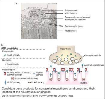
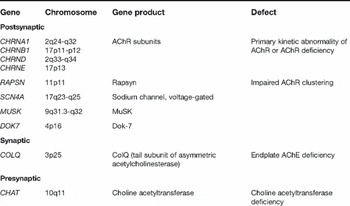
Molecular knowledge of the pathogenic mechanisms of the different types of CMSs has evolved rapidly in the past decade. Genes encoding post-, pre- or synaptic proteins at the NMJ were identified as candidate genes for CMSs, starting with the subunits of the nicotinic AChR (Refs Reference Beeson, Palace and Vincent6, Reference Vincent7, Reference Ohno8, Reference Engel9, Reference Engel10). To date, genes known to cause CMS if mutated (Fig. 1; Table 1) are the presynaptic choline acetyltransferase gene CHAT (Ref. Reference Ohno11), the gene COLQ (Refs Reference Donger12, Reference Ohno13) encoding the triple-stranded collagenic tail (ColQ) of the synaptic acetylcholinesterase (AChE), the genes encoding the different subunits of the postsynaptic AChR (CHRNA1, CHRNB1, CHRND and CHRNE), the genes for the postsynaptic proteins rapsyn (‘receptor-associated protein of the synapse’; RAPSN) (Ref. Reference Ohno14) and MuSK (MUSK) (Ref. Reference Chevessier15), and the gene for the postsynaptic sodium channel (SCN4A) (Ref. Reference Tsujino16). Recently, mutations in the DOK7 gene have been identified to cause a specific CMS form with predominant limb-girdle weakness (Ref. Reference Beeson17).
Candidate gene products for congenital myasthenic syndromes and their location at the neuromuscular junction. (a) Electron micrograph of a normal human neuromuscular junction. The lower side of the nerve terminal faces a postsynaptic region of clefts and folds. (Image reprinted with kind permission of Dr Simone Spuler, Dept of Neurology, Charite University Hospital, Berlin, Germany.) (b) Schematic showing the location of protein products of known candidate genes for congenital myasthenic syndromes (CMSs) at the neuromuscular junction. The gene names are given in italics under the protein names. Abbreviations: AChR, acetylcholine receptor; ChAT, choline acetyltransferase; ColQ, collagen-like tail of the acetlycholinesterase; Dok-7: downstream of kinase 7; MuSK, muscle-specific kinase.

Summary of genes implicated in congenital myasthenic syndromes

Abbreviations: AChR, acetylcholine receptor; Dok-7: downstream of kinase 7; MuSK, muscle-specific kinase.
Thus, CMS may be classified according to the localisation of the affected protein at the NMJ: presynaptic, synaptic (basal-lamina-associated) and postsynaptic CMS (Fig. 1; Table 1). Postsynaptic CMSs are the most frequent, accounting for ∼80% of all genetically diagnosed CMS; presynaptic CMSs are rare (∼5%) (Ref. 18; H. Lochmuller et al., unpublished). However, the underlying genetic cause of the disease has so far been established in only approximately half of the CMS patients (Ref. 18; H. Lochmuller et al., unpublished). In this article, we focus on recent developments in understanding postsynaptic CMS.
Postsynaptic CMS
A crucial step during NMJ formation is the concentration of densely packed AChR molecules at the postsynaptic membrane (for an overview see Ref. Reference Sanes and Lichtman19). The motor-nerve-derived proteoglycan agrin initiates AChR clustering via MuSK (Refs Reference DeChiara20, Reference Glass21, Reference Hoch22, Reference Colledge and Froehner23), rapsyn (Ref. Reference Froehner24) and other as yet unidentified components. In addition to the AChR itself, mutations in several components of this signalling pathway have been shown to cause CMSs.
Mutations of the AChR genes
The muscle AChR is a pentamer existing in two isoforms: an adult form comprising two α subunits and one β, one δ and one ɛ subunit; and a fetal form in which a γ subunit is expressed instead of the ɛ subunit. The subunits are homologous, each consisting of a long N-terminal extracellular domain followed by three transmembrane domains (M1–3), an intracellular cytoplasmic domain, a final transmembrane domain (M4) and an extracellular C-terminus (Ref. Reference Sine and Engel25). Between the first and the second transmembrane domain, there is a short intracellular domain, whereas a short extracellular part connects the second and the third transmembrane domain. Mutations involving subunits of the AChR fall into two major classes: kinetic mutations with or without minor AChR deficiency, and low-expressor mutations leading to a deficiency of AChR at the endplate. The kinetic mutations are further subdivided into two categories depending on their kinetic effect: ‘slow-channel’ and ‘fast-channel’ mutations. The name slow-channel originates from the abnormally slow decay of synaptic currents caused by abnormally prolonged opening events of the AChR channel. In general, slow-channel mutations are gain-of-function mutations and are inherited in a dominant pattern. Fast-channel mutations are mostly recessive loss-of-function mutations and cause abnormally fast decay of the synaptic response as a result of abnormally brief channel opening events. We do not describe these mutations in detail here, since they have been extensively reviewed elsewhere (Refs Reference Engel and Sine26, Reference Engel, Ohno and Sine27).
AChR mutations leading to receptor deficiency
Recessive mutations causing severe endplate AChR deficiency are the most common cause of CMSs and are concentrated in the ɛ subunit (encoded by CHRNE). Missense, frameshift, nonsense and splice-site mutations, as well as chromosomal microdeletions and mutations in the promoter region, have been described (Ref. Reference Ohno and Engel28). Different mechanisms may underlie the AChR deficiency state. Mutations that truncate the ɛ subunit prior to the M3–M4 cytoplasmic loop are almost certainly null alleles, whereas some missense mutations and some truncations near the C-terminus may result in low expression of functional adult AChR. The C-terminal cysteine C470 of the ɛ subunit is crucial for maturation and surface expression of the adult AChR. Thus, any mutation truncating the ɛ subunit upstream of C470 is predicted to inhibit the expression of the ɛ subunit (Ref. Reference Ealing29).
The mutation ɛ1267delG of the ɛ subunit borders the boundary of intron 11 and exon 12 and leads to a frameshift after codon 422, with 63 missense codons followed by a stop codon. The mutation disrupts the cytoplasmic loop and the fourth transmembrane region (M4) of the ɛ subunit. ɛ1267delG is an old founder mutation causing CMS in patients with Gypsy ethnic origin, dating back to ∼800 years ago. CMS is one of the most common mendelian disorders in this population, with ∼4% heterozygous carrier rates (Refs Reference Abicht30, Reference Morar31). Testing for the ɛ1267delG mutation facilitates the genetic diagnosis in Gypsy CMS patients (Ref. Reference Kalaydjieva32). Patients homozygous for the mutation ɛ1267delG usually present in the first year of life with ptosis, a weak cry and feeding difficulties. The most characteristic findings are ptosis and opthalmoparesis, and bulbar muscle weakness is frequent. Proximal muscle weakness is mild; however, restricted ambulation has been reported in some patients (Ref. Reference Beeson18). The disease course is generally benign with a tendency towards improvement and a beneficial effect of AChE inhibitors (Refs Reference Abicht30, Reference Kalaydjieva32, Reference Tournev33).
The CHRNE 1293insG mutation was found with a rate of 20% in CMS patients originating from Maghreb (especially Algeria and Tunisia). An ancient founder effect for this mutation was demonstrated by haplotyping with microsatellite markers and intragenic polymorphisms. Interestingly, 1293insG was also repeatedly found in patients originating from Spain and Portugal (Ref. Reference Beeson18). Another frequent CHRNE mutation, especially in Spanish and Portuguese patients, is 70insG; a possible founder effect for this mutation has not yet been investigated.
Splicing mutations constitute about 10% of the CHRNE mutations and cause skipping of an adjacent exon, retention of the mutant intron or both, as well as activation of a cryptic splice site (Ref. Reference Ohno and Engel34). A pathogenic intronic mutation remote from the splice acceptor site (IVS5–16G >A) has been reported, which suggests RNA analysis may be necessary in cases of intronic mutations that are not part of the consensus splice site (Ref. Reference Müller35). A recent study detected a synonymous nucleotide variant at CHRNE codon 285 that turned out to be a splicing mutation; the nucleotide change created a new aberrant donor splice site (Ref. Reference Richard36).
Furthermore, missense mutations in the signal peptide of the ɛ subunit have been identified (ɛG8R, ɛV–13D) (Ref. Reference Middleton37). These mutations lead to the replacement of hydrophobic amino acids with charged hydrophilic residues and might thereby interfere with the delivery of the nascent peptide to the endoplasmic reticulum (Ref. Reference Middleton37). Mutations in the regulatory elements of CHRNE may also cause reduced or inadequate gene expression. The promoter region of CHRNE contains E-boxes and one N-box that are crucial for the development and tissue-specific regulation of gene expression. The N-box mutations ɛ−154G>A, ɛ−155G>A, ɛ−156C>T reduce the endplate-specific expression of the ɛ subunit (Refs Reference Abicht38, Reference Nichols39).
While the symptoms of CMS patients with different underlying mutations in the ɛ subunit are similar, there is marked variability in the disease severity even in patients homozygous for identical mutations, pointing to the existence of additional factors modifying the disease phenotype. Nevertheless, the characteristic clinical features together with the ethnic background and geographic origin of the patients are important clues facilitating the genetic testing.
Prenatal myasthenic syndrome caused by disruption of the fetal γ AChR subunit
The fetal form of AChR (α2βδγ) is produced prior to the 33rd week of gestation in humans. The gradual switch from γ to ɛ subunit takes place in the late fetal and perinatal period, thereby forming the adult AChR (α2βδɛ) (Refs Reference Hesselmans40, Reference Missias41, Reference Yumoto, Wakatsuki and Sehara-Fujisawa42). The sequence and structure of the γ subunit is highly similar to the ɛ subunit, but the two receptors have different conductance and open times (Ref. Reference Mishina43). No CMS form has yet been linked to mutations in the gene CHRNG encoding the fetal γ AChR subunit. However, a specific form of arthrogryposis multiplex congenita, termed Escobar syndrome, has recently been associated with CHRNG mutations (Refs Reference Hoffmann44, Reference Morgan45). Escobar or multiple pterygia syndrome is an autosomal recessive condition characterised by excessive webbing (pterygia), congenital contractures (arthrogryposis), and scoliosis (Refs Reference Rajab46, Reference Escobar47). Variable other features include intrauterine death, congenital respiratory distress, short stature, ptosis, faciocranial dysmorphism, low-set ears, arachnodactyly, and cryptorchidism in males.
A genome-wide linkage analysis revealed significant disease linkage to a region on chromosome 2q37. Subsequently, 14 mutations within the CHRNG were identified in families with Escobar syndrome of diverse ethnic origin (Refs Reference Hoffmann44, Reference Morgan45). The mutations result in a modified, truncated or absent γ subunit. Interestingly, for one of the missense mutations, γR217C, mutations of the corresponding amino acids in the ɛ and β subunit have been identified in CMS patients [ɛR218W (Ref. Reference Abicht48) and βR220C (Ref. Reference Müller49)].
The γ subunit is required to localise the fetal AChR correctly in the cell membranes of transfected HEK 293 cells. Missense mutations underlying the Escobar syndrome are low-expressor mutations: AChRs containing a mutated γ subunit were not localised to the cell surface in transfected HEK 293 cells, leading to a lack of AChR (Ref. Reference Hoffmann44). Therefore, the observed postnatal symptoms – namely arthrogryposis, pterygia and scoliosis – are a result of a reduced AChR expression level during fetal development. Similarly, antibodies that block the fetal form of the AChR may occasionally be present in mothers and cause arthrogryposis multiplex congenita in utero (overview in Ref. Reference Vincent50). The antibodies cross the placenta and block neuromuscular transmission, leading to joint deformities and often stillbirth in these infants.
Since the γ subunit generally disappears in late fetal development and is replaced by the ɛ subunit, patients with CHRNG mutations exhibit almost no progression, and show normal muscle tone and no signs of myasthenia after birth. Thus, Escobar syndrome represents an inherited fetal myasthenic disease that also affects neuromuscular organogenesis.
Mutations of the RAPSN gene
Clinical features of CMS patients with RAPSN mutations
Ohno et al. first reported three recessive RAPSN mutations – L14P, N88K and 553ins5– in four CMS patients with AChR deficiency (Ref. Reference Ohno14). Subsequent studies in other laboratories revealed that RAPSN mutations are a frequent cause of CMSs, accounting for around 10% of all CMS cases (Refs Reference Müller51, Reference Burke52, Reference Dunne and Maselli53, Reference Richard54).
Two distinct clinical phenotypes are associated with AChR deficiency due to RAPSN mutations: a rather severe early-onset phenotype, and a mild late-onset phenotype (Refs Reference Müller51, Reference Burke52) (Table 2). The early-onset phenotype is characterised by a disease onset at birth or in the first 2 years of life. In some cases, prenatal disease onset with reduced fetal movements in utero is noted. These children might be born with multiple flexion contractures of the upper and lower limbs and/or dysmorphic facial features, such as malformed low-set ears, and an elongated face, open mouth, high-arched palate and prominent jaw (Refs Reference Müller51, Reference Banwell55, Reference Ioos56). Patients with the early-onset phenotype have a history of muscle hypotonia, poor suck and cry, bilateral ptosis, episodes of severe generalised weakness, and respiratory distress, usually triggered by febrile infections. By contrast to patients with CHRNE mutations, eye movements are not restricted. In general, treatment with esterase inhibitors leads to an improvement of strength and can prevent apneic attacks during upper respiratory infections. Interestingly, one case without any myasthenic symptoms between episodic crises has been reported recently (Ref. Reference Skeie57).
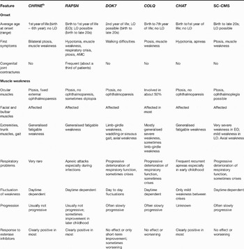
Comparison of key clinical features of congenital myasthenic syndromes arising from mutations in CHRNE, RAPSN, DOK7, COLQ and CHAT genes, and in SC-CMSa

aFor more information, see Refs Reference Beeson17, Reference Beeson18, Reference Engel and Sine26, Reference Müller76, Reference Palace78.
bAChR deficiency.
Abbreviations: AMC, arthrogryposis multiplex congenital; EO, early-onset phenotype; LO, late-onset phenotype; SC-CMS, slow-channel congenital myasthenic syndrome.
By contrast, patients with the late-onset phenotype are asymptomatic during childhood. First disease symptoms occur in adolescence or early adulthood (Refs Reference Müller51, Reference Burke52). The disease course is benign, usually with bilateral ptosis and mild muscle weakness. The late-onset phenotype is less frequent than the early-onset phenotype. However, its characterisation is of high clinical importance: because it first presents in adult life, the disease can be mistaken for seronegative myasthenia gravis. The lack of a clinical response to immunosuppressive treatments should alert the clinician to the possibility of late-onset CMS, particularly with RAPSN mutations.
RAPSN mutations: the frequent mutation N88K
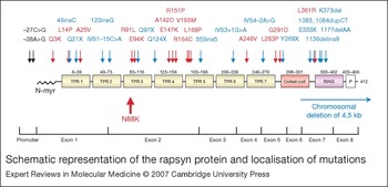
Altogether, more than 30 different RAPSN mutations have been published (Fig. 2). Surprisingly, most patients with RAPSN mutations in the translated region of the gene carry one single missense mutation, N88K, on at least one allele (Refs Reference Ohno14, Reference Müller51, Reference Burke52, Reference Dunne and Maselli53, Reference Richard54). The frequency of this mutation raised the question of whether N88K is a founder mutation or whether a mutational hotspot exists at this position. Two studies looking at microsatellite markers surrounding the RAPSN gene yielded differing haplotypes harbouring N88K, possibly indicating more than one origin of the mutation (Refs Reference Richard54, Reference Ohno and Engel58). However, a subsequent study of 21 single nucleotide polymorphisms (SNPs) in 41 mutant RAPSN N88K alleles from 21 patients of European and Indian ethnic origin revealed a short common conserved haplotype encompassing a distance of about 360 kb on all analysed N88K alleles (Ref. Reference Müller59). The identified common fragment encompassed only one of the microsatellite markers used in the previous studies; the other markers were located outside the common founder fragment. This finding supported the hypothesis that probably most RAPSN N88K alleles detected worldwide indeed derive from a single founder event that might have occurred in an ancient Indo–European population (Ref. Reference Müller59).
Schematic representation of the rapsyn protein and localisation of mutations. Missense mutations are shown in red, promoter mutations in black and null mutations in blue. The frequent missense mutation N88K is highlighted. Exon borders within the protein have been marked below; exons have not been drawn precisely to scale. Abbreviations: N-myr, myristoylation signal at the N-terminus, which targets rapsyn to the membrane; TPR, tetratricopeptide repeat; P, phosphorylation site.

Knowledge about the frequency and the prevalence of the N88K mutation of RAPSN may help to accelerate and simplify genetic analysis of European CMS patients. Only two patients of European origin lacking the common N88K allele but harbouring differing RAPSN mutations on both alleles have been reported so far: one patient is homozygous for a missense mutation (R164C); the second patient is compound heterozygous for a splice (IVS1–15C>A) and another missense mutation (L283P) (Ref. Reference Müller60). The clinical phenotype of the two patients corresponds to the previously described early-onset phenotype.
A homozygous mutation of the RAPSN promoter (−38A>G) was observed in seven phenotypically similar Jewish patients originating from the Near East (Ref. Reference Ohno61). Characteristic features in these patients were facial malformations such as mandibular prognathism, malocclusion, a high-arched palate and crowded teeth. The mutation −38A>G is localised in one of the E-box elements in the RAPSN promoter region and reduces gene expression. Haplotype analysis showed that the mutation arises from a common founder.
All RAPSN mutations described so far show a recessive inheritance pattern, although in the case of N88K – given its relatively high frequency – pseudodominant inheritance is possible (Ref. Reference Dunne and Maselli62).
Pathogenic mechanisms of RAPSN mutations
RAPSN encodes rapsyn, a 43 kDa postsynaptic peripheral membrane protein that interacts directly with AChRs and is essential for clustering the receptor molecules at the postsynaptic membrane of the NMJ (Refs Reference Froehner24, Reference Gautam63, Reference Fuhrer64). Rapsyn is a cytoplasmic protein acting downstream of agrin and MuSK; neural agrin is released from the nerve terminal to induce clustering of AChR at the endplate (Ref. Reference Ferns and Carbonetto65). Rapsyn comprises distinct structural domains: a myristoylation signal at the N-terminus targets rapsyn to the membrane (Ref. Reference Ramarao and Cohen66), seven tetratricopeptide repeats (TPRs) from codons 6–279 mediate rapsyn self-association (Refs Reference Ramarao and Cohen66, Reference Ramarao67), a coiled-coil domain at codons 363–403 interacts with the long cytoplasmic loop of AChR subunits (Ref. Reference Cartaud68), a cysteine-rich RING-H2 domain at codons 363–402 interacts with the cytoplasmic domain of β-dystroglycan (Ref. Reference Bartoli, Ramarao and Cohen69), and there is a serine phosphorylation site at codon 406 (a consensus sequence for phosphorylation by protein kinases A and C). The TPRs are α-helical repeats of 34 amino acids that are suggested to be involved in both intramolecular and intermolecular protein–protein interactions.
The pathogenic mechanisms of some of the rapsyn missense mutations detected in CMS patients were evaluated in patient endplate biopsies as well as in transfected cells. Analysis of endplate biopsies showed reduced AChR numbers, impaired development of postsynaptic folds, simplification of postsynaptic membranes, reduced NMJ areas, and also endplates fragmented into multiple small regions spread over an increased length of muscle fibre surface (Refs Reference Ohno14, Reference Maselli70, Reference Yasaki71). No decrease of ACh release or kinetic abnormality of AChR channels (Ref. Reference Ohno14) were detected, but expression levels of rapsyn at the endplates were reduced (Ref. Reference Yasaki71).
Expression studies of mutant constructs in transfected cells indicated that RAPSN mutations diminished co-clustering of AChR with rapsyn, leading to receptor deficiency and impaired neuromuscular transmission (Refs Reference Ohno14, Reference Müller60). A recent study by Cossins et al. (Ref. Reference Cossins72) uncovered different pathogenic mechanisms for some RAPSN missense mutations: R91L inhibited self-association of rapsyn in transfected cells; K373del reduced the stability of the rapsyn protein (a very rapid decline of rapsyn protein levels in transfected cells was observed); A25V prevented association of rapsyn with the AChR in co-transfected cells (rapsyn-A25V did not localise at the cell surface but appeared intracellularly); and N88K- and L361R-rapsyn formed agrin-induced clusters that were unstable and disassembled much faster compared with wild-type rapsyn clusters.
The functional effects of the mutations do not resemble the pathogenic effects seen in previous biochemical characterisation of rapsyn domains and domain-deletion experiments. For instance, TPR domains are important for self-association of rapsyn; however, the loss of a single TPR domain did not have any effect on self-clustering, as it could be compensated by the remaining TPRs (Refs Reference Ramarao and Cohen66, Reference Eckler, Kuehn and Gautam73). The results of Eckler et al. (Ref. Reference Eckler, Kuehn and Gautam73) suggested that TPRs 1 and 3 are selectively important for rapsyn self-association, as mutant rapsyns lacking TPRs 1 or 3 were unable to form clusters efficiently in transfected cells. Other studies, however, indicated that TPRs 1 and 2 were sufficient for rapsyn self-association in transfected cells (Ref. Reference Ramarao and Cohen66). Nevertheless, the mutation R91L in TPR3 dramatically inhibits rapsyn self-association; this is the first and only TPR mutation reported that influenced self-association. By contrast, the mutation N88K, only three amino acids away from R91L, does not inhibit rapsyn self-association at all.
Several other mutations within the TPRs inhibit co-clustering of rapsyn with the AChR, although TPR domains were not thought to be important for interaction of rapsyn and AChR. Previous reports have mapped the coiled-coil domain of rapsyn as the major region responsible for interaction with the AChR (Ref. Reference Ramarao67). A possible explanation for this contradiction might be that the missense mutations in the TPRs could lead to overall misfolding of the protein and thereby prevent rapsyn–AChR interaction. No missense mutation in the rapsyn coiled-coil domain has been identified and analysed yet.
Genotype–phenotype correlation
No obvious correlation of underlying RAPSN mutation and/or pathogenic mechanism and clinical symptoms of patients has been observed. There is a very broad variation of severity of symptoms even for patients homozygous for the frequent mutation N88K (Refs Reference Müller51, Reference Burke52, Reference Dunne and Maselli53, Reference Richard54). By contrast to patients with CHRNE or COLQ mutations, CMS patients with RAPSN null mutations on both alleles have not been reported. Complete loss of rapsyn expression is probably not compatible with postnatal life, similar to the situation in rapsyn-deficient mice (see section ‘Mouse models for CMS’). A significant association of appearance of arthrogryposis in patients with one RAPSN null mutation has been observed in patient cohorts analysed in Oxford, UK and at the Mayo Clinic, USA (Refs Reference Beeson18, Reference Burke52, Reference Cossins72). This association was not very pronounced in our patient cohort (Ref. Reference Müller60): of 11 out of 35 patients with RAPSN mutations born with joint contractures, only three harbour a heterozygous null mutation, whereas two patients with a heteroallelic null mutation did not have contractures.
It is difficult to establish a solid correlation of disease severity and the number of N88K alleles present in a CMS patient. Nevertheless, the second mutation of a heteroallelic N88K patient may in part determine disease severity (Refs Reference Müller51, Reference Ioos56, Reference Cossins72). Patients heteroallelic for N88K and a different missense mutation or harbouring two mutations different from N88K may be affected more severely (Refs Reference Müller51, Reference Ioos56, Reference Müller60, Reference Ohno61). All RAPSN patients presenting with the mild late-onset phenotype reported so far are homozygous for N88K; this might be attributed to the rather mild pathogenic changes of N88K compared with the effects of other missense rapsyn mutations (Ref. Reference Cossins72).
Mutations of the DOK7 gene
Neural agrin activates MuSK to establish the postsynaptic apparatus during NMJ development (Refs Reference DeChiara20, Reference Gautam63, Reference Gautam74). The precise signalling pathways downstream of MuSK, however, are poorly characterised. Recently, a MuSK-interacting cytoplasmic protein, termed Dok-7 (downstream of kinase 7) has been cloned (Ref. Reference Okada75). Previously known Dok-family proteins (Dok-1–6) play a role in signalling downstream of receptor and nonreceptor phosphotyrosine kinases. Like other Dok proteins, Dok-7 has a pleckstrin-homology (PH) domain and a phosphotyrosine-binding (PTB) domain in the N-terminal half, and multiple tyrosine residues in the C-terminal region. Okada et al. (Ref. Reference Okada75) demonstrated that Dok-7 overexpression can induce the aneural activation of MuSK and subsequent clustering of AChR in cultured myotubes.
Clinical features of patients with DOK7 mutations
DOK7 mutations were initially identified in CMS patients in which the proximal limb muscles were mainly affected (‘limb-girdle myasthenia’; LGM) (Ref. Reference Beeson17). Symptoms of this CMS form begin between birth and the third decade, with most patients displaying a characteristic ‘limb-girdle’ pattern of weakness with ptosis and a waddling gait, but no ophthalmoparesis (Table 2). Clinical onset is typically after first motor milestones are achieved at normal age, and is characterised by progressive difficulty in walking. Respiratory problems and crises are frequent. Daytime-dependent fluctuation of symptoms (worsening of symptoms in the evening) is uncommon; however, patients report good and bad periods (weeks or even months). The disease course is often progressive and may lead to loss of ambulation in adulthood. Nevertheless, the clinical picture varies from very mild late-onset cases to very severe early-onset forms. None of the cases showed a long-term positive response to esterase inhibitors; some of the patients even worsened under treatment with esterase inhibitors (Ref. Reference Müller76). No efficient treatment is yet known for this CMS form.
Pathogenic mechanisms of DOK7 mutations
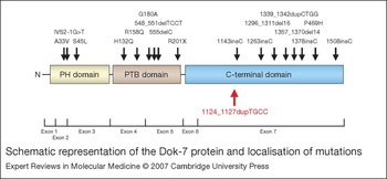
Eighteen different mutations of the DOK7 gene have been reported (Fig. 3); most of them are located in exon 7. The pathogenic mechanisms of DOK7 mutations have not been characterised in detail. Some of the patients with DOK7 mutations described by Beeson et al. were reported to have small endplates (Refs Reference Beeson17, Reference Slater77). However, it is not yet clear how exactly Dok-7 malfunction may lead to decreased endplate size.
Schematic representation of the Dok-7 protein and localisation of mutations. The frequent frameshift mutation 1124_1127dupTGCC is marked in red. Exon borders within the protein have been marked below; exons have not been drawn precisely to scale. Abbreviations: PH, pleckstrin homology; PTB, phosphotyrosine binding domain.

One of the mutations, the frequent C-terminal domain mutation 1124_1127dupTGCC, has been studied in cell culture. Interestingly, all but three out of 12 kinships in one study (Ref. Reference Müller76) and all but four of 24 kinships in another (Ref. Reference Palace78) carry at least one copy of this frameshift. Furthermore, we identified five additional CMS patients in our cohort who are heterozygous carriers of 1124_1127dupTGCC; although their clinical features are compatible with LGM, no second heteroallelic mutation has yet been detected in the coding regions of the DOK7 gene (Ref. Reference Müller76). Similarly, Palace et al. reported five kinships harbouring 1124_1127dupTGCC, including two kinships with two affected siblings, where they were unable to identify a mutation in the coding sequence of the second allele (Ref. Reference Palace78).
Analysis of AChR clusters induced by Dok-7 harbouring the mutation 1124_1127dupTGCC showed a significant reduction in the number of branched-type AChR plaques compared with wild-type Dok-7 in fully differentiated transfected C2C12 myotubes (Ref. Reference Beeson17). By contrast, the ability of the 1124_1127dupTGCC mutant to bind and induce MuSK phosphorylation was not impaired in undifferentiated myoblasts and in heterologous cells, suggesting that the C-terminal domain might play a key role in the maturation of the synaptic structure (Ref. Reference Beeson17). Other mutations of the C-terminal domain of Dok-7 might have similar effects in myotubes.
The PH domain of Dok-7, as well as the PTB domain, is indispensable for the activation and phosphorylation of MuSK in heterologous cells as well as in C2 myotubes (Ref. Reference Okada75). Dok-7 binds the PTB target motif (NPXY) in MuSK (Ref. Reference Okada75), a motif previously shown to be essential for activation of MuSK and subsequent clustering of AChRs in vitro and in vivo. Missense mutations in the PH and PTB domains might therefore impair the interaction of Dok-7 with MuSK. The mutations H132Q, R158Q and G180A are in the PTB domain, and A33V and S45L are located in the PH domain; however, no functional data are available yet on these mutations.
Similar to rapsyn, obvious correlations of clinical symptoms and the location of the mutations within the Dok-7 protein have not been established. Remarkably, our patients with late onset of symptoms are all homozygous for the common frameshift mutation 1124_1127 dupTGCC (Ref. Reference Müller76). Moreover, patients with both DOK7 mutations located in exons 1–6 have not been observed to date. Residual Dok-7 activity is probably essential for postnatal survival, since Dok7-deficient mice die shortly after birth (Ref. Reference Okada75).
Mutations of the MUSK gene
There is only one reported case of a CMS arising from mutations in MUSK (Ref. Reference Chevessier15). The female patient presented with clinical symptoms of ptosis and respiratory distress in the neonatal period. A similarly affected brother died during a respiratory crisis. The course of the disease in the sister was favourable. She complained only of slight eyelid ptosis, fatigability on walking and the inability to do sport. Symptoms fluctuated, but were aggravated during a pregnancy in adulthood. The patient responded well to a combinatory therapy of esterase inhibitors and 3,4-diaminopyridine (3,4-DAP). Molecular genetic analysis revealed a MUSK null allele in combination with the missense mutation V790M, which did not affect MuSK catalytic kinase activity but diminished expression and stability of MuSK, leading to decreased agrin-dependent AChR aggregation (Ref. Reference Chevessier15) and possibly impaired Dok-7–MuSK interaction (Ref. Reference Okada75).
Synaptic basal-lamina-associated CMS: mutations in COLQ
The enzyme AChE is responsible for rapid hydrolysis of ACh released at the cholinergic synapses. The molecular form of AChE at the endplate is an asymmetric enzyme composed of one, two or three homotetramers of globular subunits (AChET) attached to a triple-stranded collagenic tail (ColQ) (Refs Reference Massoulie79, Reference Bon, Coussen and Massoulie80, Reference Simon, Krejci and Massoulie81, Reference Deprez and Inestrosa82, Reference Ohno83). The collagenic tail subunit has an N-terminal proline-rich region attachment domain (PRAD), a collagenic central domain, and a C-terminal region enriched in charged residues and cysteines. Each ColQ strand can bind an AChET tetramer to its PRAD. Two heparan-sulphate-proteoglycan-binding domains in the collagen domain and residues in the C-terminal domain interact with perlecan and the extracellular domain of MuSK to anchor the enzyme in the synaptic space. The C-terminal region is also required for initiating the triple-helical assembly of ColQ. Recessive mutations in COLQ cause endplate AChE deficiency by preventing the attachment of AChET to ColQ (PRAD mutations), producing a short single-stranded and insertion-incompetent ColQ (mutations in the collagenic domain) or by impairing the triple-helical assembly of the collagenic domain and/or hindering insertion into the basal lamina (C-terminal mutations). More than 30 mutations have been identified to date (Ref. Reference Ohno and Engel28). The majority of them truncate the protein distally to PRAD (e.g. 275insC, Q211X, Q240X, E214X, S169X, R282X, 788insC, 1082delC, 107del215, W148X, 806insC, 375delT) and display distinct sedimentation profiles on density gradient centrifugation consistent with the absence of the asymmetric A12, A8 and A4 AChE species and a prominent ∼10.5S mutant peak composed of a single truncated ColQ strand and a catalytic tetramer (Refs Reference Ohno13, Reference Ohno83, Reference Shapira84, Reference Ohno85). Interestingly, homozygous Q240X mutations were found in six Palestinian Arab patients and in an Iraqi Jewish patient (Ref. Reference Shapira84). Similarly, three patients from two unrelated families originating from the same German geographic area were found to be homozygous for T441A and share a high degree of similarity for the T441A-carrying allele on haplotype analysis (Ref. Reference Müller86). Y430S was originally described in a Spanish kinship and found later in additional Spanish patients (Ref. Reference Donger12).
The absence of AChE prolongs the lifetime of ACh in the synaptic space. On a single nerve stimulus, the increased duration of the endplate current outlasts the refractory period of the muscle fibre and may excite a second compound muscle action potential (CMAP). Similar to the slow-channel CMS, there is overloading of the synaptic space with cations and evidence of endplate myopathy. The following clinical clues point to the diagnosis: recessive inheritance pattern, a decremental EMG response, a repetitive CMAP, sometimes a slow pupillary light response, and no effect or even worsening after administration of AChE inhibitors (Ref. Reference Ohno83). Most patients with mutations in COLQ are severely disabled from an early age with respiratory difficulties and progressive involvement of the axial muscles, leading to severe scoliosis and restrictive ventilatory deficit. However, some patients with missense mutations in the C-terminal domain of ColQ have a milder clinical course (Ref. Reference Donger12). In addition, variations in phenotypic expressivity between and within kinships harbouring the same mutation can also occur (Refs Reference Shapira84, Reference Müller86).
Presynaptic CMS
Presynaptic CMSs are rare and may be caused by either decreased synthesis, packaging in vesicles or release of the neurotransmitter ACh. So far, only mutations in the gene encoding choline acetyltransferase (CHAT) have been identified as causing presynaptic CMS. Defects in ChAT lead to reduced catalytic activity of the enzyme, and also reduced enzyme expression, and thereby diminished ACh synthesis (Ref. Reference Ohno11). CMS with underlying CHAT mutations is associated with episodes of severe respiratory distress and bulbar weakness leading to apnea (CMS with episodic apnea). These attacks are precipitated by infections, fever and excitement; between the attacks, the degree of muscle weakness may be variable. Similar respiratory crises may also be caused by RAPSN mutations (see above).
CHAT mutations are recessive and mostly missense mutations, and the majority of them are detected in only one kinship or patient. However, there are some exceptions: the missense mutation I336T has been identified in three independent kinships of Turkish origin (Ref. Reference Schmidt87), which may indicate a founder effect in the Turkish population. The compound heterozygous combination of the mutations L210P with S498L and S694C with T354M have each been found in two independent patients from Finland and Croatia, respectively (Refs Reference Ohno11, Reference Barisic88).
Except for the CMS caused by mutations in CHAT, presynaptic CMSs have not been well characterised at the molecular level. In a recent publication (Ref. Reference Milone89), Milone and colleagues describe two novel and highly disabling CMSs, one determined by presynaptic and the other determined by combined presynaptic and postsynaptic defects. Evoked quantal release was reduced to approximately 25% of normal in both patients. In one of the patients, the synaptic response to ACh was further compromised by degeneration of the junctional folds, with concomitant loss of the AChR. However, the genetic basis of these syndromes awaits discovery, as the search for mutations in components of the synaptic vesicle release complex and in other candidate proteins was not successful. The same applies to another presynaptic CMS arising from quantal release deficiency (Ref. Reference Maselli90). Some years ago, patients with Lambert–Eaton-syndrome-like presynaptic CMS or CMS due to paucity of synaptic vesicles were described (Refs Reference Bady, Chauplannaz and Carrier91, Reference Walls92); the underlying genetic defect has not yet been reported.
Treatment options for patients with CMSs
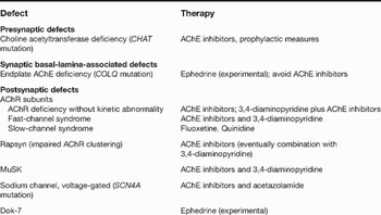
The diverse underlying genetic defects in CMSs result in different pathophysiology, and so the administration of the best therapeutic agent depends on the precise molecular diagnosis (Table 3).
Options for drug treatment of congenital myasthenic syndromes with different underlying molecular defectsa

aFor more information see the text and Refs Reference Beeson18, Reference Engel and Sine26.
Abbreviations: AChE, acetylcholinesterase; AChR, acetylcholine receptor; CHAT, choline acetyltransferase gene; COLQ, gene encoding collagen-like tail of acetlycholinesterase; MuSK, muscle-specific receptor tyrosine kinase; SCN4A, voltage-gated sodium channel; Dok-7, downstream of kinase 7.
CMSs that decrease the synaptic response to ACh are treated with drugs that augment cholinergic stimulation – namely inhibitors of AChE, and 3,4-DAP (Ref. Reference Engel and Sine26) (Table 3). Accordingly, anti-AChE medications benefit patients with presynaptic CMS (CHAT mutations) and with postsynaptic CMS caused by AChR deficiency (CHRNE and RAPSN mutations) (Refs Reference Abicht30, Reference Tournev33, Reference Banwell55, Reference Engel, Ohno and Sine93), or by a fast-channel syndrome (AChR subunit mutations) (Ref. Reference Engel, Ohno and Sine93). Some patients, especially with fast-channel syndrome or rapsyn defects, derive further benefit from AChE inhibitors in combination with 3,4-DAP (Ref. Reference Engel, Ohno and Sine93) (Ref. Reference Banwell55). In CHAT patients and maybe in RAPSN patients, oral prophylactic treatment with AChE inhibitors should be administered even in asymptomatic patients as it prevents or mitigates crises (Ref. Reference Engel and Sine26).
By contrast, inhibitors of AChE should be avoided in other CMS subtypes (Table 3) – namely in AChE deficiency (COLQ mutations), slow-channel syndromes (AChR subunit mutations), and in patients with DOK7 mutations (Table 3). In these subtypes, AChE inhibitors are ineffective or even worsen symptoms. AChE-deficient patients may be more prone to muscarinic side effects (Ref. Reference Engel, Ohno and Sine93).
If AChE inhibitors are ineffective, for example in COLQ defects (Ref. Reference Beeson18), beneficial effects may be achieved by 3,4-DAP alone. Alternative drugs are ephedrine, quinidine or fluoxetine. Ephedrine is a sympathomimetic amine and has shown beneficial effects in COLQ (Ref. Reference Bestue-Cardiel94) and DOK7 defects (Refs Reference Müller76, Reference Palace78) but its mechanism of action is not understood. The slow-channel syndromes are treated with quinidine or fluoxetine, which both act as long-lived open-channel blockers of AChR (Refs Reference Engel and Sine26, Reference Colomer95, Reference Harper, Fukodome and Engel96, Reference Harper and Engel97).
A promising molecular approach for treatment of dominantly inherited slow-channel syndromes might be the use of small interfering RNA (siRNA). Two studies demonstrated efficient suppression of the mutant but not the wild-type allele at the mRNA, protein and functional levels by introducing siRNAs into cells (Refs Reference Shen98, Reference Abdelgany, Wood and Beeson99).
Mouse models for CMS
The generation of knockout mice for NMJ proteins as models for CMS disease development has encountered difficulties of postnatal lethality or dissimilarity of the mouse model to the human counterpart.
Transgenic mice with targeted disruption of the ɛ subunit gene have been generated by two separate laboratories (Refs Reference Missias100, Reference Witzemann101). Unexpectedly, their phenotype was dissimilar to the clinical phenotype of patients with AChR deficiency caused by null mutations in CHRNE: in both reports, the homozygous ɛ-subunit-deficient mice appear normal for the first few weeks of life, but subsequently become less active, fail to put on weight, and die between 8 and 14 weeks of age, whereas CHRNE patients are affected only mildly by myasthenic syndromes (Ref. Reference Missias100). This may be caused by species differences in AChR ɛ and γ subunit expression. In mice, transcription of the AChR γ subunit mRNA – unlike in human muscle – is difficult to detect in adult muscle (Ref. Reference MacLennan102). It has been proposed that incorporation of the AChR γ subunit into the endplate AChR pentamers enables CMS patients with two ɛ-subunit null alleles to survive (Refs Reference Engel9, Reference Ohno103).
Based on this hypothesis, Cossins and coworkers generated transgenic mice lacking the AChR ɛ subunit, but constitutively expressing the human AChR γ subunit (Ref. Reference Cossins104). These mice are less robust than wild-type or heterozygous mice, show weakness and fatigue, with reduced AChR numbers, and reduced MEPP and EP (miniature endplate potential and endplate potential) amplitudes at their endplates, but survive well into adult life. Therefore, these mice represent an AChR-deficiency model that reflects well the situation in CMS patients and may be used to test novel therapeutic agents.
ColQ −/− mice completely lacked asymmetric AChE in skeletal and cardiac muscles and brain (Ref. Reference Feng105). Homozygous ColQ −/− mice were born in expected numbers, and were indistinguishable from heterozygous (ColQ +/−) and wild-type siblings at birth. Starting around 5 days after birth, however, ColQ −/− mice developed a tremor when moving that persisted throughout life. The homozygotes remained active, responsive, and capable of feeding, but failed to thrive, and grew at a slower rate than littermates. In addition, the majority had a reduced lifespan of a few weeks; 10–20% of ColQ −/− mice survived into adulthood (>3 months) but remained much smaller than littermates. Electron microscopy revealed structural abnormalities at synaptic sites in ColQ −/− muscles, such as local degeneration of the postsynaptic site and partial enwrapment of nerve terminals by processes of Schwann cells (Ref. Reference Feng105). The local degenerative response is believed to result from entry of Ca2+ through excessively activated AChRs (Ref. Reference Leonard and Salpeter106); such findings have also been seen in slow-channel syndrome patients (Refs Reference Engel10, Reference Gomez107).
CMS patients with complete loss of AChR ɛ subunit or ColQ are frequent, but no patients with complete lack of rapsyn, MuSK, Dok-7 or ChAT have been reported. This observation is in accordance with findings in mice. As the absence of MuSK and rapsyn causes profound defects in neuromuscular development, rapsyn- and MuSK-deficient mice die at birth from respiratory failure caused by aberrant innervation and lack of postsynaptic differentiation (Refs Reference DeChiara20, Reference Gautam63). Similar observations were made for ChAT- or Dok-7-deficient mice (Refs Reference Okada75, Reference Brandon108). The generation of transgenic mice harbouring rapsyn, MuSK or Dok-7 mutations (e.g. RAPSN N88K or DOK7 1124_1127dupTGCC), however, would be very interesting for evaluation of the function of these proteins in vivo.
Research in progress
Despite the fact that the NMJ has been studied for several decades, the molecular processes are far from being completely understood. The components identified so far may correspond to only a small fraction of the players at the NMJ and others remain to be characterised in detail. There is increasing interest especially in the proteins involved in the formation and maintenance of the highly specialised pre- and postsynaptic structures. CMSs have proven to be suitable models to study both neuromuscular transmission and synaptic transmission in general. Although mutations in several genes have already been identified to cause CMSs, the genetic background of many CMS forms are still unresolved. In approximately half of the CMS patients the underlying genetic defects still await identification (Ref. Reference Beeson18). Therefore, we expect that CMS research will add more exciting details to knowledge on the NMJ.
Acknowledgements and funding
We thank the peer reviewers of our manuscript for their thoughtful comments and their help in improving this article. We thank Dr Simone Spuler (Dept of Neurology, Charite University Hospital, Berlin, Germany) for providing us with an electron micrograph of the neuromuscular junction. Work in the authors' laboratory was supported by grants from the Association Francaise contre les Myopathies (AFM) to H.L., A.A. and J.S.M; A.A. and H.L are members of the German Muscular Dystrophy Network (MD-NET 01GM0601) funded by the German Ministry of Education and Research (BMBF, Bonn, Germany) [www.md-net.org: MD-NET is a partner of TREAT-NMD (EC, 6th FP, proposal # 036825; www.treat-nmd.eu)]. V.M. receives a BAYHOST fellowship from the Bavarian state.