


Introduction
Additions to the early crinoid record in the form of new taxa published over the past 20 years (Tremadocian and Floian, Early Ordovician) have dramatically increased our understanding of early crinoid evolutionary development, in turn informing efforts to reconstruct early crinoid phylogeny (Guensburg and Sprinkle, Reference Guensburg and Sprinkle2001, Reference Guensburg and Sprinkle2003; Guensburg, Reference Guensburg2012; Ausich et al., Reference Ausich, Kammer, Rhenberg and Wright2015; Wright et al., Reference Wright, Ausich, Cole, Peter and Rhenberg2017; Guensburg et al., Reference Guensburg, Sprinkle, Mooi, David, Lefebvre and Derstler2020a). The endeavor to provide a more complete database extends to restudy of previously described taxa as well (e.g., the early cladid Aethocrinus moorei Ubaghs, Reference Ubaghs1969; see Guensburg et al., Reference Guensburg, Sprinkle, Mooi, David, Lefebvre and Derstler2020a, figs. 6.1, 6.2, 6.4). Here, the early crinoid database is expanded by documentation of pinnule and CD (posterior) interray construction of the earliest known rhodocrinitid diplobathrid camerate crinoid Proexenocrinus inyoensis Strimple and McGinnis, Reference Strimple and McGinnis1972.
Proexenocrinus was originally described as a monobathrid camerate (Strimple and McGinnis, Reference Strimple and McGinnis1972), but two features—radials separated all around and a cup base concavity—indicate diplobathrid rather than monobathrid affinities. The monobathrid assignment for Proexenocrinus was questioned in the Treatise on Invertebrate Paleontology (Ubaghs, Reference Ubaghs, Moore and Teichert1978, p. T441). Later, Proexenocrinus was revised, redescribed, and reassigned to the Rhodocrinitidae Roemer, Reference Roemer and Bronn1855, within the Diplobathrida Moore and Laudon, Reference Moore and Laudon1943b (Ausich, Reference Ausich1986). This assignment is recognized here. This latter study also included cup and proximal arm plate diagrams of the two known specimens.
Materials and methods
Study material and occurrence
Both the holotype, SUI 134561a, and a single paratype, SUI 134562 (formerly McGinnis collection 522), were examined. These specimens were collected by Phillip Walker in the Inyo Mountains of eastern California. Precise locality data is provided in Ausich (Reference Ausich1986). The specimens were collected from trilobite zone J, near the top of the Al Rose Formation. These strata correlate with the Ninemile Formation, central Nevada, the upper middle Garden City Formation, northern Utah and southern Idaho, and the upper Fillmore–Lower Wahwah formations, in the classic Ibex area, west-central Utah (Ross, Reference Ross1966). Trilobite zone J correlates with the upper Floian Global Stage, just below the Early-Middle Ordovician boundary (Floian-Dapingian) (Adrain et al., Reference Adrain, McAdams and Westrop2009).
Taphonomy and preservation
The specimens are relatively complete but weathered crowns, slightly distorted by compression, preserved on micritic matrix. Each specimen is isolated on a single fragment, but similar lithology suggests they were originally associated on a single bedding plane. En echelon, spar-filled, microscale stress fractures cut diagonally through the matrix and specimens. This diagenetic alteration is most obvious on arms of the paratype crown. Uppermost exposed surfaces are partly coated by brown-orange granular siliceous case hardening, most extensively on the paratype. Syntaxial calcite infilling of stereom and subsequent dissolution etching obscure plate sutures, particularly in the cup. Other areas, particularly those in topographic lows of arms, preserve fine structures, including floor and cover plates.
Methodology
Most imagery was prepared using a Leica dms 300 digital microscope fitted with image stacking capability. One image was prepared using an SEM (scanning electron microscope). Plate outline drawings were made on tracing paper over much enlarged coated, uncoated, and immersed images on a light table, then checked for accuracy by direct observation.
Repositories and institutional abbreviations
Studied specimens of Proexenocrinus inyoensis Strimple and McGinnis, Reference Strimple and McGinnis1972, are deposited in the Paleontology Repository, University of Iowa (SUI), Iowa City. The figured edrioasterid ambulacrum interior is deposited in the invertebrate fossil type collection, Field Museum of Natural History (PE), Chicago.
Additions to Proexenocrinus morphology
Ambulacral floor and associated plating
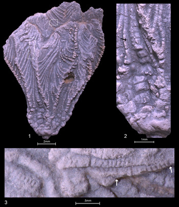
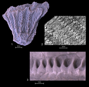
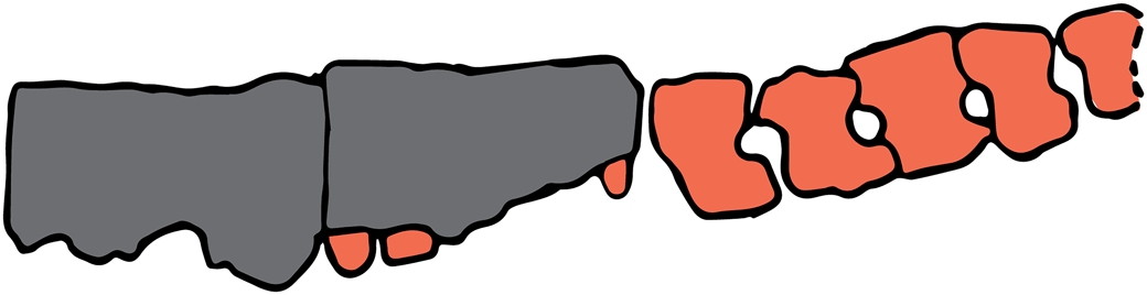
Proexenocrinus arm expressions include typical eucamerate morphology: pinnulate free arms beginning at secunidibrachial two and cuneate brachials. Proexenocrinus is the earliest-known crinoid to express pinnules. Despite this early but relatively derived ray configuration, Proexenocrinus pinnules express biserial pore-bearing floor plates. (Figs. 1.3, 3). This internal anatomy is unexposed except where brachials have been weathered. They are visible on the left branch of the holotype C arm. Here, pinnulars have, in part, been etched or broken away, revealing the floor plates beneath. Proexenocrinus floor plates are short, H-, or dumbbell-shaped plates. Podial pores are present between sequential elements. Least-weathered floor plates show pores expanding internally, with faint transverse keels on ridges separating these openings. Floor plates articulate with cover plates above. Cover plates are arranged in a single alternating biseries (Fig. 2.2). There are approximately three floor plates per pinnular.
Proexenocrinus inyoensis Strimple and McGinnis, Reference Strimple and McGinnis1972, SUI 134561a, holotype crown, C ray orientation, partly compressed, partial siliceous case-hardened coating. (1) Entire specimen with slightly depressed interrays, two fixed primibrachials, secundibrachials one and possibly two fixed, partial view of 10 pinnulate arms, pinnules branch from cuneate brachials in alternating direction, circular stalk with rounded nodal epifacets; boxes outline positions of high magnification enlargements; (2) enlargement of CD interray showing anitaxis, the mid-portion of which is crushed and offset, black arrow indicates exposed apparent lateral floorplate margins; (3) high-magnification images of box at left, rotated counterclockwise; pore-bearing floor plates visible as one half of biseries on three pinnules where pinnulars are etched away; arrows at right mark boundaries of line drawing in Figure 3 showing faint transverse keels; left side pinnule weathered, but still showing podial pores (see Fig. 2.3 for comparison with an edriosterid edrioasteroid).

(1, 2) Proexenocrinus inyoensis Strimple and McGinnis, Reference Strimple and McGinnis1972. (1) SUI 134562, paratype crown, anterior orientation, siliceous case-hardened coating over much of the specimen, shallow basal concavity, radials separated all around, interrays depressed, arms branching once per ray with primibrachial two axillary, round stalk with projecting round epifacets; (2) SUI 134561a, holotype, right hand box in Figure 1.1, high-magnification SEM image showing portions of four weathered pinnules, beveled by weathering, pinnulars more deeply etched, wedge-shaped cover plates standing in relief, best preserved cover plate arrangement on second pinnule from left showing alternating biseries; (3) PE 52690, edrioasterid sp. (Guensburg and Sprinkle, Reference Guensburg and Sprinkle1994), trilobite zone G2, early Floian, western Utah, portion of ambulacrum interior, showing large internal pockets between successive dumbbell-shaped floor plates, faint transverse keels between successive elements; comparable with much smaller Proexenocrinus floor plates in Figure 1.3 (see Bell and Sprinkle, Reference Bell and Sprinkle1978, pl. 2, fig. 10, pl. 3, fig. 1 for similar morphology in the middle Cambrian edrioasterid Totiglobus).

Proexenocrinus inyoensis Strimple and McGinnis, Reference Strimple and McGinnis1972; SUI 134561a, holotype, brachial- and floor-plate tracings, position indicated by arrows on Figure 1.3; two pinnulars on left (dark gray), floor plates exposed on right where pinnulars are stripped away (orange or lighter gray); width of segment indicated ~1.1 mm.

Posterior series
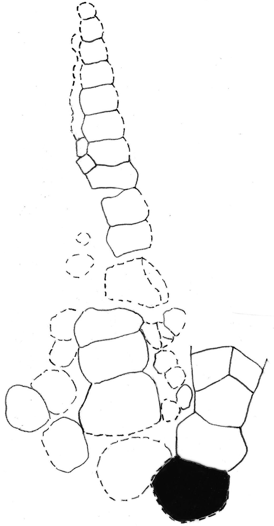
Proexenocrinus posterior plating is dominated by a linear distally tapering thick-plated, raised, sequential uniseries (Figs. 1.2, 4) (Strimple and McGinnis, Reference Strimple and McGinnis1972; Ubaghs, Reference Ubaghs, Moore and Teichert1978). This uniseries occupies most of the CD interray, and is flanked laterally on either side by smaller irregular plates. Preservation is insufficient to detect any pattern in these lateral plate fields. Varying sizes and shapes of these lateral plates suggest an irregular pattern. Plating arrangement surrounding the proximal-most plate of the uniseries appears to contact the C radial, but the exact topology is uncertain. The subsequent three plates in the series are large and thick, filling most of the CD interray. The distalmost of these three plates has a short suture on the upper left shoulder and a much longer suture on the upper right, resulting in a slight series offset toward the C ray. Distal to this asymmetrical fourth plate is a depressed area with at least one deeply embedded, poorly exposed, plate. The next more distal two plates are slightly ajar but in alignment. Beyond these, at least seven plates form a linear uniserial series of plates gradually diminishing in size. The series extends to the level of C ray secundibrachial 15, at which point it is no longer exposed.
Proexenocrinus inyoensis Strimple and McGinnis, Reference Strimple and McGinnis1972; SUI 134561a, holotype, CD interray plate tracings from much enlarged image. C ray plates on right, radial indicated in black, posterior plate dominated by anitaxis; anitaxis formed of thick-plated uniserial series; smaller, thinner, plates to either side; dashed lines indicate uncertain plate boundaries. Height of anitaxis as exposed ~5.8 mm.

Discussion
Floor plates
Proexenocrinus expresses the only known example of calcified floor plates in crinoid pinnules. These plesiomorphic axial expressions commonly occur in apinnulate earliest-known crinoid arms (Guensburg and Sprinkle, Reference Guensburg and Sprinkle2003, Reference Guensburg and Sprinkle2009; Guensburg et al., Reference Guensburg, Blake, Sprinkle and Mooi2016, Reference Guensburg, Sprinkle, Mooi, David, Lefebvre and Derstler2020a). The origins of this type of floor plate trace back to pentaradiate edrioasteroid-like (e.g., the middle Cambrian Stromatocystites pentangularis Pompeckj, Reference Pompeckj1896) and edrioasterid edrioasteroid ambulacra, where similar plates are embedded into the body wall of the primary thecal cavity (e.g., the middle Cambrian Totiglobus nimius Bell and Sprinkle, Reference Bell and Sprinkle1978, and later Ordovician taxa, such as edriosterid sp. of Guensburg and Sprinkle, Reference Guensburg and Sprinkle1994; see Fig. 2.3, for an example).
The finding of floor plates in pinnules fortifies previous evidence indicating that these constructs echo the pattern seen in arms where larger supporting skeletal structures, brachials, also derive from the extraxial body wall (Mooi ad David, Reference Mooi1997, Reference Mooi and David1998; Guensburg and Sprinkle, Reference Guensburg and Sprinkle2001; Guensburg et al., Reference Guensburg, Blake, Sprinkle and Mooi2016, Reference Guensburg, Sprinkle, Mooi and Lefebvre2020b). Floor and cover plates in both locations represent conserved plesiomorphic morphology. Interestingly, a similar but different continuity characterizes blastozoan (eocrinoids, diploporans, rhombiferans) feeding appendages, where only axial elements occur throughout (most recently Guensburg et al., Reference Guensburg, Sprinkle, Mooi, David, Lefebvre and Derstler2020a, b). Brachioles are similar in size to pinnules, but the supporting skeletal structures are axial homologs of floor plates. Thus, floor plates in blastozoans and crinoids are constructed in fundamentally different ways.
As stated above, floor plates of Proexenocrinus are exposed from below (i.e., floor plate surfaces that bounded the presumed coelomic cavity below the ambulacrum are seen). This view shows that each pore opens up into an interior cavity or pocket. Similar, although larger scale morphology can be seen in edrioasterid edrioasteroid and other early pentaradiate ambulacra (Fig. 2.3). Proexenocrinus floor plates should not be confused with lateral plating, which is extraxial expressions lying above pinnulars in crinoids of various ages, including modern comatulids, such as Poecilometra acoela (Carpenter, Reference Carpenter1888) (Clark, Reference Clark1915; see Guensburg et al., Reference Guensburg, Sprinkle, Mooi, David, Lefebvre and Derstler2020a, for further discussion).
The case for an anitaxis in Proexenocrinus
Previous studies of Proexenocrinus do not use the term anitaxis in describing the CD interray. The original description noted a posterior interray “occupied by one large plate followed in series by two large plates which are flanked by smaller plates” (Strimple and McGinnis, Reference Strimple and McGinnis1972, p. 72), but a later study reported a “CD interray probably undifferentiated” from other interrays (Ausich, Reference Ausich1986, p. 218).
A third early interpretation of Proexenocrinus taken from Ubaghs (Reference Ubaghs, Moore and Teichert1978, p. T441) explicitly described a “CD (posterior) interray with median series of plates accompanied by small series of plates on either side.” The Treatise glossary uses slightly different language for an anitaxis, “a linear succession of anal plates; commonly raised above laterally adjacent plates of interray” (Moore, Reference Moore, Moore and Teichert1978, p. T 231), but the meaning is essentially the same. Therefore, both the original description and a Treatise author describe anatomy that, while not specifically identified as such, agrees with a finding for an anitaxis. This is exactly what is described here in more detail (above), with the added finding that series plates are relatively thick and raised (Figs. 1.3, 4). Based on these multiple information sources, Proexenocrinus is concluded to have an anitaxis.
Among diplobathrid camerates, it is interesting that Trichinocrinus terranovicus Moore and Laudon, Reference Moore and Laudon1943a, which is also an early rhodocinitid (Dapingian, early Middle Ordovician), expresses an anitaxis as well (Moore and Laudon, Reference Moore and Laudon1943a; Ausich, Reference Ausich1998). No significance is attached to this apparent pattern pending formal analysis.
Acknowledgments
D. Quednau, Field Museum, assisted with figure preparation and T. Holstein, Field Museum assisted with SEM imagery. The reviews of C. Paul and an anonymous reviewer are appreciated. In addition, D. Blake, University of Illinois, Urbana, offered insightful manuscript suggestions and discussion. T. Adrain kindly loaned material from the University of Iowa.