




No CrossRef data available.
Article contents
Prenatal diagnosis of tetralogy of Fallot associated with a fistula from the left coronary artery to the left atrium
Published online by Cambridge University Press: 18 April 2005
Abstract
In a fetus at 20 weeks gestation, we found a large fistula from the left coronary artery to the left atrium in association with tetralogy of Fallot. Postnatally, the therapeutic issues were complex because of prematurity, low birth weight, decreased flow of blood to the lungs, and volume overload of the left ventricle because of the huge fistula. At three months, the baby underwent repair of tetralogy of Fallot, with surgical ligation of the fistula, but the baby died postoperatively.
Information
- Type
- Brief Report
- Information
- Copyright
- © 2003 Cambridge University Press
There are few reports of recognition of coronary artery fistulas in association with tetralogy of Fallot.1 To the best of our knowledge, the prenatal diagnosis of this combination has not previously been reported. In this report, we describe our recent experience in the prenatal detection and postnatal care of a baby with tetralogy of Fallot who also had a large fistula between the left coronary artery and the left atrial appendage.
Case report
A 32-year-old lady was referred for fetal echocardiography at 20 weeks gestation because of suspected congenital cardiac disease subsequent to a routine obstetric screening scan. The diagnosis of tetralogy of Fallot was confirmed, but it was noted that there was a large fistulous communication between the left coronary artery and the left atrial appendage (Figs 1 and 2). The subsequent scan at 27 weeks of gestation revealed dilation of the left atrium, cardiomegaly with a cardiothoracic ratio of 60–65 %, and shunting from the left coronary artery to the left atrium. The mother went into spontaneous labour at 33 weeks, and delivered a female infant who was small for dates with a birth weight of 0.99 kg.

Figure 1. Four chamber view of the fetal heart at 21 weeks gestation demonstrating the dilated left atrial appendage. Abbreviations: RA: right atrium; RV: right ventricle; LV: left ventricle.

Figure 2. View of the fistula from the circumflex branch of the left coronary to the left atrium at 28 weeks gestation (a). The aorta overrides a ventricular septal defect. The left coronary artery is dilated and the fistulous connection to the left atrial appendage is clearly seen. Colour flow Doppler (b) confirms the presence of flow across the fistula. Abbreviations: LAA: left atrial appendage; LCA: left coronary artery; LV: left ventricle; RV: right ventricle; VSD: ventricular septal defect.
The baby was resuscitated with intubation, and commenced on prostin at 5 ng/kg/min. All peripheral pulses were well felt, and a systolic murmur was heard at the upper left sternal edge. The chest X-ray showed cardiomegaly, and the electrocardiogram showed sinus rhythm, with the heart rate at 150 beats per min with the pattern of right bundle branch block. There was evidence for biatrial enlargement, and large equiphasic complexes were noted in the precordial leads, the so-called Katz-Wachtel phenomenon,2 suggesting biventricular hypertrophy.
Cross-sectional echocardiography showed a dilated left coronary artery, and a fistula extending from the circumflex branch of the left coronary artery to the anterior surface of the dilated left atrial appendage. The right coronary artery was normal. There was overriding of aorta, and antero-cephalad deviation of the muscular outlet septum, confirming the prenatal diagnosis of tetralogy of Fallot. Doppler interrogation revealed a pressure drop of 45 mmHG across the narrowed subpulmonary outflow tract. Left ventricular function was moderate.
The development of peripheral oedema, and low diastolic blood pressures, necessitated early withdrawal of prostin. The baby was ventilated for 10 days, and improved with regular diuretic therapy. Three emergency admissions were necessary over the next 2 months for episodes of desaturation and respiratory distress. A progressive increase in the Doppler-derived pressure drop across the right ventricular outflow tract gradient to 64 mmHG was documented.
The issues arising out of simultaneous left ventricular volume overload and reduced pulmonary blood flow were considered. The baby weighed 2.5 kg at 3 months. Repeated episodes of desaturation necessitated an early total correction of tetralogy of Fallot, and ligation of the coronary artery fistula. The echocardiographic findings were confirmed at surgery, which was successfully completed. Initially, the post-operative progress was uneventful, with good ventricular function and no residual flow across the fistulous communication. The infant was extubated successfully on the third post-operative day. On the fifth post-operative day, tachypnoea and increased inflammatory markers were treated with intravenous antibiotics. The following day, there was a sudden cardiorespiratory arrest and resuscitation was unsuccessful. A request for post-mortem examination was declined by the parents.
Discussion
Coronary arterial fistulas are rare, being found in 0.1–0.2% of patients who undergo coronary angiograms.3 Rarely, some small fistulas regress over time and undergo spontaneous closure. In published reports, fistulas from the left coronary artery account for about one-third of cases. Coronary fistulas are commonly recognised prenatally in the context of pulmonary atresia with intact ventricular septum,4 but isolated reports are rare.5 A description of the prenatal diagnosis of a fistula co-existing with tetralogy of Fallot has not, to the best of our knowledge, been reported previously. A small number of patients with coronary arterial fistulas develop symptoms of congestive cardiac failure from volume loading of the heart or myocardial ischaemia from a “coronary steal” phenomenon.6 The severity and progression of these fistulas can be monitored by serial echocardiography antenatally and postnatally.5
Surgical closure was the standard treatment in the past to treat haemodynamically significant fistulas, and to prevent myocardial ischemia, infective endocarditis and aneurysm formation.7 In the last decade, with advancement in interventional cardiology, most of these complex fistulas can be closed with various occlusion devices without the need for surgery.8–10 We elected for surgical ligation in this instance because of the associated cardiac defects and the low birth weight.
Our patient presented a number of challenges in postnatal management. She had signs of congestive cardiac failure due to the fistula, coupled with decreased saturations due to the progression of obstruction across the right ventricular outflow tract. Construction of a modified Blalock-Taussig shunt was considered inappropriate, because it would have increased aortic run-off to the pulmonary circulation. Balloon dilation of the right ventricular outflow tract was also excluded, because it would not have dealt with the coronary fistula. We elected, therefore, for total repair, despite a weight of only 2.5 kg.
References
Saxena A, Sharma S, Shrivastava S. Coronary arteriovenous fistula in tetralogy of Fallot: an unusual association. Int J Cardiol 1990; 28: 373–374.Google Scholar
Schamroth L. Electrocardiogram in congenital heart disease. In: Schamroth C (ed.) Introduction to electrocardiography by Leo Schamroth 7th edition. Blackwell Scientific Publications, Oxford, 1990, pp 261–308.
Liberthson RR, Sager K, Berkoben JP, Weintraub RM, Levine FH. Congenital coronary arteriovenous fistula. Report of 13 patients, review of the literature and delineation of management. Circulation 1979; 59: 849–854.Google Scholar
Patel CR, Shah DM, Dahms BB. Prenatal diagnosis of a coronary fistula in a fetus with pulmonary atresia and intact ventricular septum and trisomy 18. J Ultrasound Med 1999; 18: 429–431.Google Scholar
Sharland GK, Tynan M, Qureshi SA. Prenatal detection and progression of right coronary artery to right ventricle fistula. Heart 1996; 76: 79–81.Google Scholar
Dhillon R, Redington A, Kosutic J. Left atrial enlargement in infancy secondary to a fistula from the proximal left coronary artery. Cardiol Young 1999; 9: 228–229.Google Scholar
Schumacher G, Roithmaier A, Lorenz HP, et al. Congenital coronary artery fistula in infancy and childhood; Diagnostic and therapeutic aspects. Thorac Cardiovasc Surg 1997; 45: 287–294.Google Scholar
Mavroudis C, Backer CL, Rocchini AP, Muster AJ, Gevitz M. Coronary artery fistulas in infants and children: a surgical review and discussion of coil embolization. Ann Thorac Surg 1997; 63: 1235–1242.Google Scholar
Reidy JF, Anjos RT, Qureshi SA, Baker EJ, Tynan MJ. Transcatheter embolisation in the treatment of coronary artery fistulas. J Am Coll Cardiol 1991; 18: 187–192.Google Scholar
Sadiq M, Wilkinson JL, Qureshi SA. Successful occlusion of a coronary arteriovenous fistula using an Amplatzer Duct Occluder. Cardiol Young 2001; 11: 84–87.Google Scholar

Four chamber view of the fetal heart at 21 weeks gestation demonstrating the dilated left atrial appendage. Abbreviations: RA: right atrium; RV: right ventricle; LV: left ventricle.
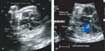
View of the fistula from the circumflex branch of the left coronary to the left atrium at 28 weeks gestation (a). The aorta overrides a ventricular septal defect. The left coronary artery is dilated and the fistulous connection to the left atrial appendage is clearly seen. Colour flow Doppler (b) confirms the presence of flow across the fistula. Abbreviations: LAA: left atrial appendage; LCA: left coronary artery; LV: left ventricle; RV: right ventricle; VSD: ventricular septal defect.
 You have
Access
- 12
- Cited by