INTRODUCTION
Changes in gastrointestinal (GI) physiology are a prominent feature of many GI disorders, which include infections by a variety of infectious agents (bacteria, viruses and parasites), chronic inflammatory bowel diseases (IBDs) and functional disorders such as irritable bowel syndrome (IBS). These changes include goblet cell hyperplasia and increased mucin secretion, increased fluid secretion and enhanced intestinal propulsive activity. However, little is known about the mechanisms of these changes in gut physiology or the precise role of these changes in host defence. Mucosal inflammation and immune activation in the gut by noxious agents may lead to changes in gut physiology, resulting in the development of an efficient defence which assists in the eviction of the noxious agent from the gut lumen.
Some parasitic nematode infections are of considerable importance in exploring the pathophysiology of many GI disorders. In the past decade, our laboratory has used primary infection of rats or mice with the nematode Trichinella spiralis as a model for studying the interface between immune and gut physiological systems in the context of host defence and the underlying mechanisms involved. The choice of this model is based on the existence of an extensive body of literature regarding the inflammatory and immune responses against this parasite (Table 1), and on the fact that many of the ingested parasites in this model are eventually expelled from the gut, forcing attention on the role of the physiological system in promoting the expulsion of these parasites from the intestine.
Advantages of the experimental model of Trichinella spiralis

This article reviews information from the studies of the immunological control of 2 major components of the GI physiological system, namely smooth muscle and goblet cells during nematode infection in the context of host defence and highlight its implication in understanding the pathophysiology of various GI disorders.
ALTERATION OF GOBLET CELL FUNCTION AND MUCIN PRODUCTION IN NEMATODE INFECTION
There is a continuous layer of mucus overlying the epithelium of the GI tract. This mucus layer has both protective and lubricative functions, protecting the mucosa from dehydration and mechanical damage, and also providing a physical barrier between the underlying epithelium and luminal contents, such as pathogenic bacteria, viruses, and parasites (Neutra and Forstner, Reference Neutra, Forstner and Johnson1987). Nevertheless, mucus is permeable to low molecular weight components, and this property is important for the intestinal absorption of nutrients. Mucins are the major macromolecular components of mucus, which are primarily synthesized and secreted by the goblet cells (Specian and Oliver, Reference Specian and Oliver1991). The concept of the mucus layer functioning as a dynamic protective barrier is suggested by studies showing an alteration in mucins under inflammatory conditions of the gut, a reduction in the numbers of goblet cells in germ-free animals and the enhanced mucus secretion in response to infections (Miller, Reference Miller1987; Kandori et al. Reference Kandori, Hirayama, Takeda and Doi1996). A selective decrease of colonic mucin subclass is observed in patients suffering from ulcerative colitis (Podolsky and Isselbacher, Reference Podolsky and Isselbacher1983). In addition, reduction in carbohydrate content and an increase in glycoprotein synthesis have been reported in colon carcinoma (Boland and Deshmukh, Reference Boland and Deshmukh1990) and coeliac disease (Crabtree et al. Reference Crabtree, Heatley and Losowsky1989), respectively. It has also been shown that enteric infection with Helicobacter pylori results in an alteration of mucin expression, which favours the attachment of the bacteria to the host's epithelia (Ota et al. Reference Ota, Nakayama, Momose, Hayama, Akamatsu, Katsuyama, Graham and Genta1998). A hyperplasia of mucin-secreting goblet cells has been described for a number of helminth infections, including that caused by T. spiralis (Ishikawa et al. Reference Ishikawa, Wakelin and Mahida1997; Khan et al. Reference Khan, Blennerhasset, Ma and Collins2001a; Sagar et al. Reference Sagar, Padol, Khan, Bonin, Blennerhassett and Hunt2004). In addition to an increase in goblet cell numbers, qualitative change in goblet cell mucin is observed during infection with another nematode, Nippostrongylus brasiliensis (Koninkx et al. Reference Koninkx, Mirck, Hendriks, Mouwen and van Dijk1988; Ishikawa et al. Reference Ishikawa, Horii and Nawa1993). The nature of the mucins in the intestinal goblet cells changes from neutral to acid (Koninkx et al. Reference Koninkx, Mirck, Hendriks, Mouwen and van Dijk1988), and an alteration of terminal sugars of goblet cell mucins (Ishikawa et al. Reference Ishikawa, Horii and Nawa1993) have been observed around the time of worm expulsion in N. brasiliensis infection.
Mucins are broadly divided into 2 groups. The first group comprises secreted mucins, which are the ‘gel-forming mucins’ found on the mucosal surface. The second group comprises the membrane-associated mucins found at the apical membranes of the epithelial cells. To date, 12 genes encoding mucin proteins have been identified (Corfield et al. Reference Corfield, Myerscough, Longman, Sylvester, Arul and Pignatell2000), of which 2 (designated MUC2 and MUC3 in humans and Muc2 and Muc3 in mice) are expressed in the intestine (Ho et al. Reference Ho, Niehans, Lyftogt, Yan, Cherwitz, Gum, Dahiya and Kim1993; Chang et al. Reference Chang, Dohrman, Basbaum, Ho, Tsuda, Toribara, Gum and Kim1994; Corfield et al. Reference Corfield, Myerscough, Longman, Sylvester, Arul and Pignatell2000). Muc2 is secretory and Muc3 is membrane-bound, both of which have been described to be upregulated in T. spiralis infection (Shekels et al. Reference Shekels, Anway, Lin, Kennedy, Garside, Lawrence and Ho2001). Increased amounts of the mucins and the Muc2 have also been reported in rats infected with N. brasiliensis (see Karlsson et al. Reference Karlsson, Olson, Jovall, Andersch, Enerback and Hansson2000; Olson et al. Reference Olson, Johansson, Klinga-Levan, Bouhours, Enerback, Hansson and Karlsson2002).
Goblet cells also produce a group of small cysteine-rich peptides, intestinal trefoil factor (ITF), which is also considered to have an important role in the mucus layer (Podolsky et al. Reference Podolsky, Lynch-Devaney, Stow, Oates, Murgue, De-Beaumont, Sand and Mahida1993; Sands and Podolsky, Reference Sands and Podolsky1996). ITF has been shown to enhance epithelial protection (Kindon et al. Reference Kindon, Pothoulakis, Thim, Lynch-Devaney and Podolsky1995), it facilitates the process of epithelial restitution and barrier function following gut injury (Dignass et al. Reference Dignass, Lynch-Devaney, Kindon, Thim and Podolsky1994; Mashimo et al. Reference Mashimo, Wu, Podolsky and Fishman1996) and its expression is upregulated in mice following T. spiralis infection (Kamal et al. Reference Kamal, Wakelin, Ouellette, Smith, Podolsky and Mahida1992).
Immunological mechanisms of goblet cell hyperplasia and mucin production
Although infection with T. spiralis leads to an enteropathy characterized by goblet cell hyperplasia and an upregulation of Muc2, Muc3 and ITF production/expression, the precise mechanisms regulating intestinal goblet cell and mucin responses and their precise role in host defence remain to be determined. Goblet cell hyperplasia in nematode (including T. spiralis) infections has been suggested to be under immunological control (Miller and Nawa, Reference Miller and Nawa1979; Khan et al. Reference Khan, Abe, Ishikawa, Nawa and Yoshimura1995; Ishikawa et al. Reference Ishikawa, Wakelin and Mahida1997). T cells are critical in many immune responses, including those associated with intestinal nematode infection. It has been shown that the treatment of T. spiralis-infected mice with cyclosporin A (which profoundly inhibits cytokine production) causes a reduction in the goblet cell hyperplasia during the infection (Garside et al. Reference Garside, Grencis and Mowat1992), implying a role for immune mediators in the control of goblet cell response in this nematode infection. In N. brasilensis infection, it has also been shown that treatment of mice 1 day before infection with a single dose of anti-CD4 antibody significantly reduced both the amount of intestinal mucus produced and the spontaneous expulsion of worms (Khan et al. Reference Khan, Abe, Ishikawa, Nawa and Yoshimura1995). In addition, activation of mucosal T lymphocytes of duodenal biopsies with monoclonal anti-CD3 antibody significantly increased both radio-isotope labelled glucosamine incorporation into glycoproteins and the secretion of radio-isotope labelled glycoproteins in vitro (Crabtree et al. Reference Crabtree, Heatley, Trejdosiewicz and Losowsky1990). These observations clearly suggest an important role for T cells in infection-induced intestinal goblet cell hyperplasia and mucus production.
Among the distinct CD4+ T helper (Th) cell subsets (Mosmann and Coffman, Reference Mosmann and Coffman1989), the Th2 type cells are important in the protective immunity in the host against many intestinal nematode infections, including that of T. spiralis, and are characterized by the expression of cytokines, such as IL-4, IL-5, IL-9, and IL-13 (Grencis, Reference Grencis1997; Else and Finkelman, Reference Else and Finkelman1998). IL-4 and IL-13 share the alpha chain of the IL-4 receptor, and their binding to this receptor results in an activation of a signalling pathway involving the signal transducer and activator of transcription factor 6 (Stat6) through the phosphorylation by Janus kinases 1 and 3. Once activated, Stat6 proteins form homodimers, translocate to the nucleus and bind to promoter regions to regulate gene transcription. In T. spiralis infection in mice, Th2 cells play the predominant role in the development of intestinal goblet cell hyperplasia (Ishikawa et al. Reference Ishikawa, Wakelin and Mahida1997). Studies from our laboratory have shown a critical role for Stat6 in the development of goblet cell hyperplasia during this nematode infection (Khan et al. Reference Khan, Blennerhasset, Ma and Collins2001a). There was no significant difference in the number of goblet cells between non-infected Stat6-deficient and non-infected wild-type mice, suggesting that there is a baseline level of goblet cells in Stat6-deficient mice (which are not regulated by immune-dependent components) (Khan et al. Reference Khan, Blennerhasset, Ma and Collins2001a). Stat6-deficient mice infected with T. spiralis did not develop an infection-induced goblet cell hyperplasia as compared with wild-type infected mice (Fig. 1). In addition, a significant attenuation of T. spiralis infection-induced goblet cell hyperplasia was observed when the Th2 immune response was ‘shifted’ towards a Th1 immune response by IL-12 gene transfer (Khan et al. Reference Khan, Blennerhasset, Deng, Gauldie, Vallance and Collins2001b). Taken together, Th2 cytokines are strong candidates for the proliferation/differentiation of intestinal goblet cells during T. spiralis infection, although we have recently shown that IL-9 is not a major contributor, because anti-IL-9 antibody treatment has no significant effect on T. spiralis infection-induced goblet cell hyperplasia (Khan et al. Reference Khan, Richard, Akiho, Blennerhasset, Humphreys, Grencis, Van Snick and Collins2003).
Intestinal goblet cell response in Trichinella spiralis-infected mice. (A) Light micrograph of small intestinal section stained with periodic acid-Schiff (PAS) from C57BL/6 wild-type (Stat6+/+) mice after T. spiralis infection on day 14 post-infection (p.i.). (B) Light micrograph of small intestinal section stained with PAS from T. spiralis-infected Stat6-deficient (Stat6−/−) mice on day 14 p.i. (C) Light micrograph of small intestinal section stained with PAS from control adenovirus (Ad5DL70-3)-treated T. spiralis-infected mice on day 10 p.i. (D) Light micrograph of PAS-stained small intestinal section from Ad5IL-12-treated and T. spiralis-infected mice on day 10 p.i.

Goblet cells are the main source of ITF, which is considered to be associated with mucin to increase the viscosity of mucus gel. A hallmark of the differentiation of intestinal goblet cells is the early expression of ITF (Thim et al. Reference Thim, Madsen and Poulsen2002). Recently, it has been demonstrated that IL-4 and IL-13 upregulate ITF from mucin-producing HT-29 CL.16E and HT-29 cells via the Stat6 pathway (Blanchard et al. Reference Blanchard, Durual, Estienne, Bouzakri, Heim, Blin and Cuber2004). In addition, it has also been shown that the transcription of MUC2 in human colon cancer cells is stimulated by IL-4 and IL-13 via a mitogen-activated protein kinase pathway (Iwashita et al. Reference Iwashita, Sato, Sugaya, Takahashi, Sasaki and Abe2003). As IL-4 receptors have been shown to be present on intestinal epithelial cells (Reinecker and Podolsky, Reference Reinecker and Podolsky1995), cultured airway epithelial cells (Dabbagh et al. Reference Dabbagh, Takeyama, Lee, Ueki, Lausier and Nadel1999) and HT-29 CL.19E cells (Blanchard et al. Reference Blanchard, Durual, Estienne, Bouzakri, Heim, Blin and Cuber2004), it is reasonable to presume that Th2 cytokines like IL-4 and IL-13 play an important role in ITF and mucin production by intestinal goblet cells.
Role of goblet cells/mucins in host defence
Mucins from goblet cells might play an important role in host defence during nematode infection in the gut by dislodging the established worms, or trapping the worms in the mucus and inhibition of parasite motility and feeding, thereby excluding them from their niche and favouring their expulsion (Miller, Reference Miller1987; Rothwell, 1989). Mucins might also mimic epithelial cell glycoproteins and thus prevent the attachment of microbes to the epithelium, or these mucins could facilitate the incorporation of mediators from inflammatory cells or of host antibody, which may have a deleterious effect on invading agents. The involvement of mucus in the expulsion of parasitic nematodes has been the subject of many investigations in various systems. Histological and perfusion studies revealed that N. brasiliensis worms failed to penetrate through mucus in primed rats (Miller, Reference Miller1987). In the same model, it has been shown that the pre-treatment of the immune intestine with mustard oil, which strips off the mucus layer, or with cysteine/papain, which is a mucolytic, allowed the establishment of worms in the intestine (Miller and Huntley, Reference Miller and Huntley1982). The entrapment of worms in the mucus and a possible barrier effect of mucus on the establishment of infective larvae were also observed during T. spiralis infection (Miller, Reference Miller1987). Recently, we showed that the inhibition of T. spiralis expulsion in Stat6-deficient mice and in mice following a switch of the immune response toward a Th1 type by IL-12 gene transfer was associated with significant reduction in the number of intestinal goblet cells (Khan et al. Reference Khan, Blennerhasset, Ma and Collins2001a, Reference Khan, Blennerhasset, Deng, Gauldie, Vallance and Collinsb). We also demonstrated that the expulsion of intestinal worms was substantially delayed in CD40 ligand-deficient (CD40L−/−) mice as compared with wild-type mice following T. spiralis infection (Khan et al. Reference Khan, Motomura, Blennerhassett, Kanbayashi, Varghese, El-Sharkawy, Gauldie and Collins2005). Consistent with the delayed worm expulsion, there was a significant impairment of intestinal goblet cell hyperplasia and Th2 cytokine responses in CD40L−/− mice (Khan et al. Reference Khan, Motomura, Blennerhassett, Kanbayashi, Varghese, El-Sharkawy, Gauldie and Collins2005). Taken together, these observations suggest that Th2 cytokines regulate the development of goblet cell hyperplasia in the gut during nematode infection via Stat6 activation, and that the increased number of goblet cells plays an important role in host protective immunity against the infection.
ALTERATION IN INTESTINAL MUSCLE FUNCTION IN NEMATODE INFECTION
In addition to changes in intestinal goblet cell function and mucin production, the intestinal phase of T. spiralis infection of mice or rats is accompanied by an increase in intestinal motility (Vermillion and Collins, Reference Vermillion and Collins1988; Vallance et al. Reference Vallance, Blennerhassett and Collins1997; Khan and Collins, Reference Khan and Collins2006). It has also been demonstrated that an enhanced propulsive activity occurs in extrinsically denervated gut segments of T. spiralis-infected guinea pigs (Alizadeh et al. Reference Alizadeh, Castro and Weems1987). This finding indicates that the enhanced, propulsive power of the motility apparatus has its origin, at least in part, in the tissues intrinsic to the gut wall, thus implicating enteric nerves and muscle. The final common pathway in the regulation of intestinal motility is the contractility of intestinal muscle, which can be evaluated in vitro using a longstanding tissue bath approach (Vallance et al. Reference Vallance, Blennerhassett and Collins1997). Clearly, changes in contractility observed in vitro cannot be extrapolated directly to the in vivo situation. Consideration must be given regarding the influences of enteric nerves, the interstitial cells of Cajal (ICC) and enteroendocrine (EE) cells. In addition, the roles of circular and longitudinal musculature differ in terms of their contribution to peristaltic activity. Nevertheless, muscle contractility is an acceptable and convenient method of assessing how inflammatory and immune cells alter this component of intestinal motility.
Studies of mice and rats have demonstrated a significant enhancement in intestinal muscle contractility in response to carbachol during primary infection with T. spiralis (see Vermillion and Collins, Reference Vermillion and Collins1988; Vallance et al. Reference Vallance, Blennerhassett and Collins1997). In applying this technique to studies of infection-induced changes in contractility, the hyperplasia and hypertrophy of muscle in this model have been observed (Blennerhassett et al. Reference Blennerhassett, Vignjevic, Vermillion and Collins1992). Thus, ‘force generation’ is expressed by unit cross-sectional area of the muscle along the axis of the contraction.
To address the question as to whether the increased muscle contractility during infection occurs as a result of the presence of parasites in the gut or the host's inflammatory response to them, T. spiralis-infected rats were treated with a corticosteroid (beta-methasone) and the myeloperoxidase (MPO) activity was investigated to evaluate the inflammatory response in the mucosa (Marzio et al. Reference Marzio, Blennerhasset, Chiverton, Vermillion, Langer and Collins1990). MPO is an enzyme contained in the azurophilic granules of neutrophils and other myeloid cells, and is commonly used as an index of neutrophil infiltration and acute inflammation (Smith and Castro, Reference Smith and Castro1978). Treatment with the corticosteroid inhibited the increase of MPO activity following infection, which was accompanied by an attenuation of intestinal muscle contractility despite the presence of worms (Marzio et al. Reference Marzio, Blennerhasset, Chiverton, Vermillion, Langer and Collins1990). This study implicated the inflammatory process rather than the parasite as the cause of the increased intestinal muscle contractility as a consequence of T. spiralis infection.
Immunological mechanisms of intestinal muscle hypercontractility
Infiltration of muscle layers by T lymphocytes during T. spiralis infection and the presence of a close communication between lymphocytes and muscle cells (as revealed by ultrastructural analysis) strongly suggest the possibility of a direct interaction between these cell types (Collins, Reference Collins1996). To investigate the role of T cells in intestinal muscle function, studies were performed using the T. spiralis model employing congenitally athymic mice, specific T cell subset-deficient (CD4+ cell or CD8+ cell-deficient) mice, and MHC II-deficient mice (Vallance et al. Reference Vallance, Croitoru and Collins1998, Reference Vallance, Galeazzi, Collins and Snider1999b). Infection-induced intestinal muscle hypercontractility is T cell-dependent, because changes were attenuated from infected athymic mice. The role of CD8+ T cells seems not to be significant, as there was no difference in the development of infection-induced muscle hypercontractility and in worm expulsion between CD8-deficient and wild-type mice (Vallance et al. Reference Vallance, Galeazzi, Collins and Snider1999b). However, CD4+ T cells appear to play a role, since CD4-deficient mice exhibited impaired muscle contractility; MHC II-deficient mice showed the greatest attenuation in the development of muscle contractility and also exhibited significant inhibition in worm expulsion (Vallance et al. Reference Vallance, Galeazzi, Collins and Snider1999b). The reconstitution of T cell function in athymic mice, by injecting splenic lymphocytes from euthymic mice prior to infection with T. spiralis, restored the profile of intestinal muscle contraction observed in euthymic mice and accelerated the expulsion of the parasite from the gut. To examine the role of CD4+ cells further, MHC II-deficient mice were reconstituted with purified CD4+ T cells and the effect on intestinal muscle contractility was assessed following infection. The results suggest partial restoration of muscle function during the infection, implying the importance of CD4+ T cells in enteric muscle function. Recently, we have shown that a Stat6-dependent Th2 immune response is critical for the development of intestinal muscle hypercontractility in T. spiralis infection (Khan et al. Reference Khan, Vallance, Blennerhasset, Deng, Verdue, Matthaei and Collins2001c) and have observed a significant reduction in infection-induced intestinal muscle contractility in Stat6−/− mice as compared with wild-type mice. The attenuated muscle contractility was associated with delayed worm expulsion from the intestine. Importantly, we were also able to demonstrate the presence of IL-4 and IL-13 in the lamina muscularis externa during the primary infection with T. spiralis in wild-type mice, whereas there no transcription was detected for IL-4 or IL-13 in infected Stat6−/− mice. It has been also shown that IL-4 and IL-13 enhances carbachol-induced contraction in dispersed smooth muscle cells isolated from the jejunum of mice (Akiho et al. Reference Akiho, Blennerhassett, Deng and Collins2002). These findings support the finding that Stat6 signalling promotes protective immunity against T. spiralis through mast cell- and T cell-dependent mechanisms (Urban et al. Reference Urban, Schopf, Morris, Orekhova, Madden, Betts, Gamble, Byrd, Donaldson, Else and Finkelman2000). These observations suggest that Th2 cytokines, IL-4 and IL-13, acting via Stat6, mediate the generation of infection-induced intestinal muscle hypercontractility. More recently, it has been reported that IL-4 and IL-13 play a key role in the generation of intestinal muscle contractility in mice infected with N. brasiliensis and Heligosomoides polygyrus (see Zhao et al. Reference Zhao, McDermott, Urban, Gause, Madden, Yeung, Morris, Finkelman and Shea-Donohue2003), further supporting our hypothesis that a Th2 response mediates muscle contractility in nematode infection.
To evaluate the role of Th1/Th2 immune responses in intestinal muscle function, muscle contractility was investigated following over-expression of IL-12 in T. spiralis-infected mice. IL-12 is a key cytokine in the differentiation of CD4+ Th cells toward Th1-associated responses by stimulating the production of IFN-γ by Th and natural killer cells (Manetti et al. Reference Manetti, Parronchi, Giudizi, Piccinni, Maggi, Trinchieri and Romagnani1993). It has been reasoned that if the Th2 response is critical for both the development of intestinal muscle hypercontractility and the expulsion of the worms, then a shift of the Th2 response toward a Th1 response should not only prolong the infection but also attenuate infection-induced muscle hypercontractility. Indeed, that is the case and a shift to Th1 response via an overexpression of IL-12 significantly altered intestinal muscle hypercontractility in this Th2-based, enteric infection (Khan et al. Reference Khan, Blennerhasset, Deng, Gauldie, Vallance and Collins2001b). In this study, IL-12 gene transfer using a single injection of the recombinant adenovirus vector expressing IL-12 (Ad5IL-12) into T. spiralis-infected mice effectively inhibited the development of infection-induced intestinal muscle hypercontractility and prolonged worm survival in the gut. This was accompanied by an upregulation of the Th1 cytokine IFN-γ and downregulation of Th2 cytokines IL-4 and IL-13 production. These findings clearly show that Th2 cytokines are prime candidates for the development of intestinal muscle hypercontractility in this infection. However, not all Th2 cytokines are critical, as it has been shown that IL-5 or IL-9 are not major contributors to the generation of T. spiralis-induced intestinal muscle hypercontractility (Vallance et al. Reference Vallance, Blennerhassett, Deng, Matthaei, Young and Collins1999a; Khan et al. Reference Khan, Richard, Akiho, Blennerhasset, Humphreys, Grencis, Van Snick and Collins2003). Deficiency in IL-5 or neutralization of IL-9 did not have a significant effect on muscle contraction in the host during this infection, which might be due to a dominant effect of IL-4 or IL-13.
To understand the initial events linking infection to the development of muscle hypercontractility, we recently examined the contribution of CD40-CD40L interaction in the development of intestinal muscle hypercontractility and in the Th2 response in CD40L-deficient (CD40L−/−) mice infected with T. spiralis (Khan et al. Reference Khan, Motomura, Blennerhassett, Kanbayashi, Varghese, El-Sharkawy, Gauldie and Collins2005). The interaction between CD40 ligand (CD40L) on T cells and CD40 on antigen-presenting cells has been shown to regulate both humoral and cellular immune responses (Grewal and Flavell, Reference Grewal and Flavell1996). Expulsion of intestinal worms was substantially delayed in CD40L−/− mice as compared with wild-type mice following T. spiralis infection (Khan et al. Reference Khan, Motomura, Blennerhassett, Kanbayashi, Varghese, El-Sharkawy, Gauldie and Collins2005). Consistent with delayed worm expulsion, there was a significant attenuation of intestinal muscle contractility in CD40L−/− mice (Khan et al. Reference Khan, Motomura, Blennerhassett, Kanbayashi, Varghese, El-Sharkawy, Gauldie and Collins2005). Infected CD40L−/− mice also exhibited marked impairment in the production of Th2 cytokines. These data suggest that the CD40-CD40 ligand interaction is an important, early event in the generation of a Th2 response and in the development of intestinal muscle hypercontractility in T. spiralis infection.
In addition to T cells, other cells such as mast cells, ICC and EE cells in the gut mucosa may play important roles in the development of intestinal muscle hypercontractility. Intestinal mucosal mastocytosis is a characteristic feature of nematode (including T. spiralis) infection (Grencis, Reference Grencis1997), and these cells are considered to have an important role in host protective immunity (Grencis et al. Reference Grencis, Else, Huntley and Nishikawa1993; Ha et al. Reference Ha, Reed and Croll1983). The activation of mast cells and release of their mediators have been implicated not only in the expulsion of the worms but also in the development of an altered physiology seen during the infection (Perdue and McKay, Reference Perdue and McKay1994; Goldhill et al. Reference Goldhill, Morris, Malizewski, Urban, Finkelman and Shea-Donohue1997; Lawrence et al. Reference Lawrence, Paterson, Higgins, MacDonald, Kennedy and Garside1998; Madden et al. Reference Madden, Young, Zhao, Gause, Finkelman, Katona, Urban and Shea-Donohue2004). Recently, an important role for mast cells has been demonstrated in epithelial cell function in T. spiralis infection, suggesting that the mast cells may contribute to worm expulsion in this infection by altering epithelial cell permeability (McDermott et al. Reference McDermott, Bartram, Knight, Miller, Garrod and Grencis2004). Studies of T. spiralis-infected c-kit-defective W/WV mice, which are mast cell-deficient, have revealed an attenuation of infection-induced intestinal muscle hypercontractility (Vallance et al. Reference Vallance, Blennerhassett, Huizinga and Collins2001). Bone marrow grafts from congenic wild-type mice restored mast cells as well as other haematopoietic cell populations to W/WV mice, but the attenuated muscle contractility persisted. A possible explanation for this observation was the demonstration that W/WV mice also lack a population of c-kit-dependent ICC. In spite of the restoration of mast cells, bone marrow reconstitution in W/WV failed to correct deficiencies in ICC, which might be a possible cause of the persistent altered muscle function in these mice (Vallance et al. Reference Vallance, Blennerhassett, Huizinga and Collins2001). ICC regulates myoelectrical slow wave activity and is important in the control of the propulsive motor activity of the small bowel (Huizinga et al. Reference Huizinga, Thunenberg, Kluppel, Malysz, Mikkelson and Bernstein1995). In the primary infection of mice with T. spiralis, structural damage has been observed in the network of ICC, which was associated with the appearance of ectopic pacemaker activity and the occurrence of retrograde peristalsis, implicating a major role for ICC in inflammation-induced gut motor disturbances (Der et al. Reference Der, Bercik, Donnelly, Jackson, Berezin, Collins and Huizinga2000). These findings were also evident in bone marrow-reconstituted W/WV mice. Together, these results indicate that non-bone marrow-derived cells such as ICC, in cooperation with mast cells, play separate but integrated roles in the function of intestinal muscle in mice with T. spiralis infection.
EE cells are specialized cells of gut epithelium and are dispersed throughout the mucosa of the gut. These cells release various biologically active compounds, such as cholecystokinin (CCK), chromo granin A (CGA) and serotonin (5-hydroxytryptamine, 5-HT), in response to various chemical and mechanical stimuli (Gershon, Reference Gershon1999; Lundgren, Reference Lundgren2002; Sharkey and Mawe, Reference Sharkey and Mawe2002). The most well-characterized subset of EE cells are the enterochromaffin (EC) cells, which synthesize and release the biogenic amine 5-HT (Cetin et al. Reference Cetin, Kuhn, Kulaksiz, Adermann, Bargsten, Grube and Forssmann1994; Sharkey and Mawe, Reference Sharkey and Mawe2002). 5-HT is an important enteric mucosal signalling molecule influencing gut physiology (motor and secretory function), and is considered important in maintaining intestinal homeostasis (Sharkey and Mawe, Reference Sharkey and Mawe2002). EC cells are located in close proximity to the mucosal sensory nerve endings and interganglionic neurons, being synaptic on motor excitatory and inhibitory neurons. An upregulation of EC cell numbers has been observed in infections of nematodes, including N. brasiliensis, T. spiralis or Trichuris muris (see Farmer and Laniyonu, Reference Farmer and Laniyonu1984; Wheatcroft et al. Reference Wheatcroft, Wakelin, Smith, Mahoney, Mawe and Spiller2005; Wang et al. Reference Wang, Steeds, Motomura, Deng, Verma-Gandhu, El-Sharkawy, McLaughlin, Grencis and Khan2007). Studies using T cell receptor (β×δ)-deficient mice demonstrated a reduced increase in EC cells in the small intestine in T. spiralis infection, suggesting a role for T cells in the development of infection-induced EC cell hyperplasia in this infection (Wheatcroft et al. Reference Wheatcroft, Wakelin, Smith, Mahoney, Mawe and Spiller2005). Recent work from our laboratory has shown an important immuno-endocrine axis in the gut, in which secretory products from CD4+ T cells interact with EC cells to enhance 5-HT production in the gut via Th2-based mechanisms (Wang et al. Reference Wang, Steeds, Motomura, Deng, Verma-Gandhu, El-Sharkawy, McLaughlin, Grencis and Khan2007). We also observed that EC cell and 5-HT responses in inflammation induced by T. muris infection are influenced by Th1 or Th2 cytokine predominance (Motomura et al. Reference Motomura, Ghia, Wang, Akiho, El-Sharkawy, Collins, Wan, McLaughlin and Khan2008). In addition, we have demonstrated that the increase in EC cell numbers in T. muris infection correlates well with an upregulation of colonic muscle hypercontractility (Motomura et al. Reference Motomura, Verma-Gandhu, El-Sharkawy, McLaughlin, Grencis and Khan2004). Recently, the 5-HT2A receptor has been shown to contribute to enteric infection-induced intestinal muscle hypercontractility, which further supports a role for 5-HT in alteration of muscle function in the gut during infection and inflammation (Zhao et al. Reference Zhao, Urban, Morimoto, Elfrey, Madden, Finkelman and Shea-Donohue2006).
Role of intestinal muscle hypercontractility in host defence
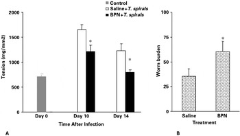
Previous work from our laboratory has shown that the inflammation-induced intestinal muscle hypercontractility is prominent in the proximal part of the intestine, and that the distal parts such as the ileum and colon exhibit reduced muscle contractility (Grossi et al. Reference Grossi, McHugh and Collins1993). Therefore, it seems likely that the distribution of changes would create an aboral gradient in muscle tension generation during infection, enhancing the aboral propulsion of luminal contents. If such forces contribute to the host's defence against parasite infection, then one might expect to see a relationship between the magnitude of intestinal muscle hypercontractility and the ability of the host to evict the parasites from the gut. This is indeed the case, with strong responders to T. spiralis infection (such as NIH Swiss mice which rapidly expel the parasite) exhibiting a greater degree of muscle hypercontractility and slow responders (such as B10.BR mice which slowly expel the parasite) exhibiting a lesser degree of hypercontractility (Vallance et al. Reference Vallance, Blennerhassett and Collins1997). These observations support the proposal for an association between host defence and changes in muscle function, and prompted a closer examination of the underlying immune-mediated mechanisms involved in the alteration of intestinal muscle contractility. Studies involving mice deficient in T cells, CD4+ cells, MHC II, CD40L and Stat6, and IL-12 gene transfer-mediated switching of the immune response towards Th1 type clearly demonstrate that infection-induced immune-mediated intestinal muscle hypercontractility in T. spiralis infection is associated with efficient worm expulsion. An attenuation of intestinal muscle contractility is associated with an inhibition of worm expulsion in this model. Recently, it was shown that worm expulsion is delayed in smooth muscle cell IL-4R-alpha-deficient mice infected with N. brasiliensis, suggesting a role for IL-4R-alpha-responsive smooth muscle cells in host defence in nematode infections (Horsnell et al. Reference Horsnell, Cutler, Hoving, Mearns, Myburgh, Arendse, Finkelman, Owens, Erle and Brombacher2007). It seems likely that cytokines, specifically IL-4 and IL-13 from the Th2 type cells, effect the secondary messenger system of muscle cells by acting either on the calcium channels or on the muscarinic receptor, which subsequently alters the intestinal muscle contractility; this alteration of contractility is associated with enhanced parasite eviction. We have investigated a causal link between these responses by administering an opiate (buprenorphine) to attenuate muscle hypercontractility in T. spiralis-infected mice, and determined whether this delayed worm expulsion occurred without attenuating the Th2 response. The results showed a marked inhibition of worm expulsion and intestinal muscle hypercontractility (Fig. 2) without impairing the IL-4 response, supporting the hypothesis that altered motility plays an important role in host defence in this model (Khan et al. Reference Khan, Motomura, Verdu, Blennerhasset, El-Sharkawy, Verma-Gandhu and Collins2004). Taken together, these findings indicate clearly that the intestinal motor apparatus acts as an extension of the immune system, aiding in the expulsion of worms through increased propulsive activity.
Intestinal muscle contractility and worm expulsion after opiate treatment. C57BL/6 mice were infected with Trichinella spiralis and were treated with opiate (buprenorphine; BPN) subcutaneously. Control mice received sterile saline. (A) Contraction of intestinal muscle. Maximum tension generated by intestinal muscle taken from non-infected and from BPN- and saline-treated mice after infection. (B) Worm recovery from BPN- and saline-treated mice after T. spiralis infection on day 14 p.i. * Significantly different (P<0·05) between BPN-treated and saline-treated infected mice.

CONCLUDING REMARKS
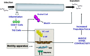
Data obtained from the studies of the mouse-T. spiralis model provide useful information on the effect of the immune system on gut physiology in the context of host defence. Physiological changes and worm expulsion in intestinal nematode infection seem to share a common immunological basis and may be causally linked. Studies have revealed that the infection-induced inflammation of the mucosa and the activation of immune cells, specifically of T cells, lead to an upregulation of intestinal mucin production and muscle function, which subsequently play an important role in host defence. A conceptual model of the interaction among mucosal inflammation, immune activation and gut physiological changes (specifically, the alteration in mucin production and muscle function) in the defence of the gut against an infectious agent is summarized in Fig. 3. The immune-mediated upregulation of mucus in response to nematode infection might trap the worms and prevent attachment to the epithelial surface; subsequently, an enhancement in propulsive activity assists the expulsion of the worms from the gut. These observations not only provide insight into the mechanisms of host defence in nematode infection but also have broader implications, considering widespread disturbances in mucus production, motility and other aspects of gut physiology which occur in a variety of clinical conditions (such as infections, IBD and IBS) associated with inflammation and immune activation. The T. spiralis-model is considered a powerful tool with which to explore visceral hypersensitivity and/or persistently altered motility following infection (Bercik et al. Reference Bercik, Wang, Verdu, Mao, Blennerhassett, Kean, Tougas and Collins2004; Kalia et al. 2008). Models of ‘post-infective IBS’ based on primary T. spiralis infection in NIH Swiss mice show that, while an initial immunological (Th2) response is required for the induction of the neuromuscular changes, it is not required for the maintenance of these changes post-infection. The state of persistent dysfunction of the neuromuscular tissue is maintained by the production of mediators, such as TGF-β and prostaglandin E2 by intestinal muscle cells (Barbara et al. Reference Barbara, Vallance and Collins1997; Akiho et al. Reference Akiho, Deng, Blennerhassett, Pelegrin, Grundy and Bardhan2005). Another important notion emerging from the studies in the mouse-T. spiralis model is that manipulation of the host immune response is able to modulate the accompanying physiological changes, which could have clinical relevance in the conditions associated with altered mucin production and intestinal motility. The information from these studies contributes significantly to the understanding of physiological changes in gastrointestinal infections. In addition to providing intriguing insight into the immunological control of intestinal homeostasis and host defence, these studies have the potential to lead to novel therapeutic strategies in gastrointestinal infections and/or inflammatory conditions.
Physiological changes in the gastrointestinal tract in the context of host defence – conceptual model. Infection with Trichinella spiralis produces mucosal inflammation and activates the Th2 type immune response. Th2 type immune response generated by the infection induces an upregulation in the intestinal mucin production and increased intestinal muscle contractility. Th2 cytokines also induce changes in other components of the motility apparatus such as EC cells. 5-HT released from EC cells act on muscle cells through 5-HT receptors. The resultant increases in mucus production and propulsive forces contribute to the expulsion of the worms from the intestine.

The author's research was supported by a grant from Canadian Institutes of Health Research (CIHR). The author thanks Dr Stephen M. Collins (McMaster University) for his valuable input; he holds a McMaster University Department of Medicine Internal Career Research Award.