

Introduction
Minerals of the pyrochlore supergroup have recently attracted considerable attention and several new members of the group have been described from various world localities (Atencio et al., Reference Atencio, Andrade, Christy, Gieré and Kartashov2010, Reference Atencio, Ciriotti and Andrade2013, Reference Atencio, Andrade, Bastos Neto and Pereira2017; Witzke et al., Reference Witzke, Steins, Doering, Schuckmann, Wegner and Pöllmann2011; Christy and Atencio, Reference Christy and Atencio2013; Biagioni et al., Reference Biagioni, Orlandi, Nestola and Bianchin2013; Hålenius and Bosi, Reference Hålenius and Bosi2013; Yang et al., Reference Yang, Li, Xiong, Pan and Yan2014; Jingwu et al., Reference Jingwu, Li, Guangming, Ge, Xu and Wang2015; Andrade et al., Reference Andrade, Atencio, Persiano and Ellena2013, Reference Andrade, Atencio, Chukanov and Ellena2015, Reference Andrade, Yang, Atencio, Downs, Chukanov, Lemée-Cailleau, Persiano, Goeta and Ellena2016; Li et al., Reference Li, Yang, Lu, Xiong, Ge, Pan and de Fourestier2016; Fan et al., Reference Fan, Ge, Li, Yu and Shen2016; Mills et al., Reference Mills, Christy, Kampf, Birch and Kasatkin2017). The pyrochlore-supergroup minerals are abundant in the Kovdor phoscorite-carbonatite pipe (Ivanyuk et al., Reference Ivanyuk, Yakovenchuk and Pakhomovsky2002, Reference Ivanyuk, Kalashnikov, Pakhomovsky, Mikhailova, Yakovenchuk, Konopleva, Sokharev, Bazai and Goryainov2016; Mikhailova et al., Reference Mikhailova, Kalashnikov, Sokharev, Pakhomovsky, Konopleva, Yakovenchuk, Bazai, Goryainov and Ivanyuk2016; Kalashnikov et al., Reference Kalashnikov, Yakovenchuk, Pakhomovsky, Bazai, Sokharev, Konopleva, Mikhailova, Goryainov and Ivanyuk2016), with an economic-level concentration in the axial zone of the pipe (Epstein et al., Reference Epstein, Basmanov, Berezina, Gol'dfurt, Zhuravleva, Nechaeva, Sokolov and Chernyshova1970). Roughly one quarter of the pyrochlore-supergroup minerals in Kovdor is the Na–Nb–OH mineral (Ivanyuk et al., Reference Ivanyuk, Yakovenchuk and Pakhomovsky2002), which is described herein as a hydroxynatropyrochlore, and the rest includes mainly hydroxycalciopyrochlore and U–Th-REE-rich hydroxykenopyrochlore (REE = rare-earth element), a recently approved mineral species (Miyawaki et al., Reference Miyawaki, Momma, Matsubara, Sano, Shigeoka and Horiuchi2017), which was known at Kovdor previously as ‘hatchettolite’. According to the previous reports (Yakovenchuk et al., Reference Yakovenchuk, Ivanyuk, Pakhomovsky and Men'shikov2005; Subbotin and Subbotina, Reference Subbotin and Subbotina2000), hydroxynatropyrochlore also occurs in Khibiny and Sebl'yavr in Murmanskaya Oblast', Russia, and other alkaline and alkaline–ultrabasic massifs. However, it has not been fully characterised (no fluorine content, structural data, etc.). Herein we report on the full characterization of the mineral, using the data obtained for the Kovdor sample.
The mineral is named in accordance with the pyrochlore supergroup nomenclature (Atencio et al., Reference Atencio, Andrade, Christy, Gieré and Kartashov2010). The new mineral and its name were approved by the Commission on New Minerals, Nomenclature and Classification of the International Mineralogical Association (IMA2017-074, Ivanyuk et al., Reference Ivanyuk, Yakovenchuk, Panikorovskii, Konoplyova, Pakhomovsky, Bazai, Bocharov and Krivovichev2017) The holotype specimen is deposited in the collections of the Mineralogical Museum of St. Petersburg State University, Russia, under catalogue number 1/19679.
Occurrence
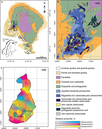
The Kovdor massif of peridotite, foidolite, melilitolite, phoscorite, carbonatites and related metasomatic rocks (diopsidite, phlogopitite, skarn-like rocks and fenite) is a Devonian (~400 Ma, Rodionov et al., Reference Rodionov, Belyatsky, Antonov, Kapitonov and Sergeev2012) volcano–plutonic complex situated within the Archean granite-gneisses in the Southwest part of the Murmansk region, Russia (67°33′N, 30°31′E). The axial part of this complex (6 km × 2 km) consists of peridotite, whereas its marginal zone (up to 1 km thick) is composed of the foidolite–melilitolite rock series (Fig. 1a). Intrusion of the alkaline melts at the contact between peridotite stock and host granite–gneisses caused intensive metasomatic alteration of both, with the formation of diopsidite, phlogopitite, melilite-, monticellite-, vesuvianite- and andradite-rich skarn-like rocks (after peridotite) and fenitised gneiss and fenite after granite–gneisses (Mikhailova et al., Reference Mikhailova, Kalashnikov, Sokharev, Pakhomovsky, Konopleva, Yakovenchuk, Bazai, Goryainov and Ivanyuk2016).
Geological maps of: (a) the Kovdor massif (after Mikhailova et al., Reference Mikhailova, Kalashnikov, Sokharev, Pakhomovsky, Konopleva, Yakovenchuk, Bazai, Goryainov and Ivanyuk2016) and (b) the phoscorite-carbonatite pipe (after Ivanyuk et al., Reference Ivanyuk, Kalashnikov, Pakhomovsky, Mikhailova, Yakovenchuk, Konopleva, Sokharev, Bazai and Goryainov2016); and (c) distribution of relative amounts of Na-, Ca-, and vacant-dominant pyrochlores within the pipe. Stars indicate localities of holotype (972/86.9) and cotype (K-017-4) hydroxynatropyrochlore.
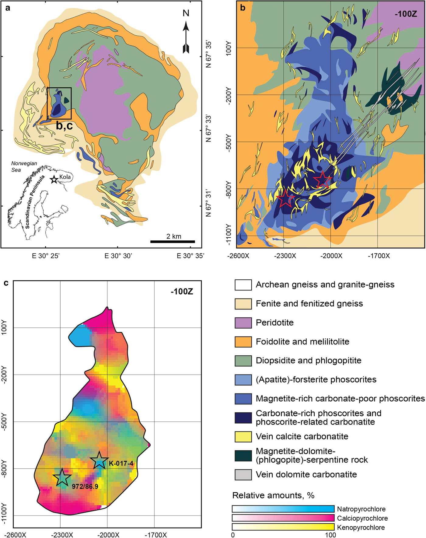
At the western contact of the peridotite and foidolite intrusions, there is a vertical concentrically zoned pipe of phoscorite–carbonatite rock series, with the later ring stockwork of vein calcite carbonatite and the linear zone of vein dolomite carbonatite (Fig. 1b). The concentric zonation of the phoscorite–carbonatite pipe is caused by the gradual transition of (apatite)–forsterite-rich phoscorite of the marginal zone into low-carbonate magnetite-rich phoscorite of the intermediate zone and then into carbonate-rich phoscorite and phoscorite-related carbonatite of the axial zone, and accented by concentration of vein calcite carbonatite approximately at the contact between the intermediate and axial zones (Mikhailova et al., Reference Mikhailova, Kalashnikov, Sokharev, Pakhomovsky, Konopleva, Yakovenchuk, Bazai, Goryainov and Ivanyuk2016; Ivanyuk et al., Reference Ivanyuk, Kalashnikov, Pakhomovsky, Mikhailova, Yakovenchuk, Konopleva, Sokharev, Bazai and Goryainov2016).
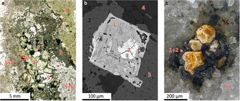
Within the phoscorite–carbonatite pipe, the content and grain sizes of the pyrochlore-group minerals gradually increase from the pipe walls towards the pipe axis (Ivanyuk et al., Reference Ivanyuk, Kalashnikov, Pakhomovsky, Mikhailova, Yakovenchuk, Konopleva, Sokharev, Bazai and Goryainov2016). In addition, there is a concentric zonation in distribution of calcium-, sodium- and vacancy-dominant pyrochlores (Fig. 1c). The marginal low-carbonate phoscorite includes only rare and tiny (up to 5 µm in diameter) grains of hydroxynatropyrochlore, while the axial carbonate-rich phoscorite and carbonatite are enriched in much larger (up to 500 µm in diameter) crystals of the same mineral. The calcite carbonatite that contains the holotype hydroxynatropyrochlore sample 972/86.9 is a fine-grained rock (Fig. 2a) consisting mainly of calcite (53 modal%), (tetraferri)phlogopite (30 modal%), hydroxylapatite (9 modal%) and magnetite (8 modal%). Characteristic accessory minerals include (in order of abundance): chamosite–clinochlore, ilmenite, pyrrhotite, valleriite, baddeleyite, U–Th-rich hydroxykenopyrochlore, hydroxynatropyrochlore (Fig. 2b), zirconolite, baryte, strontianite, chalcopyrite, galena and barytocalcite.
Crystals of hydroxynatropyrochlore (1) in calcite carbonatite 972/86.9 (a, b) and dolomite carbonatite K-017-4 (c). Photo of thin section in polarised light (a); BSE-image (b); and macroscopic photo (c). 2 – U-rich hydroxykenopyrochlore, 3 – hydroxylapatite, 4 – phlogopite, 5 – calcite, 6 – chlorite, 7 – magnetite with ilmenite inclusions and 8 – dolomite.

Hydroxynatropyrochlore also occurs in the latest dykes of dolomite carbonatite (see Fig. 1b) that cut through the axial carbonate-rich phoscorite and calcite carbonatite. In particular, dolomite carbonatite K-017-4 (see Fig. 1b,c) consists mainly of dolomite (85 modal%) and calcite (15 modal%) and contains numerous voids incrusted by the crystals of dolomite, calcite and magnesite overgrown by the hydroxynatropyrochlore grains (up to 300 µm in diameter, Fig. 2c) and compact two-zonal spherulites of gladiusite (in the core) and juonniite (in the marginal zone).
General appearance, physical and optical properties
In the phoscorite and calcite carbonatite, hydroxynatropyrochlore forms well-shaped cubic and truncated octahedral crystals (up to 700 µm in diameter) that usually contain irregularly shaped relics of amorphous U-rich hydroxykenopyrochlore (see Fig. 2b). In the veins of dolomite carbonatite, there are well-shaped truncated octahedral crystals of hydroxynatropyrochlore (up to 300 µm) with a thin (up to 50 µm) marginal zone of U-rich hydroxykenopyrochlore (see Fig. 2c).
Hydroxynatropyrochlore is brittle and has a conchoidal fracture. The cleavage is average on {111}. No twinning or parting were observed. The Mohs hardness is ~5. The density, determined by the sink-float method in Clerici solution, is 4.60(5) g cm–3, and the calculated density is 4.77 g cm–3 (using the empirical formula and single-crystal unit-cell parameters). The observed difference is most probably due to the admixtures of hydroxykenopyrochlore.
Macroscopically, hydroxynatropyrochlore is pale brown, with an adamantine to greasy lustre. The crystals are translucent to transparent, with a white streak. The mineral is isotropic, with the refractive index n = 2.10(5) at 589 nm. The mineral is non-fluorescent. A Gladstone-Dale calculation provides a compatibility index of 0.028 (with the empirical formula and single-crystal XRD data), which is regarded as excellent (Mandarino, Reference Mandarino1981).
Chemical composition
The chemical composition of hydroxynatropyrochlore was determined by wavelength-dispersive spectrometry using a Cameca MS-46 electron microprobe (at the Geological Institute, Kola Science Centre, Russian Academy of Sciences, Apatity) operating at 20 kV, 20–30 nA, with a 10 µm beam diameter. The standards used were: lorenzenite (Na and Ti), diopside (Ca), hematite (Fe), thorite (Th), synthetic LaCeS2 (Ce), and also metallic Nb, Ta and U. The fluorine content was determined using a LEO-1450 scanning electron microscope with a Quantax energy-dispersive microanalyser using fluorapatite as a standard. The water content was not determined due to the intimate intergrowth of hydroxynatropyrochlore and U–Ta-rich water-bearing hydroxykenopyrochlore. It was estimated from the weight deficit, whereas the presence of OH groups and absence of molecular water were confirmed by the Raman spectroscopy.
Analytical results for the structurally characterised holotype hydroxynatropyrochlore 972/86.9 are provided in Table 1 (see Figs 1c and 2b). The average chemical composition of the mineral corresponds to the following empirical formula (calculated on the basis of Nb + Ta + Ti = 2): (Na1.02Ca0.73Ce0.09Th0.09U0.05 ${\rm Fe}_{{\rm 0}{\rm. 02}}^{2 +} $)Σ2.00(Nb1.68Ti0.23Ta0.09)Σ2.00O6.03(OH1.04F0.01)Σ1.05. The simplified general formula is (Na,Ca,Ce)2Nb2O6(OH).
Composition of holotype hydroxynatropyrochlore.

*S.D. = standard deviation; b.d. = below detection limit.
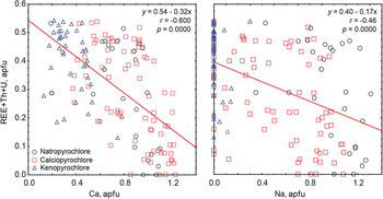
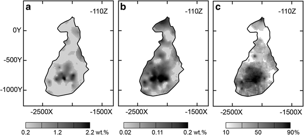
In the pyrochlore-supergroup minerals of the Kovdor phoscorite–carbonatite complex, both Na and Ca contents decrease with the increase in the total content of high-valent cations (Fig. 3), though the slope of the Ca-plot is steeper. This indicates that, most probably, natropyrochlore is a result of the complex substitution of Ca2+ by REE 3+, Th4+ and U4+ (±Na+ and □) in the Na–Ca pyrochlore series, (NaCa)Nb2O6(OH,F): NaCa ↔ NaCa0.7REE 0.2□0.1, NaCa ↔ NaCa0.5(Th,U)0.25□0.25, NaCa ↔ Na1.5REE 0.5 and NaCa ↔ Na1.6(Th,U)0.1□0.3. The increase of the total (U + Th) content above 20 wt.% results in the metamictisation of the mineral, the loss of Na and Ca, the increase of the H2O content, and the transformation of the mineral into hydroxykenopyrochlore. The F content in ‘natropyrochlores’ varies randomly from zero to 0.51 apfu. (atoms per formula unit), so a small portion of the samples investigated corresponds to fluornatropyrochlore.
Relation between contents of Ca, Na and high-valent cations in the A-position of the pyrochlore-supergroup minerals from the Kovdor phoscorite–carbonatite pipe. Legend: y is the total content of REE, Th and U, x is the content of Ca (left) or Na (right), r and p are correlation coefficient and probability value at 95% level of significance, respectively.

Hydroxynatropyrochlore dissolves slowly in hot HCl, and this process is activated by the mineral metamictisation due to the high contents of U and Th.
Raman spectroscopy
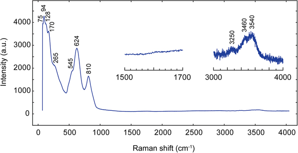
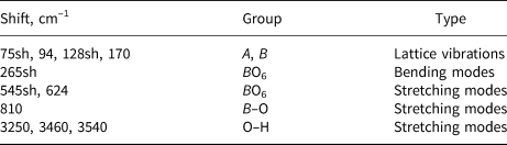
The Raman spectrum of holotype hydroxynatropyrochlore (Fig. 4) was obtained using a Horiba Jobin-Yvon LabRam HR 800 spectrometer (wavelength 514 nm). The assignments of the absorption bands have been made by analogy with other pyrochlore-like compounds (Bahfenne and Frost Reference Bahfenne and Frost2010; Mączka et al., 2012) and are given in Table 2. In the spectrum, there are weak bands of the ν1 stretching vibrations of hydroxyl groups, and there are no characteristic bands of the H2O vibrations at 1600 cm–1.
Raman spectrum of hydroxynatropyrochlore 972/86.9.
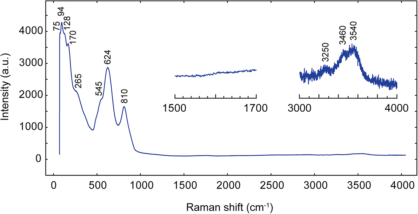
Raman shifts in the hydroxynatropyrochlore spectrum and their assignment.
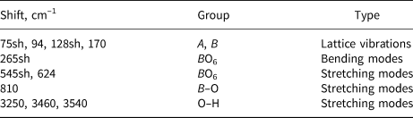
sh - shoulder.
Crystal structure
Experimental
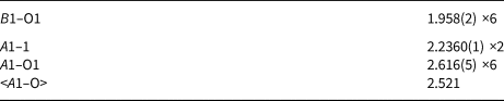
Single-crystal X-ray diffraction of the holotype hydroxynatropyrochlore 972/86.9 was performed at 293 K using a Bruker Kappa APEX DUO diffractometer equipped with an IμS microfocus source (beam size of 0.11 mm) operated at 45 kV and 0.6 mA and a CCD area detector, by means of monochromated MoKα radiation (λ = 0.71073 Å). The intensity data were reduced and corrected for Lorentz, polarisation and background effects. A multi-scan absorption-correction was applied by the Bruker software APEX2 (Bruker-AXS, 2014). The crystal structure was refined with the SHELX program (Sheldrick, Reference Sheldrick2008) in the Fd  $\bar{3}$m space group to R 1 = 0.026 (R int = 0.0222) for 80 independent reflections with F o > 4σ(F o). The crystal structure was drawn using the VESTA 3 program (Momma and Izumi, Reference Momma and Izumi2011). The scattering factors were calculated from the initial model using the Ca- and Na-scattering curves for the A site, the Nb- and Ti-scattering curves for the B site, and the O-scattering curves for other sites. All cation site occupancies are given in accordance with the electron-microprobe data (normalised on the basis of two B cations per formula unit). Crystal data, data collection information and structure refinement details are given in Table 3, atom coordinates and displacement parameters are listed in Table 4, and selected interatomic distances are provided in Table 5. The crystallographic information files have been deposited with the Principal Editor of Mineralogical Magazine and are available as Supplementary material (see below).
Data collection information and structure-refinement parameters for hydroxynatropyrochlore.

Atom coordinates, displacement parameters (Å2), site occupancies and site-scattering factors (SSFs, in epfu) for hydroxynatropyrochlore.

Selected interatomic distances in the crystal structure of hydroxynatropyrochlore.

Results
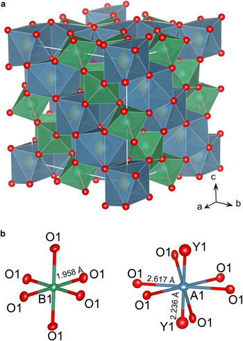
The crystal structure of hydroxynatropyrochlore (Fig. 5) belongs to the pyrochlore structure type with the ideal formula A 2B 2O6Y 1 (Atencio et al., Reference Atencio, Andrade, Christy, Gieré and Kartashov2010). It can be described as a three-dimensional octahedral framework of corner-sharing BO6 octahedra with A cations and OH groups in the interstices (Gargori et al., Reference Gargori, Galindo, Cerro, García, Llusar and Monrós2010; Yin et al., Reference Yin, Li, Yang, Ge, Xu and Wang2015).
A general view of crystal structure of hydroxynatropyrochlore: (a) in cation-centred polyhedral representation (AO8 polyhedra are blue, BO6 octahedra are green, oxygen sites shown as red spheres); and (b) coordination environments of the A and B sites. Displacement ellipsoids are drawn at the 50% probability level.

The calculated site-scattering factor for the A site is 18.4 epfu (electrons pfu), which is somewhat smaller than the value of 19.6 epfu derived from electron-microprobe data, probably due to the variable U content in different zones of the crystal studied. The A (16c) site is occupied predominately by Na, and its final occupancy can be given as (Na0.51Ca0.35□0.04U0.03Th0.035Ce0.035)Σ1.00. The octahedral B (16d) site has the site-scattering factor of 40.2 epfu, which agrees quite well with the assigned occupancy of (Nb0.84Ti0.095Ta0.065)1.00 (41.3 epfu). The O1 (48f) and Y1 (8a) sites are occupied by O and OH, respectively. The crystal chemical formula of the holotype hydroxynatropyrochlore can be written as (Na1.02Ca0.70□0.08Ce0.07Th0.07U0.06)Σ2.00(Nb1.68Ti0.19Ta0.13)Σ2.00[O5.96ОН0.04]Σ6.00(OH)1.00, which is in good agreement with the empirical formula.
Powder X-ray diffraction
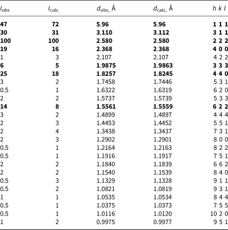
The powder X-ray diffraction pattern of the sample 972/86.9 (Table 6) was obtained using a Rigaku R-AXIS RAPID II diffractometer equipped with a cylindrical image plate detector using the Debye-Scherrer geometry (d = 127.4 mm; CoKα radiation). The data were integrated using the Osc2Tab/SQRay software (Britvin et al., Reference Britvin, Dolivo-Dobrovolsky and Krzhizhanovskaya2017). The unit-cell parameters refined from the powder data are as follows: Fd  $\bar{3}$m, a = 10.3211(3) Å, V = 1099.46(8) Å3 and Z = 8, which are in good agreement with the single-crystal data (see Table 3).
Powder X-ray diffraction data for hydroxynatropyrochlore*.

*The strongest lines are highlighted in bold.
Discussion
In the Kovdor phoscorite–carbonatite pipe, there was an intensive hydrothermal alteration of primary minerals, with the formation of numerous secondary phases: clinochlore or glagolevite after phlogopite, clinohumite and serpentine after forsterite, pyrochlore and zirconolite after baddeleyite, etc. (Mikhailova et al., Reference Mikhailova, Kalashnikov, Sokharev, Pakhomovsky, Konopleva, Yakovenchuk, Bazai, Goryainov and Ivanyuk2016; Ivanyuk et al., Reference Ivanyuk, Yakovenchuk and Pakhomovsky2002, Reference Ivanyuk, Kalashnikov, Pakhomovsky, Mikhailova, Yakovenchuk, Konopleva, Sokharev, Bazai and Goryainov2016). The higher content of Nb and Sc in baddeleyite from the axial carbonate-rich phoscorite and carbonatite due to the 2Zr4+ ↔ Sc3+ + Nb5+ substitution results in the intensive replacement of this mineral by pyrochlore-supergroup minerals (Fig. 6) accompanied by the formation of Sc phosphates (Yakovenchuk et al., Reference Yakovenchuk, Ivanyuk, Pakhomovsky, Panikorovskii, Britvin, Krivovichev, Shilovskikh and Bocharov2018):
 $$\eqalign{&24{\rm Bdy}^{\prime} + 12{\rm Dol} + 8{\rm Ap} + 56{\rm H}_2{\rm O} + 10{\rm C}{\rm O}_2 + 6{\rm N}{\rm a}^ + \cr & \quad = 6{\rm Pcl} + 34{\rm Cal} + 12{\rm Jnn} + 3{\rm H}_2,}$$
where Ap = hydroxylapatite; Bdy’ = Sc–Nb end-member of baddeleyite, Sc0.5Nb0.5O2; Cal = calcite, Dol = dolomite; Jnn = juonniite, CaMgScP2O8(OH)·4H2O; and Pcl = pyrochlore-supergroup member with formula NaCaNb2O6(OH).
Relation between the contents of Sc (a) and Nb (b) in baddeleyite and the occurrence of the pyrochlore-supergroup-bearing rocks (c) in the Kovdor phoscorite–carbonatite pipe.

Thus, in the Kovdor phoscorite–carbonatite complex, members of the pyrochlore supergroup are secondary hydrothermal minerals resulting mainly from the alteration of Nb-rich baddeleyite. Hydroxynatropyrochlore itself originates from the hypothetical Na–Ca pyrochlore, NaCaNb2O6(OH), due to preferential substitution of Ca by REE, Th and U. The increase of the U and Th contents results in its metamictisation. In addition, at the latest stage of the phoscorite–carbonatite pipe formation, an insignificant part of hydroxynatropyrochlore transforms into fluornatropyrochlore with the approximately equal contents of F and OH, by analogy with accompanied transition of hydroxylapatite into fluorapatite (Ivanyuk et al., Reference Ivanyuk, Kalashnikov, Pakhomovsky, Mikhailova, Yakovenchuk, Konopleva, Sokharev, Bazai and Goryainov2016).
Supplementary material
To view supplementary material for this article, please visit https://doi.org/10.1180/minmag.2017.081.102
Acknowledgements
We thank P. Leverett, D. Atencio and an anonymous reviewer for useful remarks. Authors are grateful to Drs A.O. Kalashnikov, J.A. Mikhailova and V.A. Sokharev for the co-operation in the geological study of the Kovdor phoscorite–carbonatite complex. The research is supported by the Russian Science Foundation, grant 16-17-10173. X-ray diffraction studies were undertaken at the XRD Research Resource Centre of St. Petersburg State University.