Introduction
Mucilage (slime) is common in different groups of plants, including algae, spore plants, gymnosperms and angiosperms. Within angiosperms, mucilage is described in both monocots and dicots (Grubert, Reference Grubert1974; Kreitschitz, Reference Kreitschitz and Gorb2009). It has been found in different plant organs, among them fruits and seeds (diaspores). Mucilage is responsible for various functions, such as water maintenance and diaspore supply, enabling the anchoring of the diaspore to the ground and rapid germination (Fahn and Werker, Reference Fahn, Werker and Kozlovsky1972; Huang and Gutterman, Reference Huang and Gutterman1999; Huang et al., Reference Huang, Gutterman and Hu2000; Ryding, Reference Ryding2001; Kreitschitz and Vallès, Reference Kreitschitz and Vallès2007; Kreitschitz, Reference Kreitschitz and Gorb2009). Due to its adhesive properties, mucilage also facilitates different methods of fruit and seed dispersal, e.g. by endo- and epizoochory, and hydrochory. Such diaspore transport, evidenced in short distances from the mother plant, also makes it possible to spread diaspores over longer distances, e.g. between islands (Sorensen, Reference Sorensen1986; Baiges et al., Reference Baiges, Espadaler and Blanché1991). The ability to produce mucilage and mucilage envelope formation differs between taxa, may depend on the diaspore morphology itself and may be induced by environmental conditions (Ryding, Reference Ryding2001; Kreitschitz and Vallès, Reference Kreitschitz and Vallès2007; Kreitschitz, Reference Kreitschitz and Gorb2009).
Mucilage is composed of polysaccharides, the main components of which are pectins. These are mostly polymers of galacturonic acid residues, with a strong ability for hydration. After wetting, pectins form a gel-like envelope around the diaspore (Frey-Wyssling, Reference Frey-Wyssling1959; Western et al., Reference Western, Debra and Haughn2000, Reference Western, Burn, Tan, Skinner, Martin-McCaffrey, Moffatt and Haughn2001; Kreitschitz and Vallès, Reference Kreitschitz and Vallès2007; Western, Reference Western2012). Thus, mucilage forms a colloidal solution that controls physical and chemical conditions (e.g. viscosity and surface tension) responsible for the proper development and functioning of the germinating diaspore (Broda, Reference Broda1948; BeMiller, Reference BeMiller1986).
Two types of mucilage have been described, i.e. ‘true slime’, characteristic of Linum sp., consisting mostly of pectins; and cellulosic mucilage, with an additional cellulose component in the form of a cellulosic skeleton. The second type is characteristic of, for example, Salvia spp., Ocimum basilicum or Artemisia spp. (Mühlethaler, Reference Mühlethaler1950; Wojciechowska, Reference Wojciechowska1966; Korobkov, Reference Korobkov1973; Mouradian, Reference Mouradian, Hind, Jeffrey and Pope1995; Yakovleva et al., Reference Yakovleva, Korobkov and Boyko2002; Kreitschitz and Vallès, Reference Kreitschitz and Vallès2007). Mucilage, particularly the cellulosic type, is important in better anchoring and establishing the seed in the soil (Gutterman et al., Reference Gutterman, Witztum and Evenari1967; Sorensen, Reference Sorensen1986). The presence of cellulosic fibrils makes mucilage more rigid and results in closer contact between the mucilage envelope and diaspore, it also prevents the pectins from being washed away from the envelope (Witztum et al., Reference Witztum, Gutterman and Evenari1969; Grubert, Reference Grubert1974; Mendu et al., Reference Mendu, Griffiths, Persson, Stork, Downie, Voiniciuc, Haughn and DeBolt2011).The ability to produce the mucilage is a particular characteristic of plants growing in dry habitats (Fahn and Werker, Reference Fahn, Werker and Kozlovsky1972; Huang and Gutterman, Reference Huang and Gutterman1999; Huang et al., Reference Huang, Gutterman and Hu2000). The genus Artemisia L. is represented by taxa distributed mainly in the steppe or semi-desert regions of the northern hemisphere. Many of them are woody shrubs or perennial herbs, but common weeds occupying mesotropic habitats are also frequent (Bremer, Reference Bremer1994; Polyakov, Reference Polyakov1995; Vallès et al., Reference Vallès, Garcia, Hidalgo, Martín, Pellicer, Sanz and Garnatje2011).
Diaspore morphology can be a reflection of habitat influences, causing different modifications which allow seeds/fruits better adaptation to a new environment (Stebbins, Reference Stebbins1971; Koch et al., Reference Koch, Bhushan and Barthlott2008). Evolutionary processes of myxospermatic diaspore adaptation may concern changes in the amount of mucilage production and may evolve very quickly, depending on habitat conditions and plant requirements (Ryding, Reference Ryding2001).
In a previous study, the patterns of distribution of mucilage cells and mucilage envelope formation in some Artemisia taxa were analysed (Kreitschitz and Vallès, Reference Kreitschitz and Vallès2007). In order to expand the present knowledge, studies were undertaken on other taxa from this genus. The results obtained should broaden knowledge about the morphology of myxocarpic diaspores, mucilage structure and its ecological function.
The main aims of this work were: (1) the examination of the presence of mucilage cells in the achene coat; (2) an analysis of mucilage envelope formation ability of different Artemisia taxa; (3) an assessment of connections between different factors, e.g. habitat type or ploidy level, on mucilage production; (4) to address other possibilities of water supply in cases of absence of mucilage cells or mucilage; (5) the characterization of the (cellulosic) mucilage in the Artemisia genus and its ecological function.
Materials and methods
Plant material
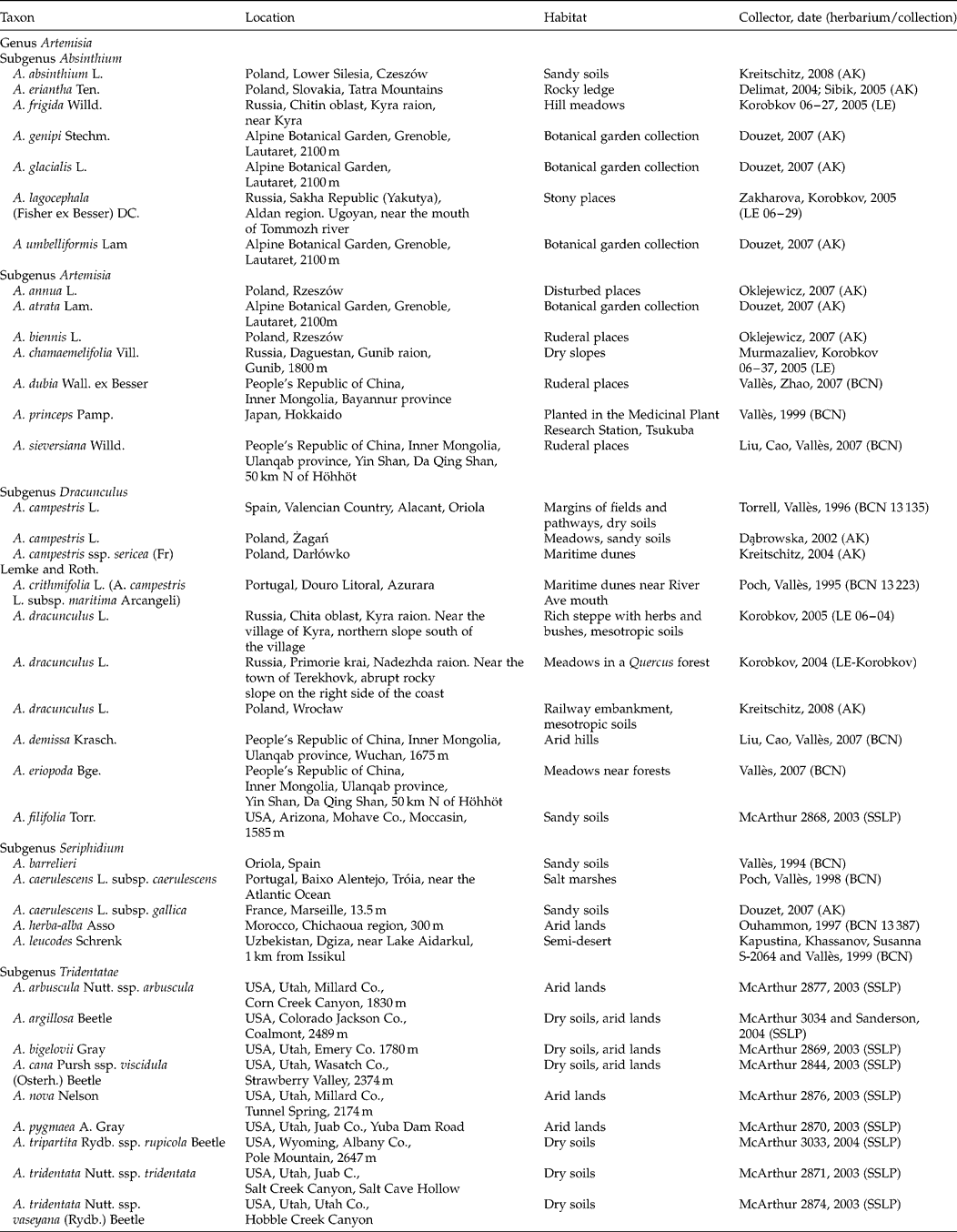
Mature achenes of 38 Artemisia taxa were collected mostly from natural populations; in four cases material was obtained from Botanical Garden collections (Table 1). Plant vouchers are deposited in the herbaria of the Centre de Documentació de Biodiversitat Vegetal de la Universitat de Barcelona, Spain (BCN); the Botanicheskyi Institut im. V.L. Komarova, Saint Petersburg, Russia (LE); the Shrub Sciences Laboratory, Provo, Utah, USA (SSLP) and the collection of A. Kreitschitz (AK).
Origin of the material studied

BCN, Centre de Documentació de Biodiversitat Vegetal de la Universitat de Barcelona, Spain; LE, the Botanicheskyi Institut im. V.L. Komarova, Saint Petersburg, Russia; SSLP, the Shrub Sciences Laboratory, Provo, Utah, USA; AK, the collection of A. Kreitschitz. In column 4, numbers indicate the herbarium number of the collected material; years indicate the year of collection.
Test for mucilage presence and mucilage type (chemical composition of mucilage)
For at least five seeds of each taxon a hydration reaction was performed to check the ability to form mucilage. Achenes were hydrated with water for 3–5 min and then the reaction was noted. When the mucilage envelope was formed, further reactions were performed, to identify the chemical composition of the mucilage. The following reagents, mostly in aqueous solution, were used to show the mucilage components: 0.1% (w/v) methylene blue (cellulose), 0.1% (w/v) ruthenium red (pectin), 1% (w/v) safranin (pectin, cellulose), 50% (w/v, alcohol solution) safranin with Alcian blue (pectin, cellulose) and 1% (w/v) crystal violet (cellulose). For the microstructural studies of the cellulose skeleton, 0.001% (w/v) calcofluor (fluorescent brightener) was used (Filutowicz and Kużdowicz, Reference Filutowicz and Kużdowicz1951; Braune et al., Reference Braune, Leman and Taubert1975, Wędzony Reference Wędzony1996). The images were taken using an Olympus BX-50 epi-fluorescent and light microscope connected to a DP71 camera with Cell B imaging software (Olympus BX50, Olympus Optical Co., Poland).
Results
Achene surface
The shape of the achenes is predominantly obovoid, obpyriform – wide in the apex part and narrower at the bottom. The apex is frequently rounded and flat or, rarely, with corona (e.g. A. umbelliformis), while the bottom is pointed with the hilum in the shape of small ring.
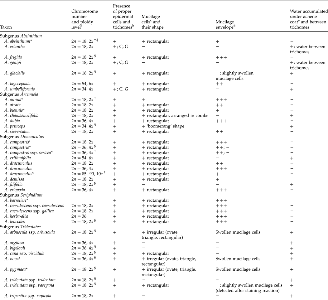
The main features of the achene studied are presented in Table 2. The achene surface is mostly glabrous and glossy. Only in subgenus Absinthium are covering and glandular trichomes present, and they are a characteristic trait for the three taxa studied, i.e. A. eriantha (Fig. 1A), A. genipi and A. umbelliformis (Table 2). The covering trichomes are T-shaped. The glandular trichomes (Fig. 1C) are two-celled, sessile and, together with clothing trichomes, sparsely cover the achene surface.
Achene characteristics and ploidy level of the studied taxa

Taxa studied previously: *Kreitschitz and Vallès (Reference Kreitschitz and Vallès2007).
a Chromosome numbers: determined by the author and/or obtained from: http://www.lib.kobe-u.ac.jp/infolib/meta_pub/G0000003asteraceae_e; †Kreitschitz and Vallès (Reference Kreitschitz and Vallès2003); §Garcia et al. (Reference Garcia, Sanz, Garnatje, Kreitschitz, McArthur and Vallès2004).
b Trichome types: C, covering; G, glandular.
c Mucilage cells: +, presence; − , absence.
d Abundance of mucilage envelope: +, weak; ++, mean; +++, abundant.
e Water accumulated under achene coat: +, presence; − , absence.
Mucilage detection in achenes of Artemisa. (A) A. eriantha: an arrow points at covering trichomes; (B) A. frigida with mucilage envelope formed; (C) A. genipi: green ‘dots’ indicated by arrows are glandular trichomes spread over the achene surface; (D) A. glacialis: white arrows point to a group of mucilage cells, while green arrows and outline indicate proper epidermal cells; (E) A. lagocephala mucilage envelope; an arrow indicates coiled cellulose threads; (F) A. umbelliformis: an individual mucilage cell is outlined and indicated by the arrows; (G) A. annua: uncoiled cellulose threads are indicated by arrows; (H) A. atrata: an arrow marks coiled cellulose threads; (I) A. chamaemelifolia: exemplary mucilage cells are outlined, white arrows indicate vertically spread strands (‘combs’) of the mucilage cells; (J) A. dubia mucilage envelope; (K) A. princeps: fragment of a row of mucilage cells, arrows and white line mark the outline of an individual mucilage cell; (L) A. sieversiana mucilage envelope; (M) A. campestris (2x, diploid) mucilage envelope; (N) A. campestris (4x, tetraploid): an arrow points to coiled cellulose threads; (O) A. crithmifolia: s, seed; ac, loosely adhered achene coat forming a space gathering water after hydration; (P) A. demissa: spirally coiled cellulose threads are indicated with an arrow; (Q) A. dracunculus mucilage envelope (2x, diploid); (R) A. dracunculus mucilage envelope (4x, tetraploid) (please note that images Q and R have the same magnification, × 10). Staining reactions: A, N, O, after hydration; B, E, J, L, Q, R, ruthenium red; D, F, I, K, safranin + Alcian blue; M, P, crystal violet; H, safranin; G, methylene blue; C, autofluorescence after excitation with green light (514 nm). Scale bars: A, B, 200 μm; C, L, O, 500 μm; D, E, G, H, Q, R, 100 μm; F, I, N, 50 μm; K, 20 μm.

Achene coat – proper epidermal cells and mucilage cells
The most external part of the fruit coat is composed of two types of cells (Fig. 1D), i.e. proper epidermal cells and mucilage cells, which together form the achene (fruit) coat. The achene coat is not integrated with the seed but loosely adheres to its surface, in some species even forming a gap/space between the seed and the coat, which fills with water after hydration (Fig. 1O). Proper epidermal cells are distributed along the long axis of the fruit. They are usually rectangular and elongated in the direction of the long axis of the seed (Fig. 1D, Fig. 2J).
Mucilage detection in achenes of Artemisia. (A) A. dracunculus (10x, decaploid; magnification × 10); (B) A. eriopoda; mucilage envelope; (C–E) A. barrelieri: (C) mucilage envelope; (D–E) fluorescence of the cellulose threads; (E) structure of the individual cellulose thread which is built of many thin, delicate microthreads; (F–H) mucilage envelope: (F) A. caerulescens; (G) A. herba-alba; (H) A. leucodes; (I, J) A. arbuscula: white arrows point to groups of mucilage cells after hydration, while a green line and arrow (J) indicate a proper epidermal cell; (K) A. pygmaea: arrows show hydrated mucilage cells with different shapes; (L) A. tridentata ssp. vaseyana: a group of mucilage cells after staining is indicated by white arrows. Staining reactions: A, F, ruthenium red; B, H, safranin; C, I–L, safranin + Alcian blue; D, E calcofluor and UV filter (fluorescence microscope). Scale bars: A, 100 μm; B, D, G, 200 μm; C, F, H, 500 μm; E, J, L, 50 μm; I, K, 20 μm.

The presence of mucilage cells was noted in the achene coat of 31 taxa out of 38 specimens studied (Table 2). In seven taxa, mucilage cells were not revealed and the achene coat was built only of proper epidermal cells (Table 2). The strands of mucilage cells are distributed along the long axis of the achene. Strips of the mucilage cells can cover almost the entire surface of the achene or they can alternate with strands of proper epidermal cells (Fig. 1D). The mucilage cells are usually rectangular (Fig. 1F, I, K). They can also be irregular, i.e. oval, ovate or triangular (Fig. 2I–K), which is a characteristic feature of three taxa: A. arbuscula, A. nova and A. pygmaea (subgenus Tridentatae), or they may also have a crescent shape, as in A. princeps (Fig. 1K). In most cases mucilage cells are arranged in ‘ladder-like’ rows (Fig. 1K, Fig. 2J) forming longitudinal strips along the long axis of the seed. The mucilage cells also form longer or shorter ‘ladder-like’ groups, which are very specific to A. arbuscula, A. nova, A. tridentata ssp. vaseyana (subgenus Tridentatae) (Fig. 2I–J, L) and A. glacialis (subgenus Absinthium) (Fig. 1D). A very special arrangement of the mucilage cells was observed in A. chamaemelifolia, where they form vertical rows (‘combs’) protruding above the achene surface (Fig. 1I). In A. umbelliformis, mucilage cells occur only at the outer and inner surface of the corona. The mucilage cells in the achene coat of A. glacialis were better detected after the staining reaction (Fig. 1D), a pattern also observed in A. tridentata ssp. vaseyana (Fig. 2L).
Mucilage type and ability to produce mucilage and/or to retain water under the achene coat and between trichomes
The mucilage components were verified based on staining reactions. The mucilage in the achenes of the studied Artemisia taxa belongs to the cellulosic type and is composed of a pectinous matrix and a cellulosic skeleton (Fig. 1B, E, G, H, L–N, P–R; Reference BeetleFig. 2A–H). Cellulose threads are immersed in the mass of pectins, forming a radial skeleton around the seed after hydration. They are coiled (Fig. 1B, E, H, J, L–N, P–R; Fig. 2B–C, F–H) or almost uncoiled under hydration and mucilage envelope formation (Fig. 1G, J). The degree of thread uncoiling depends on their lengths and the amount of pectins, in which they are embedded. It was also observed, after treating with calcofluor, that an individual (macro)thread was composed of many (several dozen) delicate, fine microthreads (Fig. 2D–E).
When considering the presence/absence of mucilage cells in the achene coat among all studied taxa, it was possible to distinguish three main groups (Table 2). The first includes achenes that have mucilage cells in the achene coat and form a distinct mucilage envelope after wetting, e.g. A. annua (Fig. 1G), A. eriopoda (Fig. 2B), A. herba-alba (Fig. 2G). The second is composed of fruits with mucilage cells which, however, are devoid of the ability to form a mucilage envelope after hydration, e.g. A. chamaemelifolia (Fig. 1I), A. crithmifolia (Fig. 1O), A. princeps (Fig. 1K), A. glacialis (Fig. 1D), A. tridentata ssp. vaseyana (Fig. 2L). A. umbelliformis also belongs to this group since it does not produce the mucilage envelope despite the presence of mucilage cells at the corona surface. The last group is formed by fruits without mucilage cells, e.g. A. eriantha (Fig. 1A), A. bigelovii or A. filifolia.
Interestingly, in seeds unable to produce mucilage, water is accumulated under the loosely adhering achene coat (Table 2), e.g. in A. crithmifolia (Fig. 1O). Furthermore, in cases of seeds covered with trichomes, the water can be additionally accumulated within or between the trichomes, as observed in A. eriantha (Fig. 1A), A. genipi and A. umbelliformis. In three taxa (A. arbuscula, A. nova, A. pygmaea) (Fig. 2I–K) water is maintained by both mechanisms: by mucilage and under the loosely adhering achene coat. However, in these species, the mucilage cells did not release their content after hydration and a mucilage envelope was not formed.
Mucilage and ploidy level
Within two taxa, representing the polyploid series (A. campestris, A. dracunculus), it was observed that the ability to form a mucilage envelope differed, most possibly in relation to the ploidy level. Achenes of diploid A. dracunculus form a not abundant, but visible, mucilage envelope (Fig. 1Q). In tetraploid accessions the mucilage envelope was the most distinctive (Fig. 1R), whereas in decaploid A. dracunculus mucilage cells swelled after hydration, but only some of them released their content (Fig. 2A). Diploid A. campestris formed a very abundant mucilage envelope (Fig. 1M). Conversely, in tetraploid A. campestris (Fig. 1N) and its coastal, also tetraploid, race A. campestris ssp. sericea, the mucilage envelope was narrow with a very limited pectin layer, and the cellulose threads stayed coiled upon wetting. In addition, some tetraploid specimens of A. campestris are completely devoid of the ability to form a mucilage envelope, although they possess typical mucilage cells at the achene surface. Out of about 45 studied specimens of A. campestris, coming from different locations, only two had the ability to form mucilage (Kreitschitz, Reference Kreitschitz2003; A. Kreitschitz, pers. comm.). In the case of A. campestris ssp. sericea, out of 10 accessions studied (data not shown) only two formed mucilage. A similar situation was observed in the hexaploid species A. crithmifolia, which belongs to the A. campestris group, as a probable vicariant taxon of this complex, and which is considered by some authors as A. campestris subsp. maritima Arcangeli. The whole achene surface in this taxon is covered with mucilage cells, but the fruits do not form a mucilage envelope, and thus water is retained under the achene coat (Fig. 1O).
Discussion
This study is the second on Artemisia achene mucilage characteristics. For most taxa the analyses were done for the first time (Table 2). The results obtained show some new details concerning the ability to form mucilage among different Artemisia taxa, as well as the micromorphological structure of the mucilage.
The main way of catching and maintaining water in the diaspores of the Artemisia taxa studied was through mucilage (slime) envelope formation. However, this ability can be attained by different strategies, which is particularly important for taxa devoid of a mucilage envelope. The strategies for water binding differ between taxa and depend on different factors, e.g. the habitat or the life form, the presence of mucilage cells (and/or their dysfunctions or absence), the occurrence of trichomes or ploidy level. The diversity observed can be a reflection of the adaptation potential of Artemisia taxa.
The mucilage envelope formation in the taxa studied was a characteristic feature of plants growing mostly in dry habitats and those occupying waste, disturbed locations. This pattern confirms observations by Fahn and Werker (Reference Fahn, Werker and Kozlovsky1972), Huang et al. (Reference Huang, Gutterman and Hu2000) and Ryding (Reference Ryding2001). Even the soil texture can be important in the case of myxocarpic seeds. Mucilage is especially beneficial in desert and other dry areas with bare ground (Ryding, Reference Ryding2001).
Mucilage may be produced under certain conditions, e.g. by plants growing in dry habitats. When its production is costly for the plant, the process may be quickly lost (Ryding, Reference Ryding2001). Such cases may be represented by an invasive taxon growing on waste ground, i.e. A. princeps, with mucilage cells but without mucilage production. The differentiation in habitat type, changing from dry to humid locations, may be a reason for the diversity of mucilage production. In humid conditions, seeds may not need additional support in water by mucilage (Kreitschitz and Vallès, Reference Kreitschitz and Vallès2007) and lose this facility (Ryding, Reference Ryding2001). The very intensive vegetative reproduction in A. princeps (J. Vallès, pers. comm.) could substitute for the lack of mucilage and thus rapid, effective germination. Similarly, an absence of mucilage was observed for A. vulgaris and A. verlotiorum occurring on mesotropic (humid, rich) soils, with ruderal preferences as well (Kreitschitz and Vallès, Reference Kreitschitz and Vallès2007). It was observed that Lamiaceae taxa growing in moist habitats more often had non-mucilaginous seeds as compared with those from drier areas (Ryding, Reference Ryding, Harley and Reynolds1992).
A characteristic of dry regions is the high salt concentration in the soil, which may inhibit germination. The mucilage envelope may serve here as a kind of filter or absorbent for the salt. Thus higher germination of the achenes of A. sphaerocephala with mucilage was observed in comparison to those with removed mucilage (Yang et al., Reference Yang, Dong and Huang2010).
The mucilage envelope is also an important strategy for annual plants. Among the taxa studied, A. annua, A. sieversiana and A. leucodes had abundant mucilage envelopes. In dry or waste (disturbed) habitats, myxocarpic seeds can benefit from this feature. Faster germination enables colonization of new habitats within a short time, and with great success (Young and Evans, Reference Young and Evans1973). Such areas occupied by a large number of only one taxon (A. annua, A. biennis) were observed (A. Kreitschitz, pers. comm.).
The absence of mucilage cells – alternative ways of gathering water
Glandular and covering trichomes on the achene surface were noted in three mountain taxa. All of them are closely related to each other (Gutterman, Reference Gutermann1979; Ehrendorfer, Reference Ehrendorfer and Lewis1980; Sanz et al., Reference Sanz, Vilatersana, Hidalgo, Garcia-Jacas, Susanna, Schneeweiss and Vallès2008). These taxa are devoid of mucilage formation ability. Only in A. umbelliformis were some mucilage cell strands observed at the pole, with the corona, but without mucilage production. The mucilage cells are located at the site where the root tip emerges, which may help at the first stages of germination, supplying the growing root with water and, thus, supporting germination. In addition, covering trichomes may help these taxa to catch water. They may form a ‘net-like’ structure which helps to keep the water around the seed. Trichomes do not form a hydrophobic barrier, as in many water plants (Neinhuis and Barthlott, Reference Neinhuis and Barthlott1997), but instead, after wetting, water fills the spaces between the trichomes and is kept around the achene. Such water-absorbing ability by simple trichomes is also known in other plant groups (Koch et al., Reference Koch, Bhushan and Barthlott2008). Apart from their possible role in water retention, covering hairs, in the absence of a mucilage envelope, may also be important in seed dispersal and seedling establishment (Wagner et al., Reference Wagner, Wang and Shepher2004; Koch et al., Reference Koch, Bhushan and Barthlott2008). On the other hand, the glandular trichomes, present only in these three taxa, may also be important for the seeds. It is known that such trichomes are responsible for insect immobilization, fungal toxicity, and water retention or temperature regulation (Wagner et al., Reference Wagner, Wang and Shepher2004; Koch et al., Reference Koch, Bhushan and Barthlott2008). Thus, regarding the protective abilities of mucilage (Kreitschitz, Reference Kreitschitz and Gorb2009 and references therein), the presence of glandular hairs can be relevant for achenes devoid of mucilage formation ability.
In Nepetoideae (Lamiaceae), nutlets covered with trichomes (also glandular) were not mucilaginous (Ryding, Reference Ryding, Harley and Reynolds1992, Reference Ryding2001). The same is true for A. eriantha, A. genipi and also A. umbelliformis, although the latter taxon possessed short strands of mucilage cells, but they were not fully functioning.
Another mode of water retention is the formation of an additional space between the loosely adhering achene coat and the seed. In all taxa devoid of mucilage formation ability (regardless of the presence or absence of the mucilage cells), a free space under the loose achene coat may be filled with water, and thus serve as a water reservoir, at least for a short time. This phenomenon also occurs in A. arbuscula, A. nova and A. pygmaea, in which the mucilage envelope is not fully formed, and probably functions as an additional water retention pool.
Cellulose mucilage function
The cellulose component of the mucilage was observed in the seed envelope of Arabidopsis thaliana, which consists of two domains, i.e. an inner layer which contains cellulose and the outer layer composed only of pectins (Macquet et al., Reference Macquet, Ralet, Kronenberger, Marion-Poll and North2007; Mendu et al., Reference Mendu, Griffiths, Persson, Stork, Downie, Voiniciuc, Haughn and DeBolt2011; Sullivan et al., Reference Sullivan, Ralet, Berger, Diatloff, Bischoff, Gonneau, Marion-Poll and North2011). Experiments showed that the outer layer was easy to remove in comparison to the inner, which stayed adhered. Cellulose is important for the maintenance of the inner mucilage layer attached to the seed (Macquet et al., Reference Macquet, Ralet, Kronenberger, Marion-Poll and North2007; Mendu et al., Reference Mendu, Griffiths, Persson, Stork, Downie, Voiniciuc, Haughn and DeBolt2011). In the case of the studied Artemisia taxa, such mucilage, consisting of many thin, delicate cellulose threads, may be built in a very complicated and strong network, preventing the pectins from loosening, and with them the trapped water. Such a function of the cellulose skeleton was described for Blepharis sp. (Witztum et al., Reference Witztum, Gutterman and Evenari1969) and Arabidopsis mucilage (Macquet et al., Reference Macquet, Ralet, Kronenberger, Marion-Poll and North2007; Mendu et al., Reference Mendu, Griffiths, Persson, Stork, Downie, Voiniciuc, Haughn and DeBolt2011; Sullivan et al., Reference Sullivan, Ralet, Berger, Diatloff, Bischoff, Gonneau, Marion-Poll and North2011). Cellulose mucilage can anchor strongly to the ground and adhere to animals (e.g. feathers) – a function which is important in seed distribution over a longer distance, thus myxocarpic seeds may travel further than those without mucilage (Sorensen, Reference Sorensen1986; Baiges et al., Reference Baiges, Espadaler and Blanché1991). Cellulose mucilage can also increase contact with the ground. Thus, after drying of pectins the formed cellulose skeleton may assure better adherence to the soil.
Specific types of mucilage in the Artemisia achenes, with a cellulosic skeleton, could also be a barrier against pathogens (e.g. fungi) or insects. The complicated structure of cellulosic mucilage, built of many thousands of microthreads, could be a difficult barrier to overcome. A better germination rate and absence of achenes infected by fungi were observed for some Artemisia taxa (e.g. A. annua) with a copious mucilage envelope (Kreitschitz and Vallès, Reference Kreitschitz and Vallès2007; A. Kreitschitz pers. comm.).
Mucilage and ploidy level
The presence of mucilage cells does not always guarantee mucilage envelope formation, as has already been shown here and in the previous work on Artemisia (Kreitschitz and Vallès, Reference Kreitschitz and Vallès2007). This ability (to form a mucilage envelope) may also depend on the ploidy level, as in the case of the polyploidy series of A. campestris and A. crithmifolia. A trend between mucilage production and ploidy level has been observed in three cytotypes of A. dracunculus. The decaploid variety has less mucilage and a less abundant mucilage envelope, which can be related to the high ploidy level. In such high polyploids, some abnormalities in the developmental processes are observed, e.g. disturbances in achene production and germination (Kreitschitz and Vallès, Reference Kreitschitz and Vallès2003 and references therein). It can be argued that for autopolyploids of A. campestris and A. dracunculus, there is some dysfunction within the genes responsible for mucilage production in achenes. Such differences in gene expression, resulting in morphological diversity, were observed in a potato autopolyploid series (Stupar et al., Reference Stupar, Bhaskar, Yandell, Rensink, Hart, Ouyang, Veilleux, Busse, Erhardt, Buell and Jiang2007). Autopolyploidy can alter gene expression and possibly modify plant phenotype (Pikaard, Reference Pikaard2001).
Polyploidy is an apomorphy present in the Artemisia genus in all of the groups (Torrell et al., Reference Torrell, Garcia-Jacas, Susanna and Vallès1999). It could explain the diversification of mucilage production ability between diploid and polyploid taxa. Polyploidy is a powerful factor in the speciation of the Artemisia genus (Vallès et al., Reference Vallès, Garcia, Hidalgo, Martín, Pellicer, Sanz and Garnatje2011), and it is therefore likely that some polyploids may not have stabilized genotypes and that adaptation processes are still ongoing. A good example may be A. abrotanum, probably a young, unstable polyploid (4x), which spreads only through vegetative multiplication by shoots (Kreitschitz, Reference Kreitschitz2008).
Subgenus Tridentatae
The taxa from Tridentatae have some individual features present only in this group, e.g. the irregular shape of the mucilage cells, their arrangement in groups and a swelling reaction without mucilage envelope formation. Such diaspores with enclosed mucilage were observed in different plants. The main role suggested for such diaspores was a water supply for the growing embryo. These seeds cannot adhere to animals, thus their dispersion may be limited (Ryding, Reference Ryding2001). The members of Tridentatae are endemic to North America (Beetle, Reference Beetle1960; McArthur and Sanderson, Reference McArthur and Sanderson1999; Garcia et al., Reference Garcia, McArthur, Pellicer, Sanderson, Vallès and Garnatje2011 and references therein). The taxa studied occur in dry habitats; some of them possess mucilage cells but do not form the mucilage envelope. This feature shows an additional evolutionary trend in Tridentatae which clearly separates this section from the other Artemisia subgenera. The separation of Tridentatae has previously been demonstrated on the basis of cytogenetic studies (Torrell et al., Reference Torrell, Garcia-Jacas, Susanna and Vallès1999; Sanz et al., Reference Sanz, Vilatersana, Hidalgo, Garcia-Jacas, Susanna, Schneeweiss and Vallès2008).
On the other hand, new cytogenetic data show the heterogeneity of this section, from which species such as A. pygmaea and A. argilosa were excluded (Garcia et al., Reference Garcia, McArthur, Pellicer, Sanderson, Vallès and Garnatje2011). Based on the present achene morphological studies, taxa such as A. arbuscula ssp. arbuscula, A. nova and A. pygmaea show common features in their achene coat structure (irregular shape of the mucilage cells), which group them together. These facts can support the heterogeneity of the genus Artemisia and different evolutionary tendencies concerning morphological adaptations (in the achene structure) which could develop independently from the genetic changes.
Interestingly, there is also a feature common to Tridentatae, which is shared by A. glacialis (subgenus Absinthium) which has mucilage cells organized in groups. It is supposed that the changes in mucilage cell distribution in these taxa [forming two different groups, clearly separated geographically as well as in the phylogeny of the genus (Sanz et al., Reference Sanz, Vilatersana, Hidalgo, Garcia-Jacas, Susanna, Schneeweiss and Vallès2008; Garcia et al., Reference Garcia, McArthur, Pellicer, Sanderson, Vallès and Garnatje2011)] may have evolved independently, and may be a result of adaptation to local habitats (in fact quite different: semi-desert in the North American taxa and high mountain in the European species).
Concluding remarks
The presence of mucilage in Artemisia achenes is a beneficial feature for several reasons. It provides the diaspore with water and enables faster germination and dispersion over longer distances. The additional cellulosic skeleton cements the seed more strongly to the soil and prevents pectins from being washed away from the mucilage envelope. The results obtained show that the presence of mucilage cells and mucilage envelope formation exemplify the different evolutionary tendencies in the Artemisia genus, members of which have adapted to diverse habitats. The described ability to form mucilage may have evolved independently and may be connected to the speciation of particular groups (such as the subgenus Tridentatae).
Acknowledgements
The author would like to thank Professor Joan Vallès (Universitat de Barcelona) for his comments and for supplying material; Dr Edyta Gola (University of Wrocław) for discussion and comments; Dr A.A. Korobkov, Professor E.D. McArthur, Dr K. Oklejewicz, Dr Jozef Šibík and Mr A. Delimat, for supplying material. I would also like to thank my colleagues from the Department of Plant Morphology and Development (University of Wrocław) for their critical revision of the manuscript and Mr Ian Anderson for correction of the English language.