Introduction
Isolated sperm cells and egg cells, in the absence of somatic tissue, provide useful material for the study of fertilization in higher plants under controlled conditions. In vitro fertilization (IVF) is a technique that induces sperm and egg fusion outside the plant body. In this way, it is possible to investigate mechanisms that control the processes of egg activation and fertilization in a highly monitored experimental system. Kranz et al. (Reference Kranz, Bautor and Lörz1991) first achieved IVF of maize using isolated sperm and egg cells, and later generated fully fertile plants (Kranz and Lörz, Reference Kranz and Lörz1993). Fourteen years later, Uchiumi et al. (Reference Uchiumi, Uemura and Okamoto2007) reported a second instance of IVF in higher plants in rice. The isolation of sperm, egg and zygote cells of higher plants and in vitro fusion provide a platform for experimental investigations, using cellular and molecular biology, of the development of the sperm, egg and zygote (Wang et al., Reference Wang, Kuang, Russell and Tian2006). Numerous sperm-expressed genes have been identified (von Besser et al., Reference von Besser, Frank, Johnson and Preuss2006; Berger, Reference Berger2008; Bayer et al., Reference Bayer, Nawy, Giglione, Galli, Meinnel and Lukowitz2009; Frank and Johnson Reference Frank and Johnson2009) and their transcribed products have been shown to be essential for fertilization and normal embryogenesis (Gou et al., Reference Gou, Yuan, Wei and Russell2009; Russell et al., Reference Russell, Gou, Wei and Yuan2010, Reference Russell, Gou, Wong, Wang, Yuan, Wei, Bhalla and Singh2012). Mechanisms that regulate the transition from egg to zygote are being increasingly more fully elucidated (Sauter et al., Reference Sauter, von Wiegen, Lörz and Kranz1998; Okamoto et al., Reference Okamoto, Scholten, Lörz and Kranz2005; Sprunck et al., Reference Sprunck, Baumann, Edwards, Langridge and Dresselhaus2005; Yang et al., Reference Yang, Kaur, Kiriakopolos and McCormick2006; Abiko et al., Reference Abiko, Maeda, Tamura, Hara-Nishimura and Okamoto2013; Leljak-Levanić et al., Reference Leljak-Levanić, Juranić and Sprunck2013). All results indicated that isolated gamete cells of higher plants can be analyzed directly during IVF using modern molecular probes that allow the regulation of sexual reproductive development in angiosperms to be more fully appreciated.
However, IVF of higher plants has been successfully achieved only in maize and rice, suggesting that methods for studying this process in higher plants continue to pose hidden challenges. A major obstacle appears to be the isolation of sperm cells and egg cells (Wang et al., Reference Wang, Kuang, Russell and Tian2006). To date, viable egg cells have only been successfully isolated in about 10 species, and this has limited the progress in performing IVF research in higher plants. Leek (Allium tuberosum Roxb) is a very popular vegetable crop in China and in much of the world. In the present study, we describe a protocol for the successful isolation of living sperm using an in vivo–in vitro method, and a method that combines enzymatic maceration–digestion and mechanical dissection of egg, zygote and proembryo cells from leek.
Materials and methods
Allium tuberosum Roxb var. HuangGeZi was grown under greenhouse conditions at Xiamen University. Androecium and gynoecium maturity are asynchronous in the species. At anthesis (Fig. 1A), the style is only 1 mm long, however, which is an inadequate size to accept pollen grains. The third day after anthesis, the style has reached approximately 4 mm length, the stigma inflates and produces papillae cells, which are suitable for pollen germination and success fertilization (Fig. 1B).
Inflorescence and gynoecia of A. tuberosum. (A) Inflorescence of A. tuberosum at anthesis. (B) Two gynoecia of A. tuberosum, left exhibiting a 1 mm style at anthesis, and right a 4 mm style with an inflated stigma 2 day after anthesis.

Generative cell isolation
Allium tuberosum bears bicellular pollen grains, each containing a generative and vegetative cell at anthesis. Fresh pollen grains were collected from A. tuberosum anthers at anthesis and hydrated in 1 ml of 9–20% mannitol (473–983 mOsmol/kg H2O) solution for 10 min. Following hydration, 1 ml of 4.5–10% mannitol solution was added to induce osmotic shock, which ruptured the pollen grains to release the pollen contents, including the generative cell.
Inducing in vitro pollen tube
In the mannitol solution, pollen grains do not germinate. Therefore, a suitable medium that induces pollen grain germination and subsequent tube formation must be prepared, such as polyethylene glycol (PEG) or sucrose. The following medium was prepared for in vitro pollen tube induction: 0.1% (w/v) KH2PO4, 0.05% (w/v) CaCl2, 0.01% (w/v) boric acid, 0.01% (w/v) KH2PO4 in addition to 10–20% PEG (4500), and 5–30% sucrose. Three populations containing 100 pollen grains were counted to produce the frequency of pollen germination and ruptured grains or pollen tubes in culture, and average pollen tube length was counted from 100 pollen tubes.
Inducing semi-in vitro pollen tube by an in vivo–in vitro technique
Flowers were hand pollinated and pollen tubes allowed in vivo growth within the styles for 3 h. Then, the pollinated style was excised, and the cut end was immersed in a medium containing 0.05% (w/v) CaCl2, 0.01% (w/v) boric acid, 0.01% (w/v) KH2PO4, and 15% (w/v) sucrose. Following a period of 6–8 h of incubation, some pollen tubes grew out of the cut end of the style, and were subsequently transferred to a ‘bursting’ solution to cause the pollen tubes to rupture in the solution, releasing pollen tube contents, including two sperm cells.
Egg and zygote isolation
Allium tuberosum produces six ovules with single integument per ovary. In each assay, 30 ovules were dissected from five blooming flowers and incubated in an enzymatic solution containing 0–0.8% Pectolyse Y-23, 0–1% cellulase (Onozuka RS), 6–12% mannitol and 0.01% CaCl2 with gentle shaking at 25°C for 20–40 min. The maceration solution was passed through a Pasteur pipette several times to facilitate enzyme digestion. Following incubation, ovules were transferred to the same solution but without enzymes for mechanical dissection. A dissecting needle was used to remove the outer integument and to cut the nucellus at the middle part, then the micropylar end was gently squeezed to facilitate release of the egg apparatus. Generally, the three cells comprising the egg apparatus were released from the cut end of the ovule. Zygotes were isolated 10 h after pollination following the same method.
Early proembryo isolation
Proembryos were isolated at 15 h or later after pollination using the same procedure. Each isolated proembryo was transferred into a diluted enzymatic solution containing 0.2% cellulase (Onozuka RS) and 0.2% pectinase (Serva) for 10 min. Then, proembryo cells were separated and collected using a micromanipulator.
The isolated cell viability was evaluated using fluorescein diacetate (FDA) reaction (Heslop-Harrison and Heslop-Harrison, Reference Heslop-Harrison and Heslop-Harrison1970). The osmolality of the isolation solution was measured using an osmometer (OSMOMAT 030).
Results
Isolation of generative cells
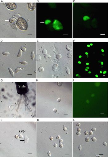
Generative cell isolation was successful using the described protocol. Pollen grains ruptured and the contents were released, including a generative cell (Fig. 2A). Each newly released generative cell displayed strong FDA fluorescence (Fig. 2B), confirming their viability. At the beginning of pollen rupturing, pollen cytoplasm content also showed some FDA fluorescence as well (Fig. 2B) but, soon, pollen grain cytoplasm fluorescence disappeared, and only the generative cell exhibited viable fluorescence (Fig. 2C). The released generative cells can be collected as a population using a micromanipulator (Fig. 1D). Pollen rupture is related to the osmolality of the isolation generative cell solution. Although most pollen grains ruptured in a 9% mannitol solution, the generative cells quickly changed to a rounded up in shape (Fig. 1E) and also ruptured. A decreased number of ruptured pollen grains were observed by increasing the mannitol solution to 20%, and the released generative cells retained a fusiform shape for approximately 10 min and still exhibited fluorescence for over 15 min, indicating that they remained viable (Fig. 1F).
Isolation of the generative cell and sperm cells of A. tuberosum. (A) A ruptured pollen grain after osmotic shock that releases its generative cell (arrow). (B) Fluorescence reaction of fluorescein diacetate (FDA) confirms the viability of the released generative cells (arrow). (C) FDA fluorescence of pollen content diminished in intensity at 10 min after pollen rupturing. (D) Population of collected generative cells that still retain their fusiform in shape at 10 min after release from pollen grains. (E) Population of collected generative cells that become spherical in shape by 20 min after release from pollen grains. (F) Population of collected generative cells with FDA fluorescence at 20 min after isolation. (G) Pollen tubes (arrow) growing out of the cut end of the style following in vivo–in vitro culture. (H) A pair of sperm cells released from a ruptured pollen tube. (I) FDA fluorescence reaction confirmed the viability of the released sperm cells. (J) Released male germ unit (MGU) (Russell, Reference Russell1984) with the two sperm cells (Svn: sperm associated with the vegetative nucleus) and a vegetative nucleus (arrow). (K) Population of collected Svn sperm cells. (L) Population of collected Sua sperm cells. Bar=10 µm.

Isolation of sperm cells
Mature pollen grains of A. tuberosum consist of a generative cell and a vegetative cell, and two sperm cells are formed in the growing pollen tube from mitotic division of the generative cell. Therefore, pollen tube formation must be initiated before sperm cells can be isolated. When mature pollen grains were incubated in a medium containing 0.05% (w/v) CaCl2, 0.01% (w/v) boric acid, 0.1% (w/v) KH2PO4 and 5–30% (w/v) sucrose (194–1231 mOsmol/kg H2O, pH 5.6), some grains germinated and produced pollen tubes. In medium containing 10–15% sucrose, pollen grain germination exceeded 30%, and average pollen tube length reached 127.03 µm following 1 h of growth. However, in the medium containing sucrose as an osmotic regulator, pollen tube growth ceased after 1 h of culture and many of the pollen tubes ruptured (Table 1).
The frequency of pollen germination and broken in different sucrose solutions

Note: Pollen collection was conducted 1 h after culture. n = 100.
Pollen grain germination was also effective using PEG as an osmotic regulator. In medium containing 15% PEG 4500, pollen grain germination exceeded 60%, average tube length (n = 100 tubes) was 338.67µm, and broken tube frequency was 22.33% following 1 h incubation. However, in this growth medium, pollen tube growth ceased after 3 h of culture, and average tube length was 514.33 µm. In the 15% PEG and 10–20% sucrose medium, the frequency of pollen germination sharply increased and the frequency of broken tubes decreased. In the 15% PEG and 10–15% sucrose (710 mOsmol/kg H2O) medium, over 80% pollen grain germination was detected (Table 2), and tube length averaged 704.23 µm following 3 h of culture.
The effect of different solutions of sucrose on pollen tube growth

Note: Pollen collection was conducted 1 h after culture. n = 100.
In vitro pollen tubes were transported into a ‘bursting’ solution containing 9% mannitol (473 mOsmol/kg H2O), which led to osmotic shock that ruptured the tube and released the tube content. However, in many cases, in vitro pollen tubes released just one cell, presumably the generative cell. This observation indicated that the generative cell of in vitro pollen tubes did not divide to form two sperm cells.
Flowers were emasculated before anthesis, and hand pollinated 2 days later. The pollinated styles were grown in vivo for 3 h. Each entire style was excised, and the cut ends were immersed in a medium containing 0.05% (w/v) CaCl2, 0.01% (w/v) boric acid, 0.01% (w/v) KH2PO4, and 15% (w/v) sucrose, with an osmolality of 553 mOsmol/kg H2O, at pH 5.5. Following a 6–8 h incubation, pollen tubes grew out of the cut end of the style (Fig. 2G). The cut end of the style was subsequently transferred to a ‘bursting’ solution containing only 6% (w/v) mannitol (328 mOsmol/kg H2O). Osmotic shock caused the pollen tubes to rupture in the solution, and the pollen tube contents, including two sperm cells were released (Fig. 2H), both of which also displayed viable fluorescence (Fig. 2I). The newly released sperm cells from pollen tubes were generally elongated, but quickly began to round up in 6% mannitol solution. At a suitable time, two sperm cells were easily identified, because the tube cytoplasm soon disperses in the solution and the vegetative nucleus (arrow) associated with one of the sperm cells (Fig. 2J). Two sperm cells could be separately collected as the Svn (sperm associated with the vegetative nucleus) (Fig. 2K) and the Sua (sperm unassociated with the vegetative nucleus) population (Fig. 2L) using a micromanipulator.
Isolation of egg cells
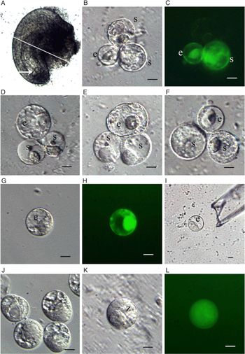
The peeled ovules of A. tuberosum were incubated in an enzymatic maceration solution for 30 min. Then the ovules were transferred to a solution without enzymes to remove their integuments. The outline of the embryo sac in the nucellus of each ovule was clearly observable (Fig. 3A). This step is critical for locating the precise position of the nucellus for dissection. Each treated nucellus was cut into two parts using a glass microneedle and egg apparatus cells were then released from the cut end of the ovule by squeezing the micropyle end of the nucellus (Fig. 3B). The isolated egg and synergid cells displayed a strong fluorescence, which suggested that both cell types remained viable following the isolation procedures. Of the two synergid cells, the larger one displayed weaker fluorescence (Fig. 3C). In the isolation solution, the egg apparatus was easily distinguishable as the egg cell and synergids because the three cells were connected and exhibited a larger size than somatic nucellus cells. The development of the egg apparatus cells includes a process that increases cell size. At anthesis, the three cells exhibited a notable difference in size, the egg cell was smaller than the two synergid cells (Fig. 3D). At 1 day after anthesis, the three cells increased in size, and displayed greater vacuolation (Fig. 3E). At 2 days after anthesis, the cells of the egg apparatus are mature, and the isolated egg cells had an average diameter of approximately 130 µm (n = 10). The two synergid cells were dimorphic, one measuring 150 µm diameter and the other 180 µm (n = 10) (Fig. 3F). In a released mature egg apparatus, the synergid cell nuclei were located on one side of each synergid cell, and displayed the same polarity, which differed from that of the egg cell. Egg cells just released from the embryo sac were pyriform, but soon changed to a rounded shape. The isolated mature egg cells exhibited a large central vacuole and a peripheral nucleus (Fig. 3G), which displayed bright fluorescence, suggesting vigorous viability (Fig. 3H). Isolated egg cells were collected using a micromanipulator (Fig. 3I), and a population of 5–8 egg cells was collected from 30 ovules within 1 h (Fig. 3J).
Isolation of egg cell and zygote of A. tuberosum. (A) The nucellar part of the ovule after the integument has been softened by enzymatic digestion and then removed from the integument by dissection, displaying its egg apparatus in the embryo sac (arrow). The line indicates the position of the cut during dissection. (B) The three egg apparatus cells released from the embryo sac. (C) FDA fluorescence reaction confirmed the release of a viable egg and a synergid cell in (B). Another synergid cell has very weak fluorescence intensity. (D) Egg apparatus isolated from the embryo sac of a flower at anthesis. (E) Egg apparatus isolated from the embryo sac of a flower at 1 day after anthesis, showing an increase in cell size. (F) Egg apparatus isolated from a flower at 2 days after anthesis, showing a decrease in cell size. At this time, the stigma is receptive to pollen and pollen germination can proceed. (G) Egg cell isolated from the egg apparatus. (H) FDA fluorescence reaction confirmed egg cell viability. (I) Isolated egg cell was collected using a micromanipulator pipette. (J) Population of five egg cells, two only partially visible in the field of view. (K) Zygote isolated from a pollinated flower at 12 h. (L) FDA fluorescence reaction confirmed zygote viability in (K). e, egg; s, synergid cell; z, zygote. Bar=10 µm.

Isolation of zygotes
The same isolation method was used to collect zygotes of A. tuberosum from pollinated flowers. At 10 h after pollination, most ovules exhibited one-celled zygotes (Fig. 3K), and compared with egg cell florescence, the zygote florescence distribution displaying great dispersion (Fig. 3L). The isolated zygotes were also collected as a population.
Isolation of proembryos and its cells
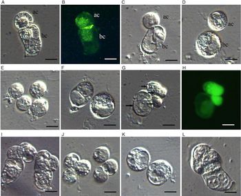
At 15 h after pollination, the zygote of A. tuberosum divides to form a two-celled proembryos. The zygote divides transversely into a small apical cell toward the interior of the embryo sac and a large basal cell toward the micropyle. Using the same isolation method as the egg cell, we could isolate various stages of early proembryos according to the pollination time. Ovules at 15 h after pollination generally released a two-celled proembryo, in which the apical cell became rounded and the basal cell remained elongate (Fig. 4A). Both proembryo cells displayed viable fluorescence with little difference in fluorescence intensity between them (Fig. 4B). Following incubation in the enzymatic solution (0.2% cellulase and 0.2% pectinase), the two proembryo cells separated (Fig. 4C) and then completely detached and became a rounded protoplast (Fig. 4D). The apical cell population (Fig. 4E) and basal cell population (Fig. 4F) were separately collected using a micromanipulator. Each ovule at 18 h after pollination generally released a four-celled proembryo (Fig. 4G), which consisted of three small cells and a large cell. Among these four cells, three small cells displayed a highly visible fluorescence, and the large cell exhibited a weak fluorescence, suggesting differences in viability or esterase activity (Fig. 4H). An ovary of A. tuberosum bears six ovules and the developmental progression of proembryos in the ovules of an ovary is relatively similar and therefore the isolated proembryos from the ovules are developmentally similar (Fig. 4I). From four-celled proembryos, the populations of embryo proper cells (Fig. 4J) and suspensor cells (Fig. 4K) were separately isolated. At 24 h after pollination the ovules generally released multicellular proembryos (Fig. 4L).
Isolation of proembryos of A. tuberosum. (A) Isolated two-celled proembryo with a small apical cell (ac) and a large basal cell (bc). (B) FDA fluorescence reaction confirmed both cells with near similar intensity, indicating the same viability. (C) Both cells of a two-celled proembryo becoming separated from one another in an enzyme solution. (D) Both cells in (C) totally separated following enzyme treatment. (E) Four collected apical cells. (F) Two collected basal cells. (G) Isolated four-celled proembryo. Arrows indicate each of the four cells. (H) Proembryo in (G) shows a different FDA fluorescence intensity among the four cells; the three smaller cells show stronger fluorescence intensity of staining than the large one. (I) Two isolated four-celled proembryos from an ovary. (J) Six isolated embryo proper cells. (K) Two isolated basal cells that will form a suspensor. (L) An isolated six-celled proembryo. Bar=10 µm.

Discussion
Isolation of sperm cells
The isolation of male gametes is a precondition for IVF in higher plants. Among tricellular pollen species, two sperm cells can be directly isolated from pollen grains by osmotic shock or physical grinding (Wang et al., Reference Wang, Kuang, Russell and Tian2006). However, the isolation of sperm cells of bicellular pollen species requires pollen germination and pollen tube growth in advance. This process can be challenging because inducing pollen tube growth is often difficult, and many generative cells do not divide during in vitro pollen tube growth unless some essential amino acids are added to the growth medium (Read et al., Reference Read, Clark and Bacic1993). Shivanna et al. (Reference Shivanna, Xu, Taylor and Knox1988) made an in vivo–in vitro technique, in which the pollen tubes were first induced in vivo, leading to generative cell division, and the two sperm cells were isolated from the pollen tubes that elongated out of the cut end of pollinated stigma/style. This method effectively results in the isolation of mature sperm cells from intact styles, and both heteromorphic sperm cells of tobacco were collected individually into two different populations (Yang et al., Reference Yang, Qiu, Xie and Tian2005). The pollen of A. tuberosum is bicellular and, at anthesis, each pollen grain maintains a generative and vegetative cell. In our assay, the generative cell was easily isolated using osmotic shock, which provides a foundation to study generative cell development and sperm genesis in angiosperms. However, using this protocol, the generative cell did not divide in the in vitro cultured pollen tubes, and limits sperm isolation in A. tuberosum. We applied an in vivo–in vitro technique to culture the entire style that had been grown 3 h in vivo; this approach resulted in pollen grain germination on the stigma and pollen tube growth in the style. Then the pollinated styles were incubated in a medium that resulted in generative cell division to form two sperm cells in the pollen tube. Following excision, these styles then were cultured 6–8 h in vitro, and pollen tubes grew out of the cut end of the style. When styles were transferred to a mannitol solution the pollen tubes ruptured, and two sperm cells were released from the burst tubes. These isolated sperm can be used for the IVF of A. tuberosum. These two released sperm cells can be collected separately as a purified population that can subsequently be used for molecular biological study of sperm cells.
Isolation of egg and zygote cells
The isolation of the egg cell from higher plants is more complex than sperm isolation because the egg cell is deeply embedded in the ovule. The principal factors required for successful egg cell isolation are: (1) the enzyme concentration; and (2) osmotic pressure in the enzyme-containing and isolation solutions. It is generally difficult to dissect ovules without enzymatic digestion and it can even be difficult to fix ovules in a solution (He et al., Reference He, Wang, Liu, Zhu and Tian2012; Lin et al., Reference Lin, Chen, Zhu, Tian and Teixeira2012; Yang et al., Reference Yang, Wei and Tian2015; Deng et al., Reference Deng, Xie and Qiu2018). However, the composition and concentration of enzymes in the digesting solution are variably effective for successful isolation of the cells of the embryo sac among different plant species and ovule structures. In Brugmansia aurea Lagerh, the egg cell can be isolated in an enzyme solution containing 1% pectinase, 1% cellulose but without Pectolyse Y-23 (He et al., Reference He, Wang, Liu, Zhu and Tian2012). In Ceiba speciosa, Pectolyse Y-23 at 0.3% was effective for isolating 38% of the egg apparatus (Lin et al., Reference Lin, Chen, Zhu, Tian and Teixeira2012). In Solanum verbascifolium, Pectolyase Y-23 was necessary to digest integument cells, with the most effective concentration at 0.07% (Yang et al., Reference Yang, Wei and Tian2015). Recently, in pepper, it was found that the most effective concentration of Pectolyse Y-23 was 0.3%. Lower Pectolyse Y-23 (0.1%) concentrations made it more difficult to peel away the integument cells and reduced the number of dissected embryo sacs. Although embryo sacs could be easily isolated using higher Pectolyse Y-23 (0.5%), the embryo sac cells adhered together, and it was more difficult to separate the egg cell and two synergid cells; consequently only a few egg cells could be isolated (Deng et al., Reference Deng, Xie and Qiu2018). In the present study, the composition and combination of enzymes in the isolation solution proved to be critical for integument removal, and later egg cell isolation. The integument could not be removed without Pectolyse Y-23. However, too high a concentration of Pectolyse Y-23 (0.8%) readily ruptured the cells comprising the egg apparatus, resulting in cells that were highly adherent and difficult to separate from each other. The most favourable Pectolyse Y-23 concentration was 0.4%, resulting in the isolation of over 23.3% egg apparatus cells from 30 ovules (Table 3). Pectolyse Y-23 in the enzyme solution mainly contributed to softening the ovule integument, which was then more easily removed, but higher concentrations of Pectolyse Y-23 impeded the separation of egg apparatus cells.
effect of enzymes on the egg apparatus isolation. Each result was obtained from 30 ovules and in 30 min

Cellulase in enzymatic solutions mainly contributed to digestion of the walls of the egg apparatus cells, helping to release egg apparatus cells from dissected ovules. However, a 1% cellulase concentration did not effectively soften the ovule integument, resulting in difficult dissections. In the absence of cellulase, the egg apparatus cells were not released. However, if cellulase content was too high, the three cells of the egg apparatus would stick together. In this latter case, the egg cell was also difficult to separate from the two synergid cells. The suitable concentration of cellulase for isolating the egg cell of A. tuberosum was 0.4% (Table 3).
The osmotic pressure of embryo sac cells is higher than that of nucellar somatic cells, and different species have varying osmotic pressures (Imre and Kristof, Reference Imre and Kristof1999). In our study, when the pressures of the enzyme and isolating solutions were low (6%), the egg apparatus cells were difficult to release and the cells were more easily broken during dissection. In higher pressure solutions (over 10%), the released egg apparatus cells shrank and their FDA fluorescence was shortened, indicating reduced cell viability. The importance of osmotic materials in enzyme-containing digestion solutions and isolation solutions needs to be considered, both in terms of frequency of egg cells released and their length of viability. In our study, an 8% mannitol solution was found to be optimum for isolating A. tuberosum egg cells (Table 4).
The effect of osmolality on egg isolation in dissecting solution. Each result was obtained from 30 ovules and in 30 min

Isolation of proembryo cells
The embryogeny of angiosperms is quite variable. Following a predetermined mode of development, the zygote gives rise to an embryo that has the potential to form a complete plant. In most dicotyledons, the zygote divides transversely, resulting in a small apical cell that will develop into the embryo proper, and a large basal cell that produces the suspensor, which plays only a minor role in subsequent development of the embryo. Based on the plane of division of the first two zygotic divisions, numerous types of embryogeny have been recognized. The regulating mechanism of embryogeny of higher plants is unclear, but is one of the most attractive topics for future research on plant reproduction. Okamoto et al. (Reference Okamoto, Scholten, Lörz and Kranz2005) established a procedure for isolating the apical and basal cells from two-celled embryos of maize. Using these two isolated cells, Okamoto’s group identified the genes that were upregulated or downregulated in the apical or basal cell of two-celled proembryos after fertilization. They classified the expression patterns of isolated genes into six groups. They also found that the genes upregulated in the apical or basal cell were already expressed in the early zygote, suggesting that the transcripts from these genes were localized to the putative apical or basal region of the zygote, or that the transcripts were rapidly degraded in one of the daughter cells after zygotic cell division (Okamoto et al., Reference Okamoto, Scholten, Lörz and Kranz2005). The embryogeny of Allium plants is special, in that these plants are monocotyledons and have a large basal cell in the two-celled proembryo that leads to the development of a suspensor, which differs from other grasses. In the present study, we successfully isolated two-celled, four-celled and multicellular proembryos from A. tuberosum, and then further isolated the cell part of the proembryos. These cells can then be used to make cDNA libraries, helpfully to identify the specific genes controlling early ontogenesis and thereby opening up a new field of research into sexual reproduction of A. tuberosum and other seed plants.
Financial support
This study was funded by the National Nature Science Foundation of China (no. 31170289).
Authors contribution
Lin YH isolated egg cells, Lin MZ isolated sperm cells, Chen YQ isolated embryo cells and Tian HQ wrote the manuscript.
Conflict of interest
The content of this manuscript is our own research work, and all authors have read and approved the submission of the manuscript without any conflict of interest.
Ethical standards
We complied with the ethical standards of the journal and no human or animal subjects were used.