Introduction
In the near future, tissue engineering will likely become a major therapeutic approach for the repair of damaged tissues and organs. Recently, nanotechnology has emerged as an important facet of tissue engineering and regenerative medicine. The application of nanotechnology tools can produce materials and scaffolds with nanometer-scale features and surface modification at the molecular level that can improve cell interactions with the material surface. Beyond the material scaffold, composites and nano-sized surface features have been shown to support cell survival by reducing levels of free radicals in the cellular microenvironment.
Among various nanoparticles, cerium oxide nanoparticles (nanoceria) can effectively regulate/scavenge reactive oxygen/nitrogen species, including superoxide radical,Reference Korsvik, Patil, Seal and Self1 hydrogen peroxideReference Pirmohamed, Dowding, Singh, Wasserman, Heckert, Karakoti, King, Seal and Self2 and hydroxyl radical,Reference Xue, Zhai, Zhou, Wang, Tan, Luan and Yao3 peroxynitrite,Reference Dowding, Seal and Self4 and nitric oxide radical.Reference Dowding, Dosani, Kumar, Seal and Self5 Oxygen defects on the surface of these nanoparticles or delocalized electron density on the cerium oxide nanoparticles surfaceReference Cafun, Kvashnina, Casals, Puntes and Glatzel6 are believed to be the catalytic centers of the nanoceria. Due to the very low reduction potential of the Ce3+/Ce4+ redox couple (∼1.52 V), nanoceria can regenerate surface and catalytically active sites for extended time periods.Reference Das, Dowding, Klump, McGinnis, Self and Seal7 Potential applications of nanoceria in regenerative medicine include supporting cell survival and/or modifying cell proliferation by regulating oxygen levels in the microenvironment or reducing oxidative stress. In this review, we focus on the applications of nanoceria related to regenerative medicine/tissue engineering as well as factors that influence nanoceria redox activity in biologically relevant environments.
Nanoceria in biologically relevant environments
It has been pointed out that when a nanoparticle is introduced into a biological system, there are multiple types of interfaces or zones of importance.Reference Nel, Madler, Velegol, Xia, Hoek, Somasundaran, Klaessig, Castranova and Thompson8 The nature of the nanoparticle will impact each of these interfaces, but the interactions at each interface can also impact the nature of the particle. Here we discuss only a few aspects of what can be a complex interaction. Cerium-based nanoparticles synthesized or processed by different routes have been observed to have varying biological effects,Reference Karakoti, Kuchibhatla, Baer, Thevuthasan, Sayle and Seal9 and the ability of particles to quench reactive oxygen species is a strong function of the redox state of the particles, which varies with the synthesis and processing conditions used. The discussion later in the text highlights the importance of particle size and the ratio of the +3/+4 oxidation state, which are influenced by the synthesis route. Local environmental conditions also influence nanoparticles in several ways by altering effective particle size (including agglomeration or dissolution), changing the distribution in solution or enhancing sedimentation, changing the chemical state, structure, and surface charge, and altering or forming surface coatings. Changes in the size of nanoceria agglomerates and chemical state are highlighted later.
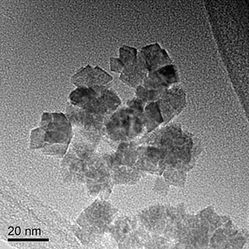
Nanoceria synthesized by a modified thermal hydrolysis precipitation procedure,Reference Zhang, Chan, Spanier, Apak, Jin, Robinson and Herman10 which are then washed and dried, have crystal sizes between 9–12 nm. When these particles are re-suspended in deionized water, they form what appear to be “loose” agglomerates between 200–350 nm in size, as shown in Figure 1. However, different buffers or bodily fluids have different pH, ions, or ionic strength, which can influence the extent of particle agglomeration. Therefore, nanoparticles need to be analyzed in biologically relevant fluids, buffers, or mediums. If nanoparticles are ingested, they will come in contact with gastric fluid (stomach) as well as intestinal fluid (intestine). Therefore, nanoparticles size has been evaluated in gastric fluid and intestinal fluid. Particles remain at similar sizes in simulated gastric fluid and simulated intestinal fluid. Phosphate buffered saline (PBS) and cell culture medium have been used in different studies or in medical applications. It is interesting to mention that they form significantly larger aggregates in PBS and CCM-RPMI (cell culture medium Roswell Park Memorial Institute), as shown in Table I. Other than buffer ions and pH, proteins present in these media also can influence the nanoparticles’ properties. As observed for other types of nanoparticles,Reference Sharma, Kodali, Gaffrey, Wang, Minard, Karin, Teeguarden and Thrall11 the addition of fetal bovine serum (source of proteins) to the medium, especially PBS and CCM, reduces the aggregate size due to formation of a protein corona (the protein absorption layer on the surface of nanoparticles).
Transmission electron microscope image of loose agglomerate formed when ceria nanoparticles are fabricated by thermal hydrolysis precipitation, dried, and re-suspended in deionized water.

Particle size: influence of liquid environment.
Characteristic size of ceria nanoparticle aggregates in assorted biologically relevant media at 25°C as determined by dynamic light scattering, with and without the addition of fetal bovine serum (FBS). Note: DI, deionized; RPMI, Roswell Park Memorial Institute medium.
The chemical state of nanoceria can be monitored by simple UV-visible spectroscopy. The sequence of colors formed during the room-temperature nucleation of cerium oxide nanoparticles in water solutionReference Kuchibhata, Karakoti, Baer, Samudrala, Engelhard, Amonette, Thevuthasan and Seal12 is shown in Figure 2a. The nanoceria start in the form of Ce(NO3)3·6H2O, to which peroxide is added to initiate the nucleation. At 1 hour, the solution is strongly yellow, characteristic of Ce4+, and transmission electron microscope (TEM) analysis of particles indicates that 3 to 5 nm particles or grains are formed into 12–20 nm agglomerates. The suspension begins to turn clear after several days, indicating that the Ce4+ is turning back into Ce3+, although TEM images of the particles’ size (Figure 2c) do not appear to change significantly. X-ray photoelectron spectra of the particles removed from suspension in an inert environment (Figure 2b) confirm the chemical state of the particles, as indicated by the solution color. In this sequence, the chemical states of the particles are changing, although based on TEM and dynamic light scattering (DLS) data, the size of the particles does not change. Laser Raman and x-ray diffraction measurements indicate that the structure of these particles made up of 3 to 5 nm crystallites is changing, as is the chemical state.Reference Kumar, Das, Munusamy, Self, Baer, Sayle and Seal13 Because peroxide is not particularly stable, it was assumed that the change in oxidation state of the particles was the result of changes in the oxygen potential of the solution. This hypothesis was confirmed by adding peroxide to the aged suspension and observing the color and chemical state change, as shown in Figure 2a.Reference Kuchibhata, Karakoti, Baer, Samudrala, Engelhard, Amonette, Thevuthasan and Seal12 These two examples highlight some of the complications that can occur in understanding and predicting biological responses to nanoceria exposures. However, it is also possible to design or tune the particles to either optimize beneficial effects or minimize any damaging effects.Reference Karakoti, Singh, Kumar, Malinska, Kuchibhatla, Wozniak, Self and Seal14
(a) Water solutions during nucleation of ceria nanoparticles and for an aging period after solution of three weeks. (b) X-ray photoelectron spectra from particles at one day and three weeks. (c) Transmission electron microscope image of the particles at one day. At one day, ceria nanoparticles have formed, and they are made up of Ce+4. After three weeks of aging, the particles are similar in size, but are mostly Ce+3. Adding H2O to the solution converts them again to Ce+4. Reprinted with permission from Reference Reference Baer, Engelhard, Johnson, Laskin, Lai, Mueller, Munusamy, Thevuthasan, Wang, Washton, Elder, Baisch, Karakoti, Kuchibhatla and Moon52. © 2013 American Vacuum Society.
Antioxidant properties of nanoceria
Though nanoceria can mimic antioxidant enzymes (i.e., superoxide dismutase,Reference Korsvik, Patil, Seal and Self1 catalaseReference Korsvik, Patil, Seal and Self1) and antioxidants (i.e., glutathione,Reference Arya, Sethy, Singh, Das and Bhargava15,Reference Kumari, Singh, Chinde, Rahman, Mahboob and Grover16 uric acidReference Dowding, Seal and Self4), there are a plethora of molecules that they can interact with, leading to unintended consequences, both positive and negative. Dowding et al. (2013) speculated that the protective effect of nanoceria in reducing reactive nitrogen species (RNS) is likely due to nanoceria’s interaction with one or more products of peroxynitrite decay.Reference Dowding, Seal and Self4 Like reactive oxygen species (ROS), RNS are a by-product of cellular respiration, and excessive generation of RNS can damage lipids, protein, as well as genetic materials. A previous review of various redox-active radical scavenging nanomaterials provides a thorough discussion of nanoceria interactions with ROS and RNS.Reference Karakoti, Singh, Dowding, Seal and Self17 In a follow-up review, Das et al. explored nanoceria and their use in nanomedicine.Reference Das, Dowding, Klump, McGinnis, Self and Seal7 This section focuses on literature that has emerged in the last two years on the “antioxidant” potential of nanoceria, with an emphasis on therapeutic applications.
Oxidative stress (OS) (excessive generation of ROS and RNS) is pervasive in biology, and it has been well established that nanoceria can reduce a prominent and biologically stable ROS, hydrogen peroxide (H2O2).Reference Pirmohamed, Dowding, Singh, Wasserman, Heckert, Karakoti, King, Seal and Self2 Numerous studies continue to explore nanoceria’s interaction with H2O2 in varied disease states. Type I diabetes is a chronic disease where OS plays a prominent role in the death of beta cells (produce insulin) in the pancreas. Pretreatment with nanoceria mitigated H2O2-mediated oxidative stress induced programmed cell death (apoptosis) in isolated rat pancreatic islets and increased their viability.Reference Hosseini, Baeeri, Rahimifard, Navaei-Nigjeh, Mohammadirad, Pourkhalili, Hassani, Kamali and Abdollahi18 Chen et al. (2013) studied endothelial cells (cells that form the interior surface of blood vessels) in a cardiovascular disease model, and again pretreatment with nanoceria prevented ROS overproduction induced by H2O2 and reduced apoptosis.Reference Chen, Hou, Cheng, Zhang, Wang and Zhang19
Neuronal cells are especially susceptible to ROS. Studies in human neuronal cells—neuroblastoma cellsReference Kumari, Singh, Chinde, Rahman, Mahboob and Grover16 and PC12 neuronal-like cellsReference Ciofani, Genchi, Liakos, Cappello, Gemmi, Athanassiou, Mazzolai and Mattoli20—both revealed that the nanoceria reduced OS in part through reduction of H2O2. Extending these studies, Arya et al. (2014), using a primary neuronal culture model, reported treatment of cells with nanoceria led to protection from H2O2 challenge through reduced oxidative stress by stabilization of the mitochondrial membrane potential.Reference Arya, Sethy, Das, Singh, Das, Ujjain, Sharma, Sharma and Bhargava21 With expansion of the number of investigators studying this unusual oxide nanomaterial, it is likely that more reports on the benefits of nanoceria in cell culture and animal models will be forthcoming.
Nanoceria in regenerative medicine
Due to their unique regenerative antioxidant property, nanoceria are finding potential use in medicine. In this section, potential applications of nanoceria in regenerative medicine are discussed. In particular, application of nanoceria in the areas of wound healing, tissue engineering, and neurodegenerative and oxidative-stress-induced eye diseases have been summarized.
Angiogenesis and wound healing
Angiogenesis is the formation of new blood vessels from the existing vasculature, and it plays a critical role in normal development and remodeling of tissues by supplying oxygen and nutrients, and clearing metabolic waste and debris.Reference Otrock, Mahfouz, Makarem and Shamseddine22 Therefore, in tissue engineering, there is a need to find new materials/molecules that can regulate angiogenesis. Recently, nanoceria was shown to promote angiogenesisReference Das, Singh, Dowding, Oommen, Kumar, Sayle, Saraf, Patra, Vlahakis, Sayle, Self and Seal23 by regulation of the intracellular oxygen environment. Nanoceria of 3–5 nm size with a high surface Ce3+/Ce4+ ratio increased in vitro vascular endothelial cell tube formation and stimulated robust growth of blood vessels in a chick chorioallantoic membrane assay, which is a model system to study new blood vessel formation. A correlation between material properties of nanoceria and their angiogenic activity was observed. Smaller, ≤15 nm, nanoceria with a higher surface Ce3+/Ce4+ ratio showed robust induction of tube or blood vessel formation. Qualitative measurement of intracellular oxygen levels by staining with pimonidazole revealed that nanoceria reduces the intracellular oxygen level transiently, which induces HIF-1α translocation to the nucleus for induction of angiogenesis. Hypoxia inducing factor 1 (HIF1)-α is a transcription regulator that is stabilized in a low oxygen environment to induce expression of several proteins that regulate new blood vessel formation.Reference Das, Singh, Dowding, Oommen, Kumar, Sayle, Saraf, Patra, Vlahakis, Sayle, Self and Seal23
The ability of nanoceria to regulate angiogenesis motivated researchers to further explore their effects on wound healing. In a punch biopsy dermal wound model in mice,Reference Chigurupati, Mughal, Okun, Das, Kumar, McCaffery, Seal and Mattson24 topical application of water-suspended nanoceria accelerated the healing of full-thickness dermal wounds by a mechanism that involved enhancement of the proliferation and migration of fibroblasts (cells), keratinocytes, and vascular endothelial cells. The nanoceria penetrated into the wound tissue and reduced oxidative damage to cellular membranes and proteins, suggesting a therapeutic potential for topical treatment of wounds with antioxidant nanoceria. On the other hand, depending on the microenvironment, nanoceria can also inhibit angiogenesis. In a cancer cell environment, a high density of blood vessels is needed to support growth of the tumor. In an in vivo ovarian cancer model, nanoceria significantly decreased the vascular density compared to the control.Reference Giri, Karakoti, Graham, Maguire, Reilly, Seal, Rattan and Shridhar25 An in vivo malignant melanoma model also revealed a similar decrease in microvascular density.Reference Alili, Sack, von Montfort, Giri, Das, Carroll, Zanger, Seal and Brenneisen26 This dual effect of nanoceria is poorly understood; however, this may be explained in light of nanoceria pH-dependent activity—at acidic pH, nanoceria accumulate H2O2, whereas in physiological pH, they scavenge H2O2 (Figure 3). Therefore, in a tumor environment (acidic pH), nanoceria may cause a high amount of hydrogen peroxide formation, which in turn may reduce tumor growth as well as inhibit blood vessel formation.Reference Wason, Colon, Das, Seal, Turkson, Zhao and Baker27
The pro-angiogenesis and anti-angiogenesis properties of nanoceria regulated by the microenvironment. Factors that influence the outcome are pH, production of reactive oxygen species, and intracellular oxygen concentration. Upper-left coordinate: nanoceria accumulate excess H2O2 at acidic pH, which inhibits angiogenesis; upper-right coordinate: neutral pH nanoceria deplete intracellular molecular oxygen and induce transient HIF1-α stabilization, which induces angiogenesis; lower-right coordinate: neutral pH nanoceria scavenge H2O2, which inhibits angiogenesis. Note: HIF1, hypoxia inducing factor 1.
Cell and tissue scaffold and tissue engineering
The previous section demonstrated that nanoceria can promote tissue remodeling by enhancing angiogenesis and stimulating wound healing properties. Nanoceria embedded in a scaffold can maintain slow and sustained release over time, which can detoxify free radicals and induce cell migration as well as proliferation, while scaffolds also provide a structural component. Nanoceria have been incorporated in bioactive glass (Na2O–CaO–SiO2–P2O5 hybrid material used as an implant) scaffolds for bone tissue regeneration.Reference Karakoti, Tsigkou, Yue, Lee, Stevens, Jones and Seal28 This scaffold showed a significant increase in osteoblastic (cell type that forms bone) differentiation compared to the control, which is critical for bone regeneration. However, a different preparation of nanoceria coated with dextran did not increase osteoblast differentiation from seeded human mesenchymal stem cells.Reference Karakoti, Tsigkou, Yue, Lee, Stevens, Jones and Seal28 These results indicate that the chemistry of nanoparticles can be tailored for specific applications. Including inorganic components in the polymer scaffold can improve functionality of the composite material by increasing biocompatibility and improving mechanical and chemical properties. Cardiac and mesenchymal stem cell differentiation on a poly(lactide-co-glycolide) (PLGA) scaffold was increased in the presence of nanoceria.Reference Mandoli, Pagliari, Pagliari, Forte, Di Nardo, Licoccia and Traversa29 Up to 10% nanoceria embedded in a scaffold increases the number of stem cells. Interestingly, 20% nanoceria incorporated in the scaffold did not show any improvement of cell differentiation over control PLGA.
It is noteworthy that a PLGA scaffold with another type of nanoparticle (TiO2) also increased the cell counts, but not to the same extent as nanoceria.Reference Mandoli, Pagliari, Pagliari, Forte, Di Nardo, Licoccia and Traversa29 The result indicates in addition to nanoparticles’ mechanical and morphological properties, chemistry plays an important role in polymer-ceramic materials. Antioxidant activity of nanoceria and improved mechanical properties were reported to be responsible for increased stem cell numbers in cultures. A similar study, with cardiac progenitor cells (CPCs, resident cardiac stem cells that are involved in repairing cardiac tissue damage and remodeling) showed increased cell numbers in the presence of nanoceria, and the presence of nanoceria did not change CPCs’ phenotype or differentiation properties.Reference Pagliari, Mandoli, Forte, Magnani, Pagliari, Nardone, Licoccia, Minieri, Di Nardo and Traversa30
It is important to understand the molecular mechanisms by which nanoceria affect cell behaviors. Two different surface chemistry nanoceria coatings were tested for their effects on cell proliferation, and a large difference was observed in cell growth and behavior.Reference Naganuma and Traversa31 Nanoceria with higher surface Ce4+ induced cell proliferation and enhanced cell spreading, migration, and adhesion, whereas cell spreading, migration, and adhesion were inhibited by a higher Ce3+ surface. On a Ce3+ surface, cell proliferation effects may be due to a different surface charge and higher hydrophobicity compared with a Ce4+ surface. This study clearly suggested that the surface chemistry of nanoceria on biomaterial surfaces can alter nanoparticle-cell interactions by changing the material physical-chemical properties in the specific microenvironment.
Neuroprotection
Neuroprotective effects of engineered nanoceria in cultured neurons include their ability to enhance the average lifespan of neuronal cells.Reference Ellison, Fry, Merchant, Kuiry, Patil, Seal and Rzigalinski32 The protective effect of nanoceria was independent of their size for 6 nm or 12 nm-sized nanoceria. In another study, the lifespan of cultured neuronal cells from spinal cords of adult rats was significantly increased in the presence of nanoceria versus control cultures.Reference Das, Patil, Bhargava, Kang, Riedel, Seal and Hickman33 This study further confirmed the protective effect of nanoceria against ROS.
The pathogenesis of Alzheimer’s disease is associated with the formation and accumulation of characteristic β-amyloid plaques. In an in vitro study using precipitation and high temperature, calcined nanoceria were found to protect neurons against β-amyloid toxicity.Reference Dangelo, Santucci, Benedetti, Loreto, Phani, Amicarelli, Paola Cerù and Cimini34 In another applied study, well-dispersed nanoceria prepared using a microemulsion method were coated with PEG acting as a spacer and conjugated with anti- β-amyloid antibodies to target the nanoceria to plaques, thereby protecting neurons associated with the β-amyloid.Reference Cimini, D’Angelo, Das, Gentile, Benedetti, Singh, Monaco, Santucci and Seal35 This study demonstrated that with proper engineering of nanoceria, it is possible to reduce the side effects and lower the required dose, which is important for patient safety when using nanoparticles for therapeutic applications.
Like other neurodegeneration diseases, stroke and multiple sclerosis pathology is associated with oxidative stress and inflammation.Reference Estevez, Pritchard, Harper, Aston, Lynch, Lucky, Ludington, Chatani, Mosenthal, Leiter, Andreescu and Erlichman36,Reference Estevez and Erlichman37 In ischemic stroke, oxidative stress causes tissue damage due to reduced oxygen and glucose supply to the brain. A recent study using a brain tissue slice model showed treatment with nanoceria at the onset of simulated ischemic stroke resulting in a reduction in cell death.Reference Estevez, Pritchard, Harper, Aston, Lynch, Lucky, Ludington, Chatani, Mosenthal, Leiter, Andreescu and Erlichman36 Another related stroke study in rats treated with well-dispersed phospholipid–PEG-coated nanoceria at concentrations of 0.5 and 0.7 mg/kg of bodyweight showed a 50% decrease in brain infarct volume, which is an index for assessing the extent of brain injury, compared to its controls. Similarly, in multiple sclerosis (MS), inflammation and oxidative stress play a key role in the pathology, which results in focal demyelination and nerve loss.Reference Estevez and Erlichman37 Using citric acid-stabilized nanoceria in preliminary studiesReference Decoteau, Estevez, Leo-Nyquist, Heckman and Reed38 in an experimental autoimmune encephalomyelitis mouse model, after seven days of MS induction, nanoceria-treated mice showed improvements in motor performance. These early studies provide a basis to further explore nanoceria in MS treatment as primary and combination therapy.
Degenerative eye diseases
Many degenerative eye diseases, including age-related macular degeneration (AMD), diabetic retinopathy, and rare orphan diseases such as retinoblastoma and inherited retinal degeneration, share a common “upstream” causative factor: the excessive production of toxic ROS that overwhelms the protective defenses of the cells.
Dry (non-vascular) macular degeneration
Intravitreal injection of less than a ½ nanogram of nanoceria has been shown to prevent light-induced blindness in a rat model of dry AMD (Figure 4).Reference Chen, Patil, Seal and McGinnis39 Uninjected or saline injected rats had almost no visual ERG (electroretinography) function following exposure to the bright light. However, nanoceria protected the retina in a concentration-dependent manner. Subsequent year-long studies showed that nanoceria had no negative effects on vision and were retained in the retina with a half-life of 417 days.Reference Wong, Hirst, Pye, Reilly, Seal and McGinnis40
Nanoceria provide concentration-dependent protection of vision from light damage. Electroretinography data from representative animals. Reproduced with permission from Reference 39. © 2006 Nature Publishing Group.
Nanoceria and inflammation
To further define how nanoceria affects inflammation and oxidative stress, we examined the effects of nanoceria on the expression of 88 major cytokines in the retinas of very low density lipoprotein receptor null (vldlr –/–) mice using a multiplexed assay that quantitates, by polymerase chain reaction (PCR) array, the mRNAs (messenger RNAs) on 96 well plates.Reference Kyosseva, Chen, Seal and McGinnis41 Within one week of nanoceria injection, several cytokines and anti-angiogenic genes were up-regulated in the vldlr –/– retina. Nanoceria also down-regulated pro-angiogenic growth factors and pro-inflammatory cytokines. These results showcase the very broad antioxidant effects of nanoceria.
Wet (neovascular) macular degeneration
The very low density lipoprotein receptor null mouse (vldlr –/–) develops intraretinal and subretinal neovascularization similar to the pathological neovascularization found in humans with a wet form of AMD.Reference Zhou, Wong, Karakoti, Seal and McGinnis42 Using multiple assays to detect ROS and ROS damage, we showed that the vldlr –/– retinas had substantial increases in ROS, nitrosylation of proteins, DNA damage, and in the amount of the ROS-producing enzyme, nicotinamide adenine dinucleotide phosphate oxidase. A single intravitreal injection of nanoceria prevented increases in each of these parameters when examined three weeks after injection. Representative images of vascular endothelial growth factor (VEGF) in a normal mouse retina and in vldlr –/– retinas with saline or nanoceria are seen in Figure 5.
Small amounts (Figure 5a) of VEGF are seen in the normal mouse in the region of the retinal pigment epithelium/rod photoreceptor outer segments, whereas excessive amounts (Figure 5b) of VEGF are seen in vldlr –/– photoreceptor cell bodies surrounding vascular lesions (not seen in these images). The retinas of the nanoceria injected eyes showed greatly reduced amounts (Figure 5c). Therapeutically more important was the discovery that nanoceria regressed almost all fully developed vascular lesions within one week of injection.Reference Zhou, Wong, Karakoti, Seal and McGinnis42 Additional studies showed that nanoceria sustained their therapeutic effects for at least six weeks and inhibited apoptosis via down-regulation of the ASK1-P38/JNK-NF-kB signaling pathway.Reference Cai, Seal and McGinnis43 Our data also suggest that nanoceria may be effective in human patients with “wet” AMD, diabetic retinopathy, or other eye diseases involving pathological neovascularizations.
Nanoceria inhibit pathological increases in retinal vascular endothelial growth factor (VEGF). Retinal nuclei (blue) and anti-VEGF (green) visualized with fluorescence microscopy. (a) Cryostat sections of PN28 retina. Normal C57Bl/6J mice and very low density lipoprotein receptor null mice injected intravitreally with (b) saline or (c) nanoceria at postnatal day 7. Reproduced with permission from Reference 42. © 2011 PLOS.
Inherited retinal degeneration
Following up on that premise, we asked if nanoceria would provide protection to mice with a recessive form of inherited retinal degeneration, called tubby. Multiple systemic injections of nanoceria slowed retinal degeneration over a two-week period by decreasing ROS, increasing the activity of neuroprotective genes, decreasing apoptosis pathways, and increasing survival signaling pathways.Reference Kong, Cai, Zhou, Wong, Karakoti, Seal and McGinnis44 A single intravitreal injection of nanoceria extended the life of photoreceptor cells and preserved vision for up to four months in the tubby mouse. Similarly, nanoceria shrunk inherited retinoblastomas and prevented their growth in the eye.Reference Das, Dowding, Klump, McGinnis, Self and Seal7 Mechanistically, it appears that each disease results in excessive increases of ROS in discrete subcellular localizations as the nanoceria modulate multiple pathways depending on the specific disease. Collectively, these data support the use of nanoceria to effectively treat multiple eye diseases both prophylactically and therapeutically, as they not only inhibit the development of eye diseases, they also dramatically reduce the pathology existing at the time of injection.
Nanoceria in tissue protection against oxidative stress
Prevention of further cell and tissue damage in the affected area helps regenerate new tissue faster, which enhances the healing process and eases the management of symptoms. Nanoceria can scavenge reactive oxygen and nitrogen species effectively in biological environments and protect cells and tissues from oxidative damage found in disease conditions, and against toxic chemical agents (monocrotaline, hydrogen peroxide, cigarette smoking) or radiation.Reference Das, Dowding, Klump, McGinnis, Self and Seal7 In cultured macrophages and an in vivo mouse model, nanoceria decreased ROS.Reference Das, Patil, Bhargava, Kang, Riedel, Seal and Hickman33,Reference Hirst, Karakoti, Tyler, Sriranganathan, Seal and Reilly45 In vivo nanoceria injection decreased the amount of lipid peroxidation, which is a measure of oxidative stress.Reference Hirst, Karakoti, Singh, Self, Tyler, Seal and Reilly46 Nanoceria were also shown to minimize apoptotic cells when cells were challenged with known agents that induce apoptosis (H2O2, puromycin, and topoisomerase 2).Reference Celardo, De Nicola, Mandoli, Pedersen, Traversa and Ghibelli47 A similar study showed that nanoceria decrease disease symptoms in an endometriosis (chronic gynecological disorder associated with pelvic pain, dysmenorrhea, and infertility) model by reducing oxidative stress and inhibiting angiogenesis.Reference Chaudhury, Babu, Das, Kumar and Seal48 Both in vitro as well as in vivo studies have shown that nanoceria can protect cells and tissues against radiation (x-rays). Nanoceria protect normal breast epithelial cellsReference Tarnuzzer, Colon, Patil and Seal49 and fibroblastsReference Colon, Herrera, Smith, Patil, Komanski, Kupelian, Seal, Jenkins and Baker50 against 10 and 20 Gy radiation exposure, respectively. In a study of nude mice, 80% of the mice survived after 33 days of radiation exposure when pretreated with nanoceria. Nanoceria also minimized complications associated with radiation treatment using a head and neck radiation model.Reference Madero-Visbal, Alvarado, Colon, Baker, Wason, Isley, Seal, Lee, Das and Mañon51
Conclusion
Reactive oxygen and nitrogen species act as secondary messengers in many physiological processes; however, their excessive production can irreversibly damage cells and tissues and contribute to the pathogenesis of many diseases. Nanoceria can regulate ROS in intracellular environments due to their broad spectrum of redox activity. Moreover, regulating ROS nanoceria supports cell survival and can enhance the proliferation or differentiation of healthy cells, which suggests nanoceria applications for tissue engineering and regenerative medicine. However, not all forms of nanoceria are equally effective, and there are unanswered questions regarding the effects of nanoceria on health. Different surface chemistries and compositions, concentration, size and stability of nanoceria, protocols for assaying the beneficial/toxicological effects, test model system/organism, and endpoints evaluated are all variables in the many published studies. Moreover, storage conditions, the biological environment, and environmentalReference Baer, Engelhard, Johnson, Laskin, Lai, Mueller, Munusamy, Thevuthasan, Wang, Washton, Elder, Baisch, Karakoti, Kuchibhatla and Moon52 factors can also alter nanoceria catalytic/redox activity and cell–nanoceria interactions. Additional studies need to be undertaken to obtain an understanding of the molecular mechanism of nanoceria action in cells.
Acknowledgments
This work was supported, in part, by the intramural research program of the National Institute on Aging. The work by J.F.M. was supported by NIH NEI Grant COBRE-P20 RR017703, P30-EY 12190, R21EY018306, R01EY18724, R01EY022111; National Science Foundation: CBET-0708172; and Research to Prevent Blindness. D.R.B. was supported by the NIEHS Center Grant U19 ES019544. Part of this work was performed using EMSL, a national scientific user facility sponsored by the US Department of Energy, Biological and Environmental Research and located at PNNL. S.S. and W.S. acknowledge NSF and NIH for various aspects of nano-biotechnology research.
Disclaimer
The contents of this article are the authors’ and do not reflect any position of the US Government or US Food and Drug Administration.