Introduction
Merkel cell carcinoma is a rare, aggressive neuroendocrine carcinoma of the skin, which affects the head and neck region in approximately 50 per cent of cases. It is mostly a disease of the elderly as incidence rates increase with age, and there is a slight male predominance.Reference Allen, Bowne, Jaques, Brennan, Busam and Coit1–Reference Allen, Zhang and Coit6 Recent evidence suggests that a Merkel cell polyomavirus is responsible for the development and maintenance of the disease.Reference Houben, Shuda, Weinkam, Schrama, Feng and Chang7 Twenty-five per cent of patients present with regional lymph node involvement. Up to 75 per cent of patients develop a locoregional recurrence. Five-year disease-specific survival rates range from 35 to 75 per cent.Reference Allen, Bowne, Jaques, Brennan, Busam and Coit1–Reference Allen, Zhang and Coit6, Reference Ott, Tanabe, Gadd, Stark, Smith and Finkelstein8–Reference Goepfert, Remmler, Silva and Wheeler14
Despite increasing awareness of this cancer, the optimal management of head and neck Merkel cell carcinoma remains unclear. For local disease, wide excision of the primary tumour is a well-established practice,Reference Allen, Bowne, Jaques, Brennan, Busam and Coit1–Reference Miller, Alam, Andersen, Berg, Bichakjian and Bowen5, Reference Brissett, Olsen, Kasperbauer, Lewis, Goellner and Spotts9–Reference Kokoska, Kokoska, Collins, Stapleton and Wade22 with opinion regarding the minimum surgical margin differing somewhat. Adjuvant radiotherapy to the surgical bed is recommended for Merkel cell carcinoma; this has been shown to reduce local recurrenceReference Medina-Franco, Urist, Fiveash, Heslin, Bland and Beenken2–Reference Eng, Boersma, Fuller, Goytia, Jones and Joyner4, Reference Ott, Tanabe, Gadd, Stark, Smith and Finkelstein8, Reference Clark, Veness, Gilbert, O'Brien and Gullane10–Reference Veness, Morgan and Gebski13, Reference Meeuwissen, Bourne and Kearsley18, Reference Jabbour, Cumming, Scolyer, Hruby, Thompson and Lee19, Reference Veness, Perera, McCourt, Shannon, Hughes and Morgan23–Reference Foote, Harvey, Porceddu, Dickie, Hewitt and Colquist28 and, more recently, to improve survival.Reference Jabbour, Cumming, Scolyer, Hruby, Thompson and Lee19, Reference Mojica, Smith and Ellenhorn29 However, local control and survival advantages are yet to be demonstrated for cases of head and neck Merkel cell carcinoma.
Treatment of nodal disease may include sentinel lymph node biopsyReference Allen, Bowne, Jaques, Brennan, Busam and Coit1, Reference Hill, Brady and Coit30–Reference Maza, Trefzer, Hofmann, Schneider, Voit and Krossin33 and lymph node dissection,Reference Allen, Bowne, Jaques, Brennan, Busam and Coit1, Reference Ott, Tanabe, Gadd, Stark, Smith and Finkelstein8, Reference Brissett, Olsen, Kasperbauer, Lewis, Goellner and Spotts9, Reference Goepfert, Remmler, Silva and Wheeler14, Reference Raaf, Urmacher, Knapper, Shiu and Cheng15, Reference Yiengpruksawan, Coit, Thaler, Urmacher and Knapper20, Reference Mehrany, Otley, Weenig, Phillips, Roenigk and Nguyen31, Reference Victor, Morton and Smith34, Reference Hitchcock, Bland, Laney, Franzini, Harris and Copeland35 with or without adjuvant radiotherapy.Reference Müller, Keus, Neumann, Lammering and Schnabel16, Reference Jabbour, Cumming, Scolyer, Hruby, Thompson and Lee19, Reference Veness, Perera, McCourt, Shannon, Hughes and Morgan23–Reference Mojica, Smith and Ellenhorn29 There is a lack of consensus regarding the management of both node-positive and node-negative disease of the neck. There is even less certainty surrounding the management of recurrent disease.
This retrospective, consecutive case review focused on the management of primary and recurrent Merkel cell carcinoma of the head and neck at a single institution. Specifically, we aimed to assess the influence of local excision, margin status, and adjuvant radiotherapy and chemotherapy on locoregional recurrence and survival.
Materials and methods
This study entailed a review of the anatomical pathology database at the Peter MacCallum Cancer Centre in Melbourne, Australia, which included all patients diagnosed with Merkel cell carcinoma from December 1998 to December 2009. In total, 116 patients were identified, 54 of which had Merkel cell carcinoma of the head and neck region, with records available for analysis. Of these 54 patients, 16 presented to the institution for primary treatment of the disease, 37 were referrals to the institution after initial excision of the primary tumour elsewhere and 1 patient was referred for ongoing follow up. All surgical and biopsy specimens had been reviewed by the anatomical pathology department at the institution, and diagnoses were made on the basis of characteristic histological features and immunohistochemical staining.Reference Poulsen3, Reference Eng, Boersma, Fuller, Goytia, Jones and Joyner4
The information gathered for each patient included demographic, tumour and treatment related variables. Data collection was approved by the Divisional Review of Retrospective Research Committee at the Peter MacCallum Cancer Centre. Demographic data collected for each patient included sex and age at diagnosis. The tumour characteristics recorded were: specific location within the head and neck, locoregional lymph node status, and the presence of metastases. Lymph node status was diagnosed clinically and radiologically; a pathological diagnosis with sentinel lymph node biopsy was not performed. Primary tumour variables, such as lymphovascular invasion, tumour size and depth, were not assessed due to insufficient data. Treatment of primary and recurrent disease was reviewed separately. The impact of treatment modality (surgery, radiotherapy and chemotherapy, either alone or in combination), radiotherapy fields and surgical margin status were analysed in terms of two survival outcomes, namely time to first recurrence and disease-specific survival. Combined treatment was defined as wide local excision and radiotherapy, with or without chemotherapy. Recurrence was defined as the disappearance of disease following initial treatment and subsequent reappearance of pathologically confirmed disease more than six weeks post primary treatment. Post-operative radiotherapy was defined as the initiation of radiation treatment within six weeks of primary treatment.
All patients were followed for a minimum of six months; follow up ceased at either the time of death or the time the patient was last seen alive. A modification of the staging system developed by Allen et al. was used.Reference Allen, Bowne, Jaques, Brennan, Busam and Coit1 Patients were thus divided into those without (stage I) and those with (stage II) regional lymph node involvement, and those with distant metastases (stage III).
Kaplan–Meier survival curves were generated using the Stata statistical software package (Statacorp LP, College Station, Texas, USA). Hypotheses concerning comparisons of survival between groups were tested using the log-rank test and a Cox proportional hazards regression model.
Results
Case characteristics and outcomes
The median age at presentation was 76 years, and there was a male predominance (37 of 54 patients, 68 per cent). Twenty-two of 50 patients (44 per cent) who initially responded to treatment developed a recurrence, with a median time to first recurrence of 24 months. The draining nodal basin was the most common site of first recurrence (18 of 50 patients, 36 per cent) (Table I). The overall survival rate at 1, 2 and 3 years was 80 per cent, 63 per cent and 55 per cent respectively, and the median follow-up time for all patients was 22 months (inter-quartile range: 13–43 months).
Case characteristics and outcomes

*Total n = 54. †Four patients were excluded from recurrence analyses because they did not respond to treatment and so could not have experienced a recurrence. ‡Location of first recurrence could involve more than one site. LN = lymph node; FU = follow up; MCC = Merkel cell carcinoma
Primary tumour and nodal basin treatment
Forty-four of the 54 patients (81 per cent) underwent surgery, 46 (85 per cent) received radiotherapy and 14 (26 per cent) received chemotherapy (Table II).
Treatment characteristics

*Total n = 54. †One patient underwent wide excision of the primary tumour and a parotidectomy. ‡The radiotherapy targets could not be accurately identified from the medical records. **Some patients received more than one treatment type. Pts = patients
In terms of surgery, all patients with an identifiable primary tumour underwent wide local excision. Of the 14 patients who presented with cervical lymph node metastases and an unknown primary tumour site, 6 underwent a limited neck dissection (excisional biopsy) and the remaining 8 underwent core biopsy (Table II).
All patients, with either node-positive or node-negative disease, were considered for adjuvant treatment; the final decision was based upon tumour size, patient age, co-morbidities and, ultimately, patient preference. Regional radiotherapy was administered more often to patients with disease stage II (13 of 14, 93 per cent) than stage I (15 of 30, 50 per cent). The most commonly used radiotherapy regimen was 50 Gy (range: 36 Gy to 60 Gy) in 25 fractions over 5 weeks. Chemotherapy was administered for palliation, and salvage of recurrent, metastatic or locally advanced disease. In the total course of their treatment, 14 patients received chemotherapy. This included 4 patients that developed a recurrence after initially presenting with stage I disease and 10 patients with stage II (nodal) disease at presentation.
Time to first recurrence
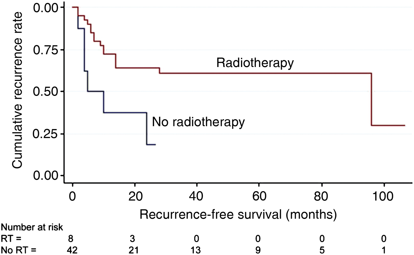
Treatment with radiotherapy was associated with a significantly prolonged time to first recurrence. The median times to recurrence were 96 months for those patients who did receive adjuvant radiotherapy versus 5 months for those who did not (log-rank test, p = 0.013) (Table III, Figure 1).
Kaplan–Meier curves for recurrence-free survival (time to first recurrence) by exposure to radiotherapy (p = 0.013). RT = radiotherapy

Time to first recurrence statistics*

* Analyses performed using the univariate Cox regression and log-rank tests. †Disease stage was controlled for in the Cox proportional hazards model. ‡Disease stage and age at diagnosis were controlled for in the Cox proportional hazards model. **Log-rank test p-value. Mth = months; HR = hazard ratio; CI = confidence interval; yr = year; M = male; F = female; LNs = lymph nodes
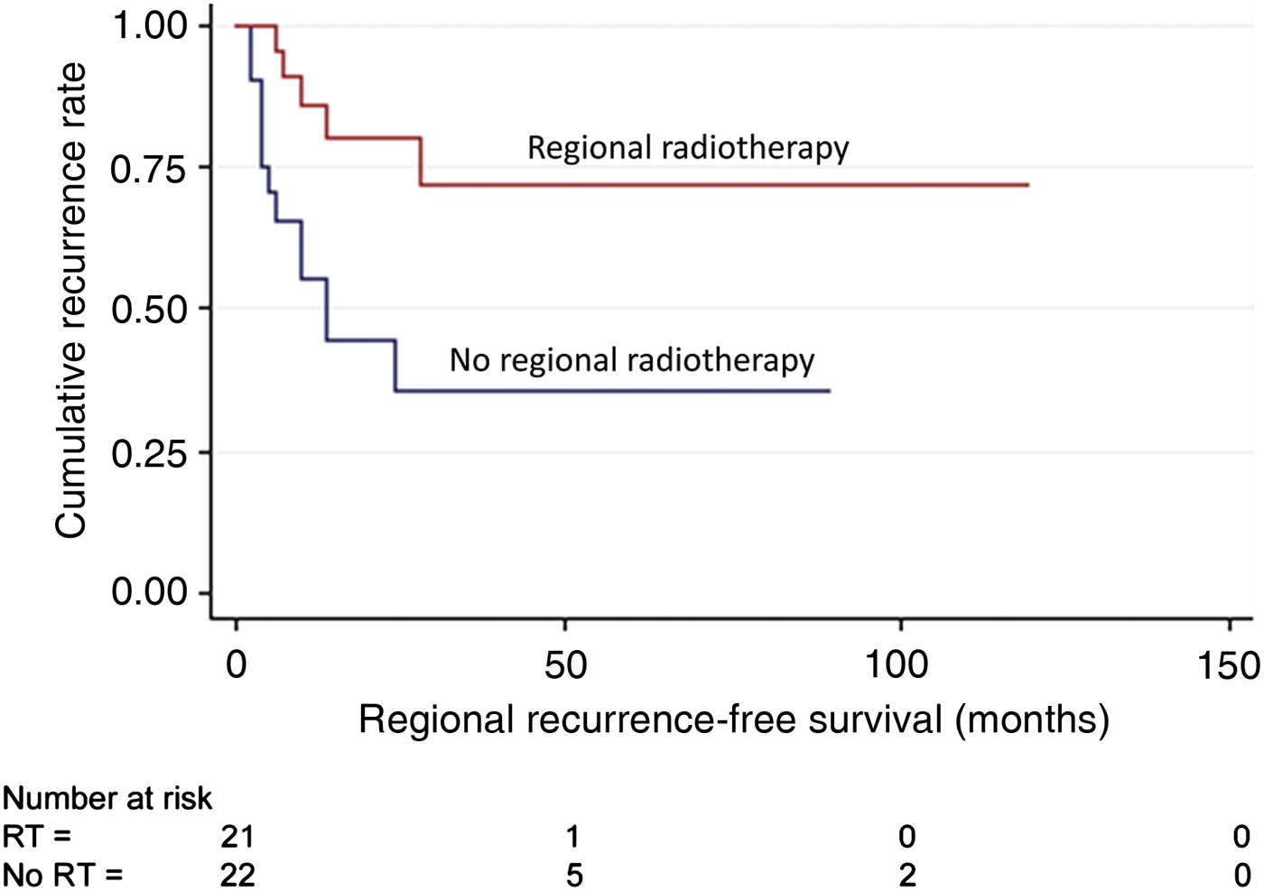
When analysed specifically, radiotherapy to the regional nodes was associated with improved time to regional recurrence (log-rank test, p = 0.01) (Figure 2).
Kaplan–Meier curves for regional recurrence-free survival by exposure to regional radiotherapy (p = 0.01). RT = radiotherapy

After controlling for disease stage at presentation, analyses revealed that (compared with single modality treatment) combined treatment prolonged the time to first recurrence. However, this was not significant at the 0.05 level (hazard ratio 0.46; 95 per cent confidence interval (CI) 0.19–1.12; p = 0.09). Note that the sample size used for this analysis was 49: 1 patient was omitted because he received neither single modality nor combined modality treatment; 4 others were excluded because they did not respond to primary treatment and hence could not experience a recurrence. There was no evidence that surgical margin status impacted on time to recurrence (univariate Cox regression hazard ratio 0.42; CI 0.09–1.86; p = 0.25). A local recurrence developed in 1 of 8 patients with positive margins after primary excision (13 per cent), and in 6 of 31 patients with negative margins (19 per cent). The only other significant predictor of outcome was the stage at presentation, which was associated with time to first recurrence (Cox regression hazard ratio 2.19; CI 1.04–4.60; p = 0.038) (Table III).
Survival
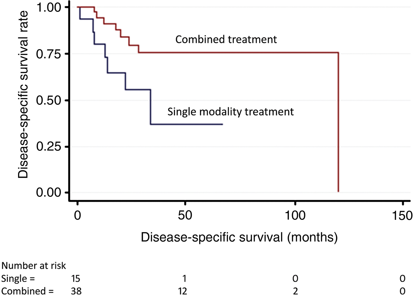
When disease stage at presentation was controlled for, the Cox regression analysis indicated that combined treatment may have had some benefit for disease-specific survival over single modality treatment. The hazard rate for death due to disease among patients receiving combined treatment was 0.37 times that of patients receiving single modality treatment (95 per cent CI 0.13–1.07; p = 0.07) (Table IV, Figure 3).
Kaplan–Meier curves for disease-specific survival by exposure to combined versus single modality treatment (p = 0.07).

Disease-specific survival statistics*

* Analyses performed using the univariate Cox regression and log-rank tests.
†Disease stage was controlled for in the Cox proportional hazards model. ‡Disease stage and age at diagnosis were controlled for in the Cox proportional hazards model. **Log-rank test p-value. Mth = months; HR = hazard ratio; CI = confidence interval; yr = year; M = male; F = female
Treatment of recurrence
In total, 22 patients developed recurrent disease, 12 of whom died as a result of Merkel cell carcinoma with a median disease-specific survival of 8 months. Of the 17 patients who developed a regional recurrence, 3 patients received surgery, which involved a modified radical neck dissection in 2 cases and a radical neck dissection in 1 case (Table V).
Treatment of recurrence

*Total n = 22. †One patient underwent local re-excision and adjuvant radiotherapy. ‡Some patients received more than one treatment type. Pts = patients
Control of recurrence was defined as no further recurrence of disease, locally or regionally, following treatment of the recurrence. Of the three patients who received combined treatment with surgery and radiotherapy, there was control of recurrence in two patients. In the 18 patients who received single modality treatment, there was control of recurrence in 8 patients (Table V). The log-rank test demonstrated no relationship between combined versus single modality treatment and survival time from recurrence (log-rank test, p = 0.38). The median follow-up time from recurrence was eight months.
In terms of other treatment factors, the use of radiotherapy, chemotherapy and surgical margin status were not associated with disease-specific survival on univariate log-rank or Cox regression analyses. In terms of other predictors, only disease stage was associated with disease-specific survival (hazard ratio 6.28; CI 2.36–16.73; p < 0.001) (Table IV).
Discussion
An important finding of this head and neck Merkel cell carcinoma study was the demonstration of a statistically significant association between radiotherapy and time to recurrence (Figure 1). In contrast to previously published studies, this result is likely to be related to: our larger sample size; the long median duration of follow up (22 months), during which a significant proportion of patients developed a recurrence (22 of 50, 44 per cent); and the fact that a large proportion of patients received radiation treatment (46 of 54, 85 per cent). The relationship between radiotherapy and disease control is well established in whole-body Merkel cell carcinoma,Reference Medina-Franco, Urist, Fiveash, Heslin, Bland and Beenken2–Reference Eng, Boersma, Fuller, Goytia, Jones and Joyner4, Reference Ott, Tanabe, Gadd, Stark, Smith and Finkelstein8, Reference Meeuwissen, Bourne and Kearsley18, Reference Jabbour, Cumming, Scolyer, Hruby, Thompson and Lee19, Reference Veness, Perera, McCourt, Shannon, Hughes and Morgan23–Reference Foote, Harvey, Porceddu, Dickie, Hewitt and Colquist28 and more recently in Merkel cell carcinoma of the head and neck.Reference Clark, Veness, Gilbert, O'Brien and Gullane10–Reference Lawenda, Arnold, Tokarz, Silverstein, Busse and McIntyre12 In a literature review of 1024 patients, Medina-Franco et al. found that local recurrence was reduced by almost 40 per cent with the introduction of radiotherapy (p < 0.00001).Reference Medina-Franco, Urist, Fiveash, Heslin, Bland and Beenken2 When analysed specifically, the current study showed that radiation to the primary tumour site was associated with a three-fold increase in the median time to local recurrence (Table III). Considered collectively, these findings support the use of adjuvant radiation treatment to improve locoregional control.
Using multivariate Cox regression analysis, we demonstrated a trend towards improved survival with combined surgery and radiotherapy compared with single modality treatment (Table IV). Previous studies on head and neck Merkel cell carcinoma have been unable to find an association between combined treatment and survival,Reference Gillenwater, Hessel, Morrison, Burgess, Silva and Roberts11–Reference Veness, Morgan and Gebski13 or have demonstrated a non-significant association.Reference Clark, Veness, Gilbert, O'Brien and Gullane10 Clarke et al. performed the largest study on head and neck Merkel cell carcinoma, and found a non-significant trend (p = 0.089) towards improved survival with combined treatment.Reference Clark, Veness, Gilbert, O'Brien and Gullane10 Only two studies on whole-body Merkel cell carcinoma have demonstrated improved survival with combination treatment.Reference Jabbour, Cumming, Scolyer, Hruby, Thompson and Lee19, Reference Mojica, Smith and Ellenhorn29 A review of 1665 cases of Merkel cell carcinoma found in the Surveillance, Epidemiology and End Results registry revealed that median survival was 63 months versus 45 months (p = 0.0002) for patients treated with or without combined treatment respectively.Reference Mojica, Smith and Ellenhorn29 An inability to consistently demonstrate a survival benefit is not unexpected given the relatively smaller cohorts of most studies and the fact that Merkel cell carcinoma is a disease of older patients, many of whom succumb to co-morbid disease. The findings of this study and other research suggest that patients with head and neck Merkel cell carcinoma should receive a combination of wide local excision followed by adjuvant radiation.
Merkel cell carcinoma has a high propensity for early lymphatic spread, and most of the published literature supports treating the first echelon lymph nodes.Reference Poulsen3, Reference Eng, Boersma, Fuller, Goytia, Jones and Joyner4, Reference Gillenwater, Hessel, Morrison, Burgess, Silva and Roberts11, Reference Raaf, Urmacher, Knapper, Shiu and Cheng15, Reference Jabbour, Cumming, Scolyer, Hruby, Thompson and Lee19, Reference Kokoska, Kokoska, Collins, Stapleton and Wade22, Reference Morrison, Peters, Silva, Wendt, Ang and Goepfert26, Reference Eich, Eich, Staar, Mauch, Stutzer and Groth27, Reference Shaw and Rumball36–Reference Veness, Palme and Morgan38 Surgical lymph node dissection has been shown to reduce nodal recurrence and enable identification of pathological stage.Reference Allen, Bowne, Jaques, Brennan, Busam and Coit1, Reference Ott, Tanabe, Gadd, Stark, Smith and Finkelstein8, Reference Brissett, Olsen, Kasperbauer, Lewis, Goellner and Spotts9, Reference Goepfert, Remmler, Silva and Wheeler14, Reference Raaf, Urmacher, Knapper, Shiu and Cheng15, Reference Yiengpruksawan, Coit, Thaler, Urmacher and Knapper20, Reference Mehrany, Otley, Weenig, Phillips, Roenigk and Nguyen31, Reference Victor, Morton and Smith34, Reference Hitchcock, Bland, Laney, Franzini, Harris and Copeland35 Merkel cell carcinoma is a radiosensitive tumour, and radiation offers regional control without the morbidity associated with surgical neck dissection.Reference Müller, Keus, Neumann, Lammering and Schnabel16, Reference Jabbour, Cumming, Scolyer, Hruby, Thompson and Lee19, Reference Veness, Perera, McCourt, Shannon, Hughes and Morgan23–Reference Mojica, Smith and Ellenhorn29 In the past, most centres have offered both therapies to patients with node-positive disease; they may or may not have offered elective regional radiotherapy for those with node-negative disease.Reference Poulsen3, Reference Eng, Boersma, Fuller, Goytia, Jones and Joyner4, Reference Ott, Tanabe, Gadd, Stark, Smith and Finkelstein8, Reference Brissett, Olsen, Kasperbauer, Lewis, Goellner and Spotts9, Reference Gillenwater, Hessel, Morrison, Burgess, Silva and Roberts11, Reference Goepfert, Remmler, Silva and Wheeler14, Reference Müller, Keus, Neumann, Lammering and Schnabel16, Reference Jabbour, Cumming, Scolyer, Hruby, Thompson and Lee19, Reference Kokoska, Kokoska, Collins, Stapleton and Wade22–Reference Mojica, Smith and Ellenhorn29, Reference Shaw and Rumball36, Reference Suntharalingam, Rudoltz, Mendenhall, Parsons, Stringer and Million37 In this series, 93 per cent of patients with node-positive disease received regional radiotherapy, but only 33 per cent of patients underwent lymph node dissection. Fifty per cent of patients with node-negative disease received prophylactic nodal irradiation. As such, the use of lymph node dissection at our institution is significantly less than that described in the literature. Our overall regional control rate was 67 per cent, which is comparable to most other centres.Reference Allen, Bowne, Jaques, Brennan, Busam and Coit1, Reference Clark, Veness, Gilbert, O'Brien and Gullane10–Reference Müller, Keus, Neumann, Lammering and Schnabel16, Reference Jabbour, Cumming, Scolyer, Hruby, Thompson and Lee19, Reference Bajetta, Celio, Platania, Lo Vullo, Patuzzo and Maurichi21, Reference Eich, Eich, Staar, Mauch, Stutzer and Groth27, Reference Hitchcock, Bland, Laney, Franzini, Harris and Copeland35, Reference Fang, Lemos, Douglas, Iyer and Nghiem39 In our study, the median time to nodal recurrence was twice as long for patients receiving nodal irradiation compared with those who did not receive radiation treatment (p = 0.01) (Figure 2 and Table III). These findings support the growing consensus that all patients should be considered for adjuvant nodal radiation therapy, regardless of whether the disease is node-positive or node-negative.
We did not find any association between clear margins following excision and recurrence-free survival or disease-specific survival on univariate analysis (Table III and IV). This is an important finding, particularly for head and neck Merkel cell carcinoma where cosmesis and function are important considerations. More than 20 per cent of patients in our series presented with anatomically awkward sites that included the nose, and perioral and periorbital regions. There is no consensus in the literature on the optimal width of surgical margins. Whilst most authors recommend wide or at least clear margins, only a handful have shown a statistically significant association between margin status, local recurrence and survival;Reference Allen, Zhang and Coit6, Reference Bajetta, Celio, Platania, Lo Vullo, Patuzzo and Maurichi21 most authors have shown no association at all.Reference Allen, Bowne, Jaques, Brennan, Busam and Coit1, Reference Clark, Veness, Gilbert, O'Brien and Gullane10–Reference Lawenda, Arnold, Tokarz, Silverstein, Busse and McIntyre12, Reference Foote, Harvey, Porceddu, Dickie, Hewitt and Colquist28 The lack of association in our study could be explained by the fact that most patients received adjuvant radiotherapy to the surgical bed (82 per cent), irrespective of margin status. Mohs micrographic surgery has been reported as being an improved method for achieving clear margins, but as yet its role remains unproven.Reference Boyer, Zitelli, Brodland and D'Angelo40
Twenty-six per cent of patients in this study received chemotherapy as part of their primary treatment, and this was administered as an adjuvant to surgery and radiotherapy. Merkel cell carcinoma has been shown to be chemosensitive,Reference Veness, Palme and Morgan38, Reference Tai, Yu, Winquist, Hammond, Stitt and Tonita41–Reference Fenig, Brenner, Katz, Rakovsky, Hana and Sulkes44 and response rates to carboplatin and etoposide, the two agents used most commonly at our institute, have been reported at 60 per cent.Reference Eng, Boersma, Fuller, Goytia, Jones and Joyner4, Reference Veness, Palme and Morgan38, Reference Tai, Yu, Winquist, Hammond, Stitt and Tonita41 Our study demonstrated no improvements in locoregional control (Table III) or survival (Table IV) in the cohort of patients who received chemotherapy, even after correcting for disease-specific factors. A variety of toxicities are associated with the currently used regimens,Reference Voog, Biron, Martin and Blay43, Reference Poulsen, Rischin, Walpole, Harvey, Macintosh and Ainslie45 and the deaths of 2 out of 14 patients in this series were secondary to immunosuppression. Our study does not seem to support the use of chemotherapy in Merkel cell carcinoma of the head and neck.
Recurrent disease poses a significant problem in head and neck Merkel cell carcinoma, with very little certainty surrounding its management. Our study showed that survival rates halved with recurrence. The overall survival rates at 2 years for those patients who did and did not develop a recurrence were 41 per cent and 79 per cent respectively. Of the 22 patients who developed a recurrence in this study, 5 underwent salvage surgery. Consistent with the trend at our institution towards adjuvant therapies, 82 per cent of patients with recurrent disease received radiotherapy, with 89 per cent of local recurrences and 76 per cent of nodal recurrences irradiated. We demonstrated an overall remission rate of 41 per cent following salvage radiotherapy for recurrent disease; this is comparable to the rates of 46 per centReference Ott, Tanabe, Gadd, Stark, Smith and Finkelstein8 and 60 per centReference Hitchcock, Bland, Laney, Franzini, Harris and Copeland35 reported in the literature. The tendency in the past has been to prescribe aggressive multimodality treatment;Reference Eng, Boersma, Fuller, Goytia, Jones and Joyner4, Reference Tai, Yu, Assouline, Lian, Joseph and Miale42 however, there have been no formal recommendations. In this study, there was a four-fold increase in the median overall survival time from the recurrence in patients who received combined treatment compared with those treated with a single modality, but because of the small patient numbers this was not statistically significant (p = 0.38). Although treatment of recurrent disease needs to be individualised in a multidisciplinary setting, with patient factors, location of recurrence and the efficacy of prior treatments taken into consideration, our data highlights the potential benefit of an aggressive multimodality approach.
The overall recurrence rate in our study was 44 per cent. This is similar to that reported at other centres,Reference Allen, Bowne, Jaques, Brennan, Busam and Coit1, Reference Clark, Veness, Gilbert, O'Brien and Gullane10–Reference Müller, Keus, Neumann, Lammering and Schnabel16, Reference Jabbour, Cumming, Scolyer, Hruby, Thompson and Lee19, Reference Bajetta, Celio, Platania, Lo Vullo, Patuzzo and Maurichi21, Reference Eich, Eich, Staar, Mauch, Stutzer and Groth27, Reference Hitchcock, Bland, Laney, Franzini, Harris and Copeland35, Reference Fang, Lemos, Douglas, Iyer and Nghiem39 and is almost identical to the recurrence rate (of 43 per cent) reported in the largest study performed on whole-body Merkel cell carcinoma by Allen and colleagues.Reference Allen, Bowne, Jaques, Brennan, Busam and Coit1 The 5-year survival rates reported in the literature range between 35 and 75 per cent,Reference Allen, Bowne, Jaques, Brennan, Busam and Coit1–Reference Allen, Zhang and Coit6, Reference Ott, Tanabe, Gadd, Stark, Smith and Finkelstein8–Reference Goepfert, Remmler, Silva and Wheeler14, Reference Müller, Keus, Neumann, Lammering and Schnabel16, Reference Jabbour, Cumming, Scolyer, Hruby, Thompson and Lee19, Reference Bajetta, Celio, Platania, Lo Vullo, Patuzzo and Maurichi21, Reference Eich, Eich, Staar, Mauch, Stutzer and Groth27 and the overall survival rate in the current study, with 22 months follow up, was 50 per cent. Stage at presentation was significantly associated with time to recurrence and disease-specific survival on multivariate Cox regression analysis; this is consistent with other studies.Reference Allen, Bowne, Jaques, Brennan, Busam and Coit1, Reference Allen, Zhang and Coit6, Reference Ott, Tanabe, Gadd, Stark, Smith and Finkelstein8, Reference Meeuwissen, Bourne and Kearsley18–Reference Yiengpruksawan, Coit, Thaler, Urmacher and Knapper20 The prevalence of nodal disease at presentation in head and neck patients has been reported to range from 11 to 32 per cent,Reference Brissett, Olsen, Kasperbauer, Lewis, Goellner and Spotts9–Reference Veness, Morgan and Gebski13 and was revealed by the current study to be 33 per cent at our institute. Nodal relapse was the most common sign of first recurrence, most frequently involving cervical levels I–II and the parotid nodes. One-quarter of patients in this study presented with clinically palpable nodes and an unknown primary site, which is higher than previously reported.Reference Allen, Bowne, Jaques, Brennan, Busam and Coit1, Reference Brissett, Olsen, Kasperbauer, Lewis, Goellner and Spotts9 Brisett et al. found that 1 out of 22 patients with head and neck Merkel cell carcinoma had an unidentifiable primary tumour site,Reference Brissett, Olsen, Kasperbauer, Lewis, Goellner and Spotts9 and Allen et al. reported a 12 per cent rate of unknown primary sites.Reference Allen, Bowne, Jaques, Brennan, Busam and Coit1 An inability to locate the primary tumour site rendered diagnosis and treatment decisions more difficult. The higher rate in our study may be a reflection of the tertiary nature of referrals to our institution: complex cases or those with clinically apparent neck disease are more likely to be referred to a multidisciplinary clinic.
We acknowledge the usual limitations of a retrospective review. However, any disease of such low incidence will never lend itself to prospective, randomised study. Our necessarily small sample reflects the rareness of this disease. Nevertheless, the numbers reported in this study are similar to those of other published multi-institutional studies, whilst maintaining the benefits of a single institution study. We have achieved sufficient numbers to be able to make some statistically significant conclusions, and where this has not been possible we have been able to comment on obvious trends. Due to referral patterns, our population of patients is biased towards those with more advanced disease; however, we feel that this strengthens the study because our treatment conclusions are made as a result of treating more complex and advanced pathology.
As we do not perform elective neck surgery in the node-negative neck, our patients were clinically and not pathologically staged. Sentinel lymph node biopsy is advocated by othersReference Allen, Bowne, Jaques, Brennan, Busam and Coit1, Reference Hill, Brady and Coit30–Reference Maza, Trefzer, Hofmann, Schneider, Voit and Krossin33 on the premise that 25–100 per cent of patients with clinically negative nodes harbour subclinical micrometastatic disease.Reference Allen, Bowne, Jaques, Brennan, Busam and Coit1, Reference Poulsen3, Reference Goepfert, Remmler, Silva and Wheeler14, Reference Victor, Morton and Smith34, Reference Gupta, Wang, Peñas, Gellenthin, Lee and Nghiem46 However, at our institution we have elected to treat the regional nodes independent of nodal status. In the current study, 81 per cent of patients received some form of treatment to their regional nodes, despite only 33 per cent of patients having clinically evident nodal disease. Regardless of whether pathologically positive nodes are present in 25 per cent or 100 per cent of patients,Reference Allen, Bowne, Jaques, Brennan, Busam and Coit1, Reference Poulsen3, Reference Goepfert, Remmler, Silva and Wheeler14, Reference Victor, Morton and Smith34, Reference Gupta, Wang, Peñas, Gellenthin, Lee and Nghiem46 we would argue that elective neck treatment should be undertaken. One of the purposes of our study was to validate this approach. The fact that we achieved similar locoregional control rates and survival data to those reported in other studies, in spite of treating a population with more advanced disease, would support our approach. Sentinel lymph node biopsy may improve the allotment of patients to adjuvant therapies, but our approach renders this intervention unnecessary. Indeed, the findings in this study support the growing consensus that even patients with node-negative (sentinel lymph node biopsy negative) disease should be considered for nodal irradiation. Studies on head and neck cancer, particularly melanoma, have shown that the outcome of cervical lymphatic drainage is variable and unpredictable,Reference Stadelmann, Cobbins and Lentsch47, Reference Balkissoon, Rasgon and Schweitzer48 with a non-localisation rate of 5–7 per centReference Stadelmann, Cobbins and Lentsch47, Reference Alex49 and a false negative rate of 4 per centReference Ellis, Weerasinghe, Corless and Vetto50 using sentinel lymph node biopsy. To date, there has been no evidence to demonstrate that sentinel lymph node biopsy improves survival or recurrence.
• This study reviewed the management of primary and recurrent head and neck Merkel cell carcinoma
• Combined treatment with surgery and locoregional radiotherapy resulted in improved disease-specific survival
• Radiotherapy was associated with improved time to recurrence and regional recurrence
• Irradiation of regional nodes resulted in improved regional control, irrespective of clinical status
• Margin-negative excision was not associated with improved local control
A significant proportion of patients in our study (37 of 54, 69 per cent) were referred from other centres following excision of the primary lesion. Hence, we have a high incidence of nodal disease at presentation (33 per cent), and a large proportion of patients presenting with nodal disease and an unknown primary tumour site (26 per cent). In spite of this, we achieved a survival rate similar to that published elsewhere. We would further argue that in this complex population of patients, we were able to provide clear and statistically significant support for the use of combined treatment. The results also indicated: the lack of benefit of adjuvant chemotherapy, the questionable role of sentinel lymph node biopsy, and the doubtful utility of wide surgical margins in treating Merkel cell carcinoma of the head and neck.
Conclusion
Merkel cell carcinoma of the head and neck is a potentially curable, but rapidly progressive and life threatening disease. Treatment of the primary tumour should include excision, with clear margins if possible, and adjuvant radiotherapy to the surgical bed. Our analysis demonstrated improved time to recurrence with the use of adjuvant radiotherapy (p = 0.013), and a trend towards improved survival with combined modality treatment (p = 0.07) on regression analysis. Based on the results of this study, which has a 2c level of evidence, we recommend that treatment of the draining nodal basin should comprise radiotherapy, irrespective of the nodal status, as this was independently associated with improved regional control (p < 0.01). We would also recommend combined treatment as the best option for recurrent disease.