Syncope occurs in up to 15% of children before the age of 18 years and is a common reason for evaluation.Reference Driscoll, Jacobsen, Porter and Wollan 1 Syncopal episodes associated with hot showers, standing up quickly, during micturition, or while hair brushing are commonly benign and are often managed by primary-care physicians; however, when children collapse in the midst of active exercise, this generally raises a higher level of concern and often warrants further evaluation. Although syncope during exercise has been associated with cardiac disease,Reference Zhang, Zhu and Wang 2 – Reference Del Rosso, Ungar and Maggi 4 the incidence among children presenting with syncope in the midst of physical activity is unknown, and to date there have been no studies evaluating children with mid-exertional syncope. The purpose of this study was to evaluate children with true syncope in the midst of exertion who underwent thorough evaluation to determine whether the diagnosis was cardiac or non-cardiac in origin and to define potential predictors to differentiate cardiac from non-cardiac causes.
Materials and methods
We performed a single-centre retrospective review of all patients ⩽18 years of age who presented for cardiac evaluation by a paediatric cardiologist due to exertional syncope between 1999 and 2012. Clinical records including all exercise stress testing, cardiac catheterisations, electrophysiology studies, pacemaker or implantable cardioverter defibrillator implants, and databases for ion channel disorders and cardiomyopathy were reviewed for diagnostic coding of syncope and/or history of exertional syncope. Patient records were then fully reviewed for exertional symptoms. Exertional syncope was defined as a transient loss of consciousness with loss of postural tone in the midst of activity, and was differentiated from syncope occurring before or after termination of exercise. Patients with peri-exertional syncope were excluded for purposes of this study. Syncope must have occurred during the active phase – for example during mid stride while running, boxing, cycling, swimming, etc. – and those events that occurred immediately after termination of exercise – for example, while standing during a boxing match – were not considered mid-exertional syncope for the purposes of this study – for example, syncope that occurred after running but while walking off the field was not considered exertional syncope. Inclusion criteria included the following: (1) age ⩽18 years; (2) mid-exertional syncope; (3) an electrocardiogram, echocardiogram, and at least one of the following: exercise stress test, electrophysiology study, or tilt test; and (4) evaluation by a paediatric cardiologist. Patients with QT prolongation with a diagnosis of Long QT syndrome did not require additional testing to meet the inclusion criteria. Patients with known structural heart defects, known arrhythmia disorders, and those who received any form of resuscitation following collapse were excluded. Patients who received any form of cardiopulmonary resuscitation or had a defibrillator or automated external defibrillator placed were excluded. Patients were evaluated in the emergency room, as an inpatient admission, or in the outpatient clinic. A cardiac diagnosis was defined as abnormal electrical conduction – high-grade atrioventricular block, channelopathy, supraventricular re-entrant, or ventricular tachycardias – structural heart disease, or cardiac myopathy. Vasovagal or neurally mediated syncope were considered non-cardiac in aetiology. This study was conducted with the internal review board’s approval.
Data were collected on prodromal and recovery symptoms surrounding the presenting syncopal event and previous history. Diagnostic testing, family history, presence of injury, and type of exertional activity according to peak static and dynamic cardiovascular demand – Classification of sports, Task force 8, 36th Bethesda Conference – were collected.Reference Mitchell, Haskell, Snell and Van Camp 5 Follow-up was defined as the last identifiable encounter in our system in which we could verify whether the patient was alive. Genetic testing was performed for some patients; however, a majority of patients did not undergo genetic testing due to lack of insurance coverage.
This study was a descriptive, exploratory study, and comparative variables were chosen a priori as candidates to be associated with cardiac disease. To evaluate intensity of exertion, levels of exertion were grouped into binary variables by grouping low, low-moderate, and moderate in one group and high-moderate and high in another group. Given the large number of comparisons and the small sample size, we are reporting both the raw p-value and the Bonferroni step-down (Holm)-adjusted p-values. Adjusted values <0.05 were considered statistically significant. Multivariate analysis could not be performed because the model experienced quasi-complete separation due to the small number of children being analysed. Statistical analysis was performed using SAS version 9.3 (SAS Institute, Inc., Cary, North Carolina, United States of America). Continuous variables are presented as medians with ranges and were analysed using the Wilcoxon rank sum test. Categorical variables are presented as counts with percentages and were analysed using the Fischer Exact test or the χ 2 test.
Results
Patient characteristics
Among patients who underwent cardiac evaluation of syncope by a paediatric cardiologist, 60 (26 male, 43%) were definitively noted to have syncope during mid-exercise and met the inclusion criteria for this study. Among these 60 patients, those with cardiac syncope were younger (median age 11 years, range 4–18 years) than those with non-cardiac syncope (median age 14 years, range 7–18 years) (p=NS). Although patients with cardiac disease had a family history of sudden cardiac death 2.5 times higher than those without a cardiac diagnosis, there was no statistically significant difference (p=1.00).
Cardiac versus non-cardiac diagnosis
Among the 60 patients with exertional syncope, 32 had a cardiac diagnosis (47% male), accounting for 53% of the total cohort, and 28 (39% male) had a non-cardiac or unknown diagnosis. Vasovagal syncope as the aetiology of the exertional syncope was diagnosed in 11/28 (39%) non-cardiac cases. In a majority of patients determined to not have definitive cardiac disease, a diagnosis could not be identified (16/28, 57%), despite work-up; one patient in the non-cardiac group was diagnosed with psychogenic syncope.
Patients diagnosed with cardiac disease were most likely to have an electrical myopathy or rhythm disorder, which accounted for 91% of all patients with cardiac-related exertional syncope – ten Long QT syndrome, six catecholaminergic polymorphic ventricular tachycardia, five supraventricular tachycardia – non-Wolff–Parkinson–White syndrome – two Wolff–Parkinson–White syndrome, two ventricular tachycardia, two idiopathic ventricular fibrillation, two atrioventricular block. Other cardiac diagnoses included two hypertrophic cardiomyopathy and one left ventricular non-compaction.
Diagnostic testing
Diagnostic testing varied by patient and included electrocardiogram, echocardiogram, exercise stress test, cardiac catheterisation, electrophysiology study, intravenous drug challenge using procainamide, epinephrine, or isoproterenol, cardiac MRI or CT, Holter or event monitor, implantable loop recorder, tilt-table testing, and cardiac genetic testing for inheritable arrhythmias and cardiomyopathies (Fig 1). Neurology work-up was performed in some patients and included an electroencephalogram, head CT, and/or head MRI. Although some patients underwent tilt-table testing, this method of diagnosis was not commonly used during cardiac evaluation. There was no difference in the total number of diagnostic tests performed among patients with or without a cardiac diagnosis – median five tests for both groups, range 2–12 for cardiac diagnosis, and range 3–9 for a non-cardiac diagnosis. At least eight diagnostic tests were performed in 16% of cardiac patients and 18% of non-cardiac patients. At least three diagnostic tests were performed in 97% of all patients, most commonly an electrocardiogram, echocardiogram, and an exercise stress test. In addition to cardiac work-up, neurological evaluation was performed in 34% of cardiac patients and 43% of non-cardiac patients.
This figure represents the testing used to help diagnosis. Among 60 patients with exertional syncope, 32 were found to have cardiac diagnosis; 13 patients had abnormal ECGs, three had abnormal echocardiograms (*two with HCM already had an abnormal ECG). The 18 remaining cardiac patients had normal ECG and echocardiograms. Abnormal test results that helped in making a diagnosis are listed. Patients may have had other testings before the indicated abnormal test, and therefore the total testing performed is listed. **Mobitz type II AVB; ***infrahisian block. Abnl=abnormal; AVB=atrioventricular block; cath=catheterisation; CPVT=catecholaminergic polymorphic ventricular tachycardia; ECG=electrocardiogram; Echo=echocardiogram; EEG=electroencephalogram; EPS=electrophysiological study; EST=exercise stress test; HCM=hypertrophic cardiomyopathy; ILR=implantable loop recorder; LQT=Long QT syndrome; LVNC=left ventricular non-compaction; Outpt monitor=outpatient monitor (Holter or event monitor); SVT=supraventricular tachycardia; VF=ventricular fibrillation; Vstim=ventricular stimulation protocol; VT=ventricular tachycardia; WPW=Wolff–Parkinson–White.

Among patients with a cardiac diagnosis, an abnormal electrocardiogram was observed in 41 versus 7% of non-cardiac patients (p=0.003, OR 8.9, adjusted p=0.036). Among the Long QT syndrome patients, the corrected QT interval ranged between 480 and 530 ms; two patients with borderline corrected QT intervals in the 460–470 ms range underwent drug challenges that demonstrated markedly abnormal corrected QT prolongation. Genetic testing was positive for cardiac ryanodine receptor mutations in two patients with catecholaminergic polymorphic ventricular tachycardia and in three patients diagnosed with Long QT type 1 (KCNQ1 mutations). Genetic testing was not performed in several patients due to insurance limitations. Abnormal electrocardiograms seen in two patients with non-cardiac diagnosis demonstrated a single premature ventricular contraction in one patient and biventricular hypertrophy with a normal echocardiogram in the other. Echocardiogram confirmed the diagnosis of cardiomyopathy in three patients – two hypertrophic cardiomyopathy and one left ventricular non-compaction; however, all these patients had abnormal electrocardiogram findings.
Symptoms
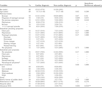
Past medical history was reviewed for a previous history of chest pain, palpitations, seizures, number of previous syncopal events, and whether these events were consistent with a diagnosis of vasovagal syncope (Table 1).
Comparison of patients with mid-exertional syncope and cardiac versus non-cardiac diagnosis.

ECG=electrocardiogram; ER=emergency room; n=number of patients with available information
Data are presented with count (%)
* Owing to the large numbers of comparison, data for statistical analysis were chosen a priori, and those selected are shown with p-values and adjusted p-values. If no p-value is listed a (–) is shown, which indicated that the variable was not statistically assessed
** Adjusted p-values <0.05 were considered statistically significant
Previous history
A previous history of syncope, whether single or multiple episodes, did not distinguish those with or without a cardiac diagnosis –25% in both groups had more than three episodes of syncope in the past. Patients with a previous history of non-exertional vasovagal syncope were more likely to receive a non-cardiac diagnosis, although not statistically significant after the Bonferroni correction (p=0.005, adjusted p=0.055).
Symptoms immediately preceding syncope
Symptoms immediately preceding syncope did not differ between the cardiac and non-cardiac patients (Table 1). In both the groups, the most commonly reported symptom was dizziness, followed by palpitations, and then chest pain. Chest pain and/or palpitations were reported in 48% of cardiac patients and 28% of non-cardiac patients. Prodromal symptoms such as dizziness, diaphoresis, vision or hearing changes, and nausea or vomiting were reported in 52% of cardiac patients and 68% of non-cardiac patients. The absence of immediately preceding symptoms did not differentiate the two groups (26% cardiac group, 20% non-cardiac group, p=0.75).
Symptoms immediately following syncopal episode
There were no significant differences in post-syncopal symptoms between the cardiac and non-cardiac groups. No symptoms were reported in 70% of cardiac and 50% of non-cardiac patients (p=0.17).
Sport classification and secondary injury
Type of sports activity during exertional syncope was classified as (1) low to moderate or (2) high-moderate to high, based on the Task force 8 classification of sports, 36th Bethesda Conference. Exertional syncope occurred primarily during three sports activities: running (53%), basketball (27%), and swimming (12%). There was no difference in the level of exertional activity or occurrence of secondary injury between the cardiac and non-cardiac patients (p=0.17).
Emergency room visit and hospital admissions
Emergency room visits and hospital admissions were more common among patients who eventually received a cardiac diagnosis. Cardiac patients were more likely to seek evaluation in the emergency room following their syncopal episode versus non-cardiac patients (68 versus 37%, p=0.03, OR 3.6); however, after Bonferroni correction, this was not statistically significant (p=0.27). The majority of cardiac patients evaluated in the emergency room (17/19, 90%) were subsequently admitted to the hospital for further evaluation, whereas 3/10 (30%) non-cardiac patients were felt to warrant hospital admission. Among the 17 cardiac syncope patients admitted to the hospital, only one patient was recognised to have cardiac disease during work-up in the emergency room. Among all, five patients with prolonged QT and one patient with hypertrophic cardiomyopathy had abnormal electrocardiograms that were missed in the emergency room but were identified after admission.
Follow-up
Median total follow-up for cardiac patients was 5 years (range 1 day–21 years). Median total follow-up for non-cardiac patients was 3 years (range 8 days–21 years). None of the non-cardiac patients received a subsequent cardiac diagnosis during follow-up. There have been no known deaths.
Discussion
True mid-exertional syncope in children is uncommon, but often raises concerns. This study focused on those patients who received a full evaluation by a paediatric cardiologist, and therefore does not reflect the overall incidence in the general population, because not all children who have syncope during exertion are evaluated by a cardiologist. Nevertheless, to our knowledge, there have been no previous studies focussing on exertional syncope in children. Our goal was to evaluate not only cardiac disease among these patients but also determine how often a diagnosis is unable to be determined under expert evaluation. Among the 60 patients with mid-exertional syncope who underwent evaluation by a paediatric cardiologist, approximately half were diagnosed with cardiac disease; however, in ~50% of the cases, the aetiology was felt to be vasovagal or could not be determined. Symptoms preceding syncope did not distinguish those with or without cardiac disease.
This was a descriptive study that evaluated specific variables chosen a priori. After adjusting for multiple comparisons using the step-down Bonferroni equation, cardiac patients having abnormal electrocardiograms was the only statistically significant difference between the cardiac and non-cardiac groups. This is not surprising and suggests that electrocardiograms are a reasonable initial diagnostic test. Nevertheless, the few variables that met statistical significance before this conservative adjustment merit discussion as they are clinically significant.
Patients with cardiac diagnosis were younger than those with non-cardiac diagnosis (median 11.5 years versus 14 years, p=0.02, adjusted p=0.200). This may be explained by the fact that some arrhythmia disorders such as catecholaminergic polymorphic ventricular tachycardia present earlier in childhood, whereas vasovagal syncope is more commonly seen in the adolescent years.Reference Napolitano and Priori 6 , Reference Sheldon, Sheldon and Connolly 7 Patients with cardiac disorders in this study were more likely to have an arrhythmia disorder rather than a cardiomyopathy. This study did not include those patients who underwent any form of resuscitation, and therefore may explain the lack of cardiomyopathy patients who are at risk for both obstruction and arrhythmias during exertion.
Cardiac syncope patients were also more likely to present to the emergency room and be admitted to the hospital. Of the 19 patients with cardiac syncope evaluated in the emergency room, all had life-threatening conditions. Of these 19 children, 17 were admitted, but in only one case was the cardiac diagnosis made while in the emergency room. In some cases, the diagnosis was missed – that is, prolonged QT on electrocardiogram and abnormal T waves suggestive of cardiomyopathy; however, in most cases, work-up required further diagnostic testing that could not be obtained while in the emergency room – for example, exercise treadmill or electrophysiology study. The difference between the two groups seeking care in the emergency room may be explained by previous history. A majority of patients (14/17, 82%) with non-cardiac diagnosis who did not seek treatment in the emergency room had a previous history of syncope, six of whom had been previously diagnosed with non-exertional vasovagal syncope. Among cardiac patients, three of the nine patients (33%) not evaluated in the emergency room had a previous syncopal episode, but none of them had a diagnosis of vasovagal syncope.
Despite a high rate of cardiac diagnosis among patients with exertional syncope, nearly half were diagnosed with potentially benign conditions. To our knowledge, none of the patients with a non-cardiac diagnosis subsequently received a cardiac diagnosis and there have been no deaths. We suspect either exhaustion or physiological changes during exercise to account for a majority of the patients with a non-cardiac aetiology. Physiological changes may include reflex-induced hypotension and bradycardia secondary to the Bezold Jarisch reflex or significant skeletal muscle vasodilation and hypotension secondary to decreased sympathetic vasoconstriction.Reference Hastings and Levine 8 Unfortunately, there is no definitive and confirmatory diagnostic test for reflex-mediated or neurally mediated syncope.
In this study, we found that the presence or absence of preceding symptoms was not a reliable method of distinguishing those with and without cardiac disease. Detailed history regarding past medical history and symptoms surrounding the syncopal event did not distinguish those children with cardiac and non-cardiac syncope. Perhaps more worrisome is the fact that patients with cardiac diagnosis can describe symptoms that are consistent with vasovagal syncope and yet have life-threatening disease. Of the 27 patients with potentially life-threatening conditions, eight (30%) reported prodromal symptoms that may have been confused with vasovagal syncope. In this study, we could not determine whether syncope resulted from life-threatening arrhythmias or other benign causes. Nevertheless, similar to our findings, both MacCormick et al and Colman et al found that among patients with known gene-positive cardiac channelopathies and syncope, symptoms typically associated with vasovagal syncope were not uncommon.Reference MacCormick, Crawford and Chung 9 , Reference Colman, Bakker, Linzer, Reitsma, Wieling and Wilde 10
As reported symptoms may not distinguish cardiac versus non-cardiac diagnosis, all children with mid-exertional syncope warrant thorough cardiac evaluation and work-up. In this cohort of patients, electrical disease was the most common cause of cardiac syncope. An abnormal electrocardiogram was found in one-third of cardiac syncope patients, and it was the only statistically significant difference between the two groups after adjusting for multiple comparisons. The electrocardiogram should be the first diagnostic test, and an exercise stress test is highly considered in the setting of a normal echocardiogram. Outpatient monitoring – Holter, event monitor, or loop recorder – and electrophysiology studies or drug challenges were also helpful diagnostic tools, although these may involve invasive procedures and should be used after careful evaluation in selected patients. Although echocardiograms are important, particularly to rule out cardiomyopathy, in patients with exertional syncope, further testing is likely warranted even when electrocardiograms and echocardiograms are normal, as 18 of 32 patients (56%) with a cardiac diagnosis had a normal electrocardiogram and echocardiogram at presentation.
Limitations
The main limitation of this study is its retrospective nature and the referral bias for exertional syncope. Patients with exertional syncope may not always be referred for cardiac evaluation. Therefore, those patients referred and evaluated in this study may be inherently a higher-risk group as they either sought care in the emergency room or were referred for further evaluation. In addition, those who were ultimately diagnosed with a significant disease may have been captured more frequently than those with benign findings. We also acknowledge that patients with supraventricular tachycardia listed under cardiac disease may have had vasovagal events rather than arrhythmia-related collapse, as we cannot determine the exact cause of syncope. We may have missed patients who were not referred for evaluation, those whose history or other factors resulted in minimal work-up not meeting the inclusion criteria for this study, particularly those with cardiomyopathy, as we required at least three diagnostic tests. Nevertheless, at our institution, a patient with exertional syncope and cardiomyopathy would have warranted further work-up to determine whether the aetiology of syncope was arrhythmic. If further tests such as an exercise stress test or electrophysiology study were not performed, this study may have underestimated the number of cardiomyopathy patients. This is a retrospective study limited to available information in chart review and limited by recall bias. Reported symptoms may be inaccurate and are dependent on the depth and detail of the assessing physician. In particular, symptoms after the syncopal event may have been limited and not reported in full detail. The study period also spanned the course of 14 years. Diagnostic work-up may have changed over time, and therefore may have affected our results. In particular, diagnosis of catecholaminergic polymorphic ventricular tachycardia would have been limited in 1999 and the early 2000s. Nevertheless, among this group of patients, there was a young child who initially presented in 1999 and was diagnosed with catecholaminergic polymorphic ventricular tachycardia. Some patients received more comprehensive testing than others. Among those patients with a negative work-up but limited cardiac testing, we cannot exclude the possibility that we missed a cardiac diagnosis. To our knowledge, there have been no late adverse events or sudden deaths.
Acknowledgements
None.
Financial Support
This research received no specific grant from any funding agency, commercial, or not-for-profit sectors.
Conflicts of Interest
None.
Ethical Standards
The authors assert that all procedures contributing to this work comply with the ethical standards of the relevant national guidelines on human experimentation and with the Helsinki Declaration of 1975, as revised in 2008, and has been approved by the institutional review board at Stanford University.