The soluble intercellular and vascular cell adhesion molecule-1 are commonly located on the endothelium. They are described as the mediators responsible for leukocyte adhesion, penetration into the vessel wall, and transendothelial migration to the tissue.Reference Springer 1 , Reference Gearing and Newman 2 These molecules also play an important role in the pathogenesis of tissue injury secondary to hypertension, cardiac failure, atherosclerosis, myocardial ischaemia, autoimmune diseases, transplant rejection, and ischaemia reperfusion injury.Reference Wang, Nawata and Kakudo 3 – Reference Rabinovitch 8 In several studies, significantly high soluble intercellular adhesion molecule-1 concentrations were found in critically ill patients suffering from trauma or post-operative complications, systemic inflammatory response syndrome, organ dysfunction, and after cardiopulmonary bypass.Reference Boldt, Wollbruck, Kuhn, Linke and Hempelmann 9 – Reference Eikemo, Sellevold and Videm 11
Very few studies have been conducted regarding the role of adhesion molecules in congenital heart diseases. Only one study is available showing a positive correlation between pulmonary hypertension and soluble intercellular adhesion molecule-1 level in paediatric cardiac patients.Reference Sungprem, Khongphatthanayothin, Kiettisanpipop, Chotivitayatarakorn, Poovorawan and Lertsapcharoen 12 However, neither the role of adhesion molecules in congenital heart diseases nor the effect of pulmonary blood flow on the level of adhesion molecules has been clearly understood yet. It is crucial to maintain normal cardiac functions in patients until the operation in order to obtain more successful results. Some new treatment modalities may be developed by assessing the relationship between the adhesion molecules and congenital heart diseases for this purpose. We conducted this study to determine whether there is a correlation between the blood levels of soluble intercellular and vascular cell adhesion molecule-1 in children with various congenital heart diseases and pulmonary blood flow rates.
Methods
Study patients
A total of 65 children hospitalised for operation with left-to-right shunt, cyanotic congenital heart diseases, left ventricle outflow tract obstruction, and dysplastic aortic valve with aortic stenosis were evaluated in this study. These patients were divided into four groups according to their pulmonary blood flow: the first group had increased pulmonary blood flow with pulmonary arterial hypertension and left-to-right shunt; the second group had increased pulmonary blood flow without pulmonary arterial hypertension and left-to-right shunt; the third group had decreased pulmonary blood flow with cyanotic congenital heart disease; and the fourth group had normal pulmonary blood flow with left ventricle outflow tract obstruction and dysplastic aortic valve with aortic stenosis. The first group had 25 patients, of whom 14 were female and 11 were male with a mean age of 8.5 months and mean weight of 6.2 kilograms. The second group had 13 patients, of whom seven were female and six were male with a mean age of 10.7 months and mean weight of 8.4 kilograms. The third group had 14 patients, of whom nine were female and five were male with a mean age of 9.2 months and mean weight of 10.3 kilograms. The fourth group had 12 patients, of whom eight were male and four were female with mean age of 14.8 months and mean weight of 10.1 kilograms. The diagnoses were made by physical examination, chest roentgenogram, electrocardiography, echocardiography, and angiography. The plasma levels of soluble intercellular and vascular cell adhesion molecule-1 were evaluated. While taking the blood samples for measurement of adhesion molecules, the patients were not receiving oxygen or antihypertensive treatment, for example, sildenafil, bosentan, or iloprost. These patients were evaluated for the presence of infection, ischaemia, hypoxia, anaemia, and renal function abnormality via some laboratory studies such as white blood cell, haemoglobin and platelet count, C-reactive protein level, erythrocyte sedimentation rate, urea, creatinine, troponin I, and cutaneous oxygen saturation. Pulmonary and aortic pressure levels of the patients were measured by catheter angiography or during the surgery. Mean pulmonary artery pressure levels above 25 millimetres of mercury were accepted as pulmonary hypertension. The study was approved by the Ethics Review Committee. Informed consent was obtained from the parents as appropriate.
Determination of soluble intercellular and vascular cell adhesion molecule-1
Clotted blood samples were obtained from the right brachial vein of the patients. The serum was separated promptly and stored at −20°C until analysis. The quantitative sandwich enzyme-linked immunosorbent assay technique was used for the determination of serum-soluble intercellular and vascular cell adhesion molecule-1 (eBioscience, Human sICAM-1 and sVCAM-1; Platinum Elisa, Bender Med System, GmbH, Vienna, Austria) levels. Tests were performed according to the manufacturer's recommendations. The results were reported in nanogram per millilitre.
Statistical analysis
SPSS 15.0 for Windows (SPSS Inc., Chicago, Illinois, United States of America) was used for statistical analysis. Quantitative parameters were given as the mean and standard deviation. Qualitative parameters were given as frequency distribution and percentage. Mann–Whitney U test was used to assess the differences between demographic factors and laboratory data of the four groups. Analysis of variance and Post-hoc tests were used to compare the levels of soluble intercellular and vascular cell adhesion molecule-1 and all other laboratory and clinical parameters of the four groups. Pearson correlation test was used in order to find the correlations between the level of adhesion molecules and factors including age, weight, haemoglobin, white blood cell and platelet count, C-reactive protein levels, erythrocyte sedimentation rate, urea, creatinine, troponin I, cutaneous oxygen saturation, and mean aortic and pulmonary arterial pressure. A p-value of less than 0.05 was considered as statistically significant.
Results
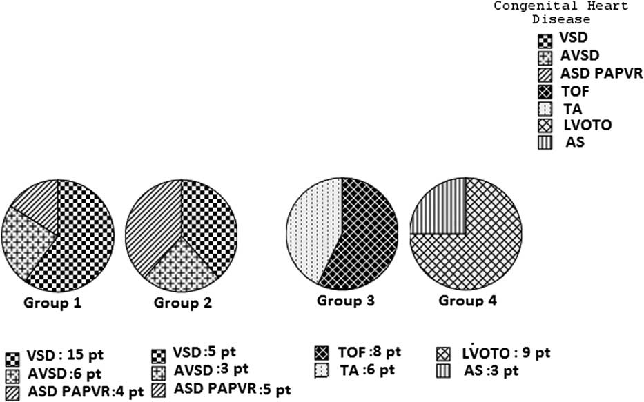
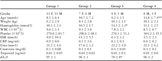
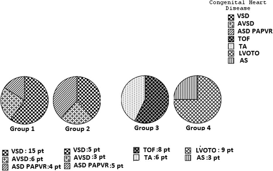
The diagnoses of the patients are seen on Figure 1. Out of the cardiac patients hospitalised for operation, only the patients with left-to-right shunt had congestive heart failure, although they were using digoxin, diuretic, and angiotensin-converting enzyme inhibitors. These patients were hospitalised for the closure of left-to-right shunt. The patients with cyanotic heart disease were hospitalised for shunt operation or total correction, and the patients with left ventricle outflow obstruction or dysplastic aortic valve for resection of the fibromuscular ridge and aortic valve comissurotomy. The patients in the third and fourth groups were not receiving any medication. The distribution of the patients according to their gender, age, weight, and laboratory parameters is shown in Table 1. The age of the patients in the first group was lower than that in the fourth group (p < 0.05). There was no age difference between other groups. The difference in weight distribution of the groups was not significant. The laboratory data of all groups are also seen in Table 1. Haemoglobin levels of the third group were higher and the oxygen saturation levels were lower than the other groups (p < 0.05). There were no significant differences in white blood cell and platelet count, C-reactive protein levels, erythrocyte sedimentation rate, urea, creatinine, and troponin I levels among all groups.
Diagnoses of patients in all groups. AS = aortic valve stenosis; ASD PAPVR = atrial septal defect and partial anomalous pulmonary venous return; AVSD = atrioventricular septal defect; LVOTO = left ventricle outflow tract obstruction; pt = patient; TA = Tricuspid atresia; TOF = tetralogy of Fallot; VSD = Ventricular septal defect.
Patient characteristics and laboratory parameters.

CRP = C-reactive protein; ESR = erythrocyte sedimentation rate; F = female; M = male; sO2% = oxygen saturation; WBC = white blood cell
Group 1: left-to-right shunt with pulmonary hypertension
Group 2: left-to-right shunt without pulmonary hypertension
Group 3: decreased pulmonary blood flow with cyanotic heart disease
Group 4: normal pulmonary blood flow with left ventricle outflow tract obstruction and dysplastic aortic valve aortic
*p < 0.05 for all groups
**p < 0.05 for Group 1 and Group 4
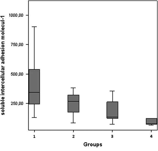
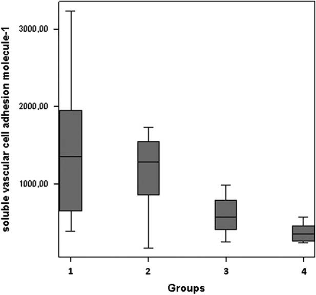
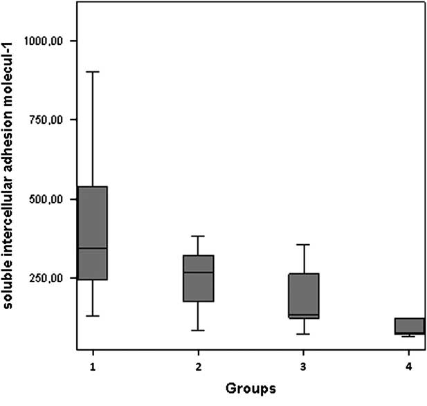
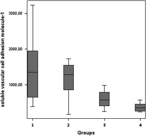
The soluble intercellular and vascular cell adhesion molecule-1 levels of all groups are shown in Figures 2 and 3. The highest soluble intercellular and vascular cell adhesion molecule-1 levels were measured in the first group of patients with left-to-right shunt with pulmonary arterial hypertension. In this group, the mean soluble intercellular adhesion molecule-1 level was measured as 420.2 plus or minus 215.3 (median: 270; range: 128–1083) nanograms per millilitre and the mean soluble vascular cell adhesion molecule-1 level was measured as 1382.1 plus or minus 564.7 (median: 1354; range: 385–2941) nanograms per millilitre. The fourth group of patients with left ventricle outflow tract obstruction and dysplastic aortic valve had lower soluble intercellular and vascular cell adhesion molecule-1 levels. In this group, the mean soluble intercellular adhesion molecule-1 level was measured as 104.4 plus or minus 49.3 (median: 78; range: 65–234) nanograms per millilitre and the mean vascular cell adhesion molecule-1 level was measured as 358.6 plus or minus 113.8 (median: 345; range: 228–564) nanograms per millilitre. In the second group of patients with left-to-right shunt without pulmonary hypertension, the mean soluble intercellular adhesion molecule-1 level was measured as 251.6 plus or minus 96.4 (median: 268; range: 83–381) nanograms per millilitre and the mean soluble vascular cell adhesion molecule-1 level was measured as 1129.1 plus or minus 516 (median: 1128; range: 167–1728) nanograms per millilitre. In the third group of patients with decreased pulmonary blood flow with cyanotic congenital heart disease, the mean soluble intercellular adhesion molecule-1 level was measured as 190.7 plus or minus 125.6 (median: 136.2; range: 74–554) nanograms per millilitre and the mean soluble vascular cell adhesion molecule-1 level was measured as 577.8 plus or minus 217.8 (median: 565; range: 246–965) nanograms per millilitre.
Plasma level of soluble intercellular adhesion molecule-1 in all groups. 1: Patients with left-to-right shunt and pulmonary arterial hypertension; 2: patients with left-to-right shunt without pulmonary arterial hypertension; 3: patients with decreased pulmonary blood flow and cyanotic heart disease; and 4: patients with normal pulmonary blood flow and left ventricle outflow tract obstruction and dysplastic aortic valve.
Plasma level of soluble vascular cell adhesion molecule-1 in all groups. 1: Patients with left-to-right shunt and pulmonary arterial hypertension; 2: patients with left-to-right shunt without pulmonary arterial hypertension; 3: patients with decreased pulmonary blood flow and cyanotic heart disease; and 4: patients with normal pulmonary blood flow and left ventricle outflow tract obstruction and dysplastic aortic valve.
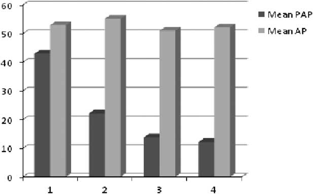
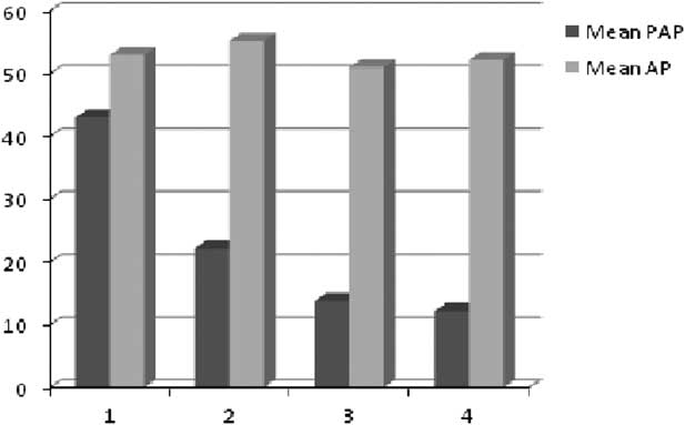
The aortic and pulmonary arterial pressure levels of all patients are shown in Figure 4. There was no aortic pressure difference among the four groups (p > 0.05). The mean pulmonary arterial pressure levels of the first group were higher than the other groups (p < 0.05).
Mean aortic and pulmonary arterial pressures in all groups. 1: Patients with left-to-right shunt and pulmonary arterial hypertension; 2: patients with left-to-right shunt without pulmonary arterial hypertension; 3. patients with decreased pulmonary blood flow and cyanotic heart disease; and 4: patients with normal pulmonary blood flow and left ventricle outflow tract obstruction and dysplastic aortic valve.
Correlation between soluble markers, clinical properties, and laboratory variables
In Pearson correlation analysis, a positive correlation was found between serum-soluble intercellular adhesion molecule-1, mean pulmonary arterial pressure (r: 0.39, p: 0.00), erythrocyte sedimentation rate (r: 0.6, p: 0.02), and soluble vascular cell adhesion molecule-1 level (r: 0.3, p: 0.00). The correlations between soluble vascular cell adhesion molecule-1 level, mean pulmonary arterial pressure (r: 0.56, p: 0.00), and soluble intercellular adhesion molecule-1 level (r: 0.32, p: 0.00) were positive. We could not find any correlation between soluble intercellular and vascular cell adhesion molecule-1 levels and the laboratory parameters, weight, age, and sex of the patients. Although the ages in the first group of patients were lower than the fourth group of patients, the effect of age difference on adhesion molecules was insignificant.
Discussion
Endothelial cells regulate leukocyte movement into tissues via adhesion molecules that mediate the adhesion of leukocytes to the endothelium.Reference Muller 13 The expression of adhesion molecules can be increased by cytokines such as tumour necrosis factor alpha, interleukin 6 and 8, and endotoxic activation.Reference Galley and Webster 14 Upon endothelial stimulation, the endothelial cells exhibit increased adhesiveness for monocytes, lymphocytes, and granulocytes. Adhesion molecules seem to be of particular importance for the attachment and transendothelial migration of leukocytes into the tissues.Reference Springer 1
Endothelial cell dysfunction results from the inflammatory processes in response to pulmonary hypertension, autoimmune diseases, acute rejection, atherosclerosis, cardiopulmonary bypass, trauma, sepsis, multi-organ failure, hypoxia, and ischaemia.Reference Wang, Nawata and Kakudo 3 – Reference Eikemo, Sellevold and Videm 11 Several studies have been performed with regard to the adhesion molecules in children with different congenital heart diseases so far. However, they have mainly focused on the role of adhesion molecules in maintaining the endothelial activity in cardiac surgery and cardiopulmonary bypass.Reference Mendoza-Carrera, Ojeda-Durán and Angulo 7 , Reference Sungprem, Khongphatthanayothin, Kiettisanpipop, Chotivitayatarakorn, Poovorawan and Lertsapcharoen 12 , Reference Blume, Nelson and Gauvreau 15 Therefore, it has not yet been clearly understood as to which mediators regulate the adhesion molecules and how the various congenital heart diseases and pulmonary blood flow rates affect the adhesion molecules. In our study, we divided our patients into four groups according to their congenital heart diseases and pulmonary blood flow rates to investigate their relationship with the adhesion molecules. We found the highest soluble intercellular and vascular cell adhesion molecule-1 levels in patients with left-to-right shunt with pulmonary hypertension and the lowest soluble intercellular and vascular cell adhesion molecule-1 levels in patients with left ventricle outflow tract obstruction and dysplastic aortic valve. We could not find correlation between the adhesion molecules and the oxygen saturation and haemoglobin levels. Therefore, we thought that the higher adhesion molecule levels of the patients with cyanotic heart disease than the patients with left ventricle outflow tract obstruction and dysplastic aortic valve is not due to the low oxygen saturation or high haemoglobin levels. This difference may be due to the decreased blood flow. Endothelial activity is influenced not only by increased flow rate and pulmonary hypertension, but also by decreased pulmonary blood flow in cyanotic congenital heart diseases.
Paediatric pulmonary hypertension is associated with endothelial dysfunction and consequently increased adhesion molecule levels. In their study, Sungprem et alReference Sungprem, Khongphatthanayothin, Kiettisanpipop, Chotivitayatarakorn, Poovorawan and Lertsapcharoen 12 demonstrated that the children with congenital heart disease and pulmonary hypertension had a tendency to raise the level of soluble intercellular adhesion molecule-1 and they found a linear correlation between the mean pulmonary arterial pressure and soluble intercellular adhesion molecule-1 levels. In our study, we found a positive correlation between the level of soluble intercellular and vascular cell adhesion molecules and pulmonary arterial pressure. The highest adhesion molecule levels were found in patients with pulmonary hypertension. Increased pulmonary blood flow and pulmonary hypertension have been thought to be responsible for increased endothelial activity.
Endothelial activity is also influenced by hypoxia. Cordina et alReference Cordina and Celermajer 16 demonstrated that chronic hypoxaemia leads to important changes in blood vessel structure and endothelial functions in patients with cyanotic congenital heart disease. In our study, we found higher adhesion molecule levels in children with cyanotic heart diseases with decreased pulmonary blood flow than the children with normal pulmonary blood flow. We concluded that decreased pulmonary blood flow affects the endothelial activity. In addition, thromboembolic events seen in cyanotic heart diseases have been thought to contribute to the endothelial dysfunction.
In some studies including the patients with sickle cell disease and pulmonary hypertension, adhesion molecules were found to be high at low haemoglobin levels.Reference Setty, Stuart, Dampier, Brodecki and Allen 17 , Reference Rother, Bell, Hillmen and Gladwin 18 They found a close linkage between the level of haemoglobin-oxygen saturation and the severity of haemolytic anaemia, proved by low haemoglobin and high reticulocyte count. They also found that low oxygen saturation is related to abnormal endothelial activation, as shown by elevated plasma levels of soluble vascular cell adhesion molecule-1, L-selectin and P-selectin, and by red cell adhesion to cultured endothelial cells.Reference Setty, Stuart, Dampier, Brodecki and Allen 17 In our study, there was no correlation between the oxygen saturation, haemoglobin concentration, and adhesion molecule levels. Although we could not reach a correlation between the levels of haemoglobin and the adhesion molecules, patients with cyanotic heart diseases who have high haemoglobin levels with high tendency to pulmonary vascular stasis and thromboembolic events may present with endothelial dysfunction and high adhesion molecule levels.
In patients waiting for cardiac operation, it is crucial to save the cardiac functions in order to increase the success rate of the operation. Digoxin, diuretics, and angiotensin-converting enzyme inhibitors are used to prevent congestive heart failure in these patients. Despite the medication, the operation has to be earlier in many cases due to low weight gain, elevated pulmonary arterial pressure levels and ventricular dysfunction. The suppression of endothelial activity by medical treatment may improve cardiac functions. Especially in patients with left-to-right shunt, we believe that the use of inhibitors of adhesion molecules in addition to anticongestive therapy will decrease the surgical morbidity and mortality by improving the cardiac functions. Endothelial activity changes take place in patients with cyanotic heart disease; however, it is not clear as to how adhesion molecules affect the clinic of these patients. Advanced studies should be conducted to highlight this issue.
Conclusion
Adhesion molecules are valuable indicators of the endothelial activity, and they are mainly affected by the changes in pulmonary blood flow and pulmonary hypertension. The level of adhesion molecules is found to be higher in patients with increased or decreased pulmonary blood flow than the patients with normal pulmonary blood flow.
Limitation of the study
The aim of this study was to determine the level of adhesion molecules and the factors affecting the adhesion molecules in patients with various congenital heart diseases. However, the number of patients in the study is not adequate to make definite comments.
Acknowledgement
We wish to acknowledge the patients who contributed to our study and the biochemistry staff in the lab.