

Introduction
Motor imagery (MI) refers to the imagination of movement in the absence of overt motor output. The use of MI training as a means to improve motor and functional performance in neurological populations has received increased interest in recent years, with promising outcomes found in patients recovering from a stroke (Carrasco & Cantalapiedra, Reference Carrasco and Cantalapiedra2016; Dijkerman, Ietswaart, & Johnston, Reference Dijkerman, Ietswaart, Johnston, Guillot and Collet2010; Malouin, Jackson, & Richards, Reference Malouin, Jackson and Richards2013; Zimmerman-Schlatter et al., Reference Zimmermann-Schlatter, Schuster, Puhan, Siekierka and Steurer2008). However, comparatively less research has investigated MI training in Parkinson’s Disease (PKD) (Dijkerman et al., Reference Dijkerman, Ietswaart, Johnston, Guillot and Collet2010; Malouin et al., Reference Malouin, Jackson and Richards2013). Of the existing research, results are mixed with comparisons between studies limited by differing interventions and outcome measures (Caligiore, Mustile, Spalletta, & Baldassarre, Reference Caligiore, Mustile, Spallette and Baldassarre2017; Abraham, Hart, Andrade, & Hackney, Reference Abraham, Hart, Andrade and Hackney2018).
PKD is considered a movement disorder, its characteristic motor symptoms resulting from a deterioration of dopaminergic neurons in the basal ganglia (Jankovic, Reference Jankovic2008). The basal ganglia are the brain’s center for motor control and have been implicated in the learning of motor tasks (Lanciego et al., Reference Lanciego, Luquin and Obeso2012). Individuals with PKD typically take longer to learn motor tasks than healthy controls, with the motor learning that does occur requiring both significantly more brain activity and engagement of extensive compensatory networks (for a review, see Nieuwboer et al., Reference Nieuwboer, Rochester, Muncks and Swinnen2009). It has been proposed that impaired motor learning in PKD may limit the benefits of MI training (Caligiore et al., Reference Caligiore, Mustile, Spallette and Baldassarre2017). In an early study, Yaguez, Canavan, Lange, and Homberg (Reference Yaguez, Canavan, Lange and Homberg1999) found that MI training of a graphomotor task improved task performance\in individuals with Huntington’s disease, but not PKD. It was thus speculated that dopaminergic input to the basal ganglia is key to translating motor representations into motor execution (Yaguez et al., Reference Yaguez, Canavan, Lange and Homberg1999).
The networks activated in PKD during MI also differ from healthy controls, indicating cerebral compensation in the disease (Di Rienzo et al., Reference Di Renzio, Collet, Guillot and Hoyek2014; Dickstein & Tamir, Reference Dickstein, Tamir, Guillot and Collet2010). However, the vividness and accuracy of MI are preserved in at least the earlier stages of the disease (Heremans et al., Reference Heremans, Feys, Nieuwboer, Vercruysse, Vandenberghe, Sharma and Helsen2011). Much like in motor performance, individuals with PKD imagine movements slower than healthy controls, a difference reduced through external cueing (Heremans et al., Reference Heremans, Nieuwboer, Feys, Vercruysse, Vandenberghe, Sharma and Helsen2012). Individuals with PKD also report higher levels of imagery vividness following MI training (Abraham et al., Reference Abraham, Hart, Andrade and Hackney2018). These latter two results suggest MI ability in PKD is modifiable to some extent.
MI training is thought to improve motor performance by inducing plastic changes in their shared neural networks, with ample evidence suggesting the two processes activate overlapping brain regions (Hétu et al., Reference Hétu, Gregoire, Saimpont, Coll, Eugene, Michon and Jackson2013; Holmes et al., Reference Holmes, Cumming, Edwards, Guillot and Collet2010; Page, Reference Page, Guillot and Collet2010; Ruffino, Papaxanthis, & Lebon, Reference Ruffino, Papaxanthis and Lebon2017). MI and execution also share electroencephalographic (EEG) correlates. The mu rhythm is a variant of the alpha band (8–13 Hz) that suppresses during motor-related tasks (Neuper & Pfurtscheller, Reference Neuper, Pfurtscheller, Guillot and Collet2010). This suppression is most evident during motor execution, but is also present during MI, action observation and motor preparation (McFarland et al., Reference McFarland, Minar, Vaughan and Wolpaw2000; Pfurtscheller, Neuper, Flotzinger, & Pregenzer, Reference Pfurtscheller, Neuper, Flotzinger and Pregenzer1997). It has been speculated that suppression of the mu rhythm is involved in motor learning, with enhanced suppression occurring alongside improvements in motor task performance (Kiefer et al., Reference Kiefer, Cremades and Myer2014; Nakano et al., Reference Nakano, Osumi, Ueta, Kodama and Morioka2013). Further, greater mu suppression has also been associated with increased MI accuracy (Chen et al., Reference Chen, Lambert, Madan and Singhal2020; Ono et al., Reference Ono, Shindo, Kawashima, Ota, Ito, Ota and Ushiba2014; Pichiorri et al., Reference Pichiorri, Morone, Petti, Toppi, Pisotta, Molinari and Mattia2015).
To date, the examination of mu activity in PKD is limited. Muller et al. (Reference Muller, Lutzenberger, Pulvermuller, Mohr and Birbaumer2000) reported that suppression in the mu rhythm was less pronounced in PKD as compared to healthy controls during both motor execution and MI tasks. Outside of MI, mu suppression was found to be largely absent in PKD during a motor observation task (Heida, Poppe, de Vos, van Putten, & van Vugt, Reference Heida, Poppe, de Vos, van Putten and van Vugt2014). As a result of this, Heida et al. (Reference Heida, Poppe, de Vos, van Putten and van Vugt2014) proposed that impairments in the mu rhythm may explain some of the motor learning difficulties associated with PKD. At present, it is unclear how mu activity in individuals with PKD would respond to MI training designed to induce learning and improve MI ability.
Objective
This case series investigates the effects of MI training on MI ability in PKD using a combination of subjective, objective and neurophysiological methods, with particular emphasis on activity in the mu rhythm.
Background
Three participants diagnosed with PKD were recruited through a local PKD organization. Participants were all in the early stages of the disease, as indicated by a score of < 22 on the PDQ-8 (Jenkinson et al., Reference Jenkinson, Clarke, Gray, Hewitson, Ives, Morley and Williams2015). Cognitive deficits were screened using the Cognitive Failures Questionnaire (CFQ), with all three reporting less cognitive failures than average for the PKD without dementia population (Poliakoff & Smith-Spark, Reference Poliakoff and Smith-Spark2008). Each participant provided written informed consent. The study was approved by the Health Research Ethics Board of the University of Alberta.
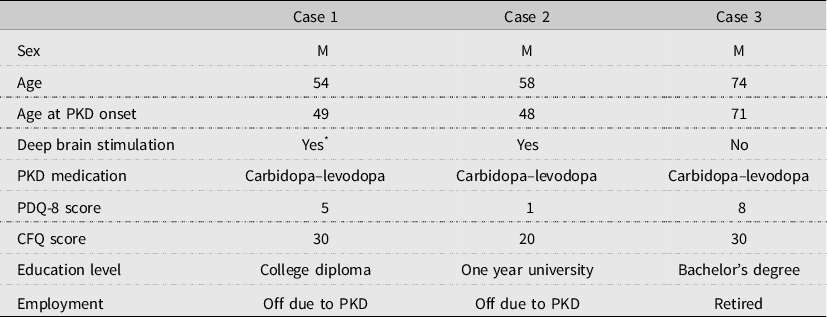
Demographic characteristics of each participant are displayed in Table 1.
Case series characteristics

Note: M = male; PKD = Parkinson’s disease.
* Case 1 had undergone deep brain stimulation, but reported discontinuing it due to depressive side effects.
Overall procedure
The study followed guidelines set out by Carey and Boden (Reference Carey and Boden2003) for the appropriate conduct and reporting of case series studies. Participants completed the screening tests, questionnaires and EEG task following an intake interview in a quiet room at the university. Participants then returned within the week for an introductory MI session and were provided with training instructions. Regular contact was kept with participants via email over the training period. After training, participants returned to repeat all questionnaires and the EEG task.
Questionnaires
In total, three questionnaires were administered to participants. No indicators were assessed aside from imagery ability. Participants completed two self-report questionnaires asking them to rate the vividness of their imagery on a Likert scale, the Vividness of Visual Imagery Questionnaire (VVIQ) and the Movement Imagery Questionnaire – Revised Version (MIQ-RS) (Campos & Pérez-Fabello, Reference Campos and Pérez-Fabello2009; Gregg et al., Reference Gregg, Hall and Butler2010). The VVIQ focuses on imagery of a static scene or object, and the MIQ-RS the imagery of movements. Higher scores are indicative of more vivid imagery.
The Test of Ability in Movement Imagery (TAMI) is an objective measure of imagery ability with established validity and reliability (Madan & Singhal, Reference Madan and Singhal2013). The test consists of 10 questions. For each question, participants are presented with a series of four body movements and asked to imagine them sequentially. They are then presented with various body positions and asked to select the one their body should now be in, given the previously imagined sequence.
MI ability is thought to be comprised of three dimensions, including vividness, accuracy and manipulability (Guillot, Louis, & Collet, Reference Guillot, Louis, Collet, Guillot and Collet2010). Improvement in one dimension does not guarantee the improvement in another and vice versa (Guillot et al., Reference Guillot, Louis, Collet, Guillot and Collet2010). The selection of multiple instruments was thus intended to account for this multidimensionality, as well as any possible discrepancies between subjective and objective imagery performance.
Electrophysiological assessment
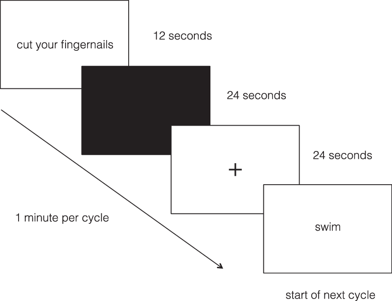
The MI task used during EEG recording was presented using E-prime (Psychology Software Tools) and adapted from Szameitat et al. (Reference Szamietat, Shen and Sterr2007). Participants were asked to imagine 14 different everyday movements. They were instructed to use the first-person perspective, or imagine performing the movements within their own body and observing through their own eyes. Sitting at a computer, participants were presented with the movement to be imagined and given time to prepare an action plan for their imagery. Once the screen turned black, participants closed their eyes and imagined performing the movement. They were instructed to continue imagining the movement until the screen turned white, which was easy to recognize due to the change in luminance in the darkened room. Participants then focused on a fixation cross until they were presented with the next movement. Each movement cycle took 1 min. Figure 1 shows one movement cycle.
Electroencephalographic motor imagery task.

EEG signals were recorded using EGI Net Station (Electrical Geodesics, Inc., Eugene, OR, USA). A high-density 64-channel Geodesic Sensor Net was used to collect the signals, which was amplified at a gain of 1000, with a sampling rate of 250 Hz. Electrode impedance was kept below 50 kΩ. The recording was conducted in an electrically shielded and sound-attenuated room at the University of Alberta.
MI training protocol
Participants attended a single, in-person introductory session to MI adapted from Wondrusch and Schuster-Amft (Reference Wondrusch and Schuster-Amft2013). There, participants learned about the motivation behind MI research and different MI techniques before being introduced to the training program. Training occurred 5 days a week, for 4 weeks in total. For each session, participants listened to an audio recording guiding MI that was created following an extensive literature review (Bovend’Eerdt et al., Reference Bovend’Eerdt, Dawes, Sackley and Wade2012; Braun, Beurskens, Borm, Schack, & Wade, Reference Braun, Beurskens, Borm, Schack and Wade2006; Malouin et al., Reference Malouin, Jackson and Richards2013; Schuster-Amft et al., Reference Schuster-Amft, Hilfiker, Amft, Scheidhauer, Andrews, Butler and Ettlin2011). There was a different audio recording for each week, with all four recordings lasting approximately 10 min.
A 2-minute relaxation task, adapted from Hallsson (Reference Hallsson2013), introduced each recording. The MI portion began with 1 min of imagining a basic locomotor action. The rest of the recording was dedicated to MI of three functional tasks typically difficult to perform in PKD, which were combined to create a recognizable pattern of daily activity (Hariz & Forsgren, Reference Hariz and Forsgren2010). The recordings aimed to place these activities in a realistic setting, with a natural progression between each task. Participants were instructed to use the first-person perspective during imagery. The training protocol resulted in 2 h of MI training, a duration sufficient to generate changes in motor performance (Braun et al., Reference Braun, Beurskens, Borm, Schack and Wade2006).
Participants were provided with a manual featuring training scripts and supplemental images to assist MI as needed, alongside space for participant comments. The manual also contained an imagery log where participants tracked their training and rated each session’s general MI success. No other rehabilitation was performed within the study protocol aside from MI training. Participants were encouraged to continue their daily activities as normal during the study period.
Analysis of questionnaire data
Total pre and post-training scores on the VVIQ, MIQ-RS and TAMI were reported. The mean scores for each participant’s imagery log were calculated for each week, giving each participant a total of four weekly imagery success scores.
Analysis of EEG data
Data were preprocessed using the open-source EEGlab toolbox in MATLAB. The data were first bandpass filtered from 0.1 to 50 Hz. Bad channel components were removed and the data were re-referenced to the vertex electrode (Cz). Artifacts were corrected using independent component analysis and data interpolated to the earlier removed channels.
The preprocessed data were analyzed for oscillatory activity in MATLAB using the duration measure the Better OSCillation detection (BOSC) (Whitten et al., Reference Whitten, Hughes, Dickson and Caplan2011). Using BOSC, segments of signal are only classified as oscillatory when they exceed a set power threshold for a set duration of time. For this study, the power threshold was set to the 95th percentile at each frequency, meaning 95% of the signal background was excluded from the analysis. The duration threshold was set at three cycles to prevent fleeting increases in power from being classified as oscillatory. This procedure computed a measure known as a P episode , which refers to the amount of oscillatory activity detected at a given frequency.
The electrodes selected for analysis were those located at the motor cortex: Cz (the vertex), C3 (left side of scalp) and C4 (right side of scalp). These are the principle motor electrodes and reflect the site of mu activity in previous studies (Formaggio et al., Reference Formaggio, Storti, Cerini, Fiaschi and Manganotti2010; McFarland et al., Reference McFarland, Minar, Vaughan and Wolpaw2000; Pfurtscheller et al., Reference Pfurtscheller, Neuper, Flotzinger and Pregenzer1997). Mu activity, in terms of P episode , was plotted to compare the change in activity from the pre-training to post-training recording for each participant.
Results
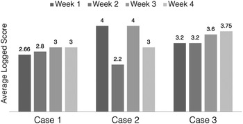
All three participants completed the MI training and tracked session success in their imagery log. The mean success scores from each week are presented in Fig. 2. Both Case 1 and Case 3’s mean success scores increased from week 1 to week 4. In contrast, Case 2’s scores fluctuated every week. It should be noted that Case 2 reported a significant family event during the second week that disrupted his focus.
Mean imagery log success scores.

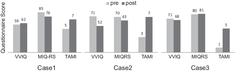
Imagery ability was further evaluated using a combination of subjective and objective questionnaires pre and post-training, the results of which are displayed in Fig. 3. Following training, all participants improved on the TAMI, but none reported significantly higher scores on the MIQ-RS or the VVIQ. This suggests participants improved in MI accuracy, but not vividness.
Questionnaire scores pre and post-training.

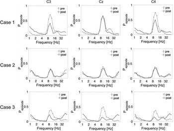
Beyond questionnaire data, we also investigated the neurophysiological effects of MI training. Figure 4 shows plots of each participant’s EEG activity at the electrodes C3, Cz and C4 pre and post-training. Upon visual data inspection, all three participants exhibited greater suppression in the mu rhythm post-training. The details of this suppression varied according to participant and electrode. Case 1 exhibited prominent mu suppression at C3 and C4, but not at Cz, while in Case 3, it occurred in all three electrodes. Case 2’s pattern of change in the mu rhythm was smaller than both Cases 1 and 2, but slight mu suppression was still observed at C4 and Cz.
Mu activity pre and post-training.

Discussion
The presented study investigated the effects of MI training on MI ability and neurophysiological activity in individuals with PKD. All three participants improved in MI accuracy following training as measured by the TAMI. It is unlikely that familiarity with the TAMI accounts solely for these changes given its high test–retest reliability (Madan & Singhal, Reference Madan and Singhal2013). While the basic motor movements assessed in the TAMI were not the focus of training, our protocol did combine and contextualize basic movements to create functional tasks. Research has indicated that MI training of functional tasks can lead to improvements in the performance of non-trained tasks (Liu et al., Reference Liu, Chan, Wong, Kwan, Yau, Li and Lee2009). Our results further indicate that the generalization of learning occurs following MI training.
In addition to the TAMI, the average success score provided at the end of each training session increased in two participants from week 1 to week 4. However, none reported notably higher levels of imagery vividness following training. While this result is in contrast with Abraham et al. (Reference Abraham, Hart, Andrade and Hackney2018); Wondrusch and Schuster-Amft (Reference Wondrusch and Schuster-Amft2013) reported that scores of MI vividness did not improve following participation in their introductory MI program. It was speculated that this change may be due to the participants’ increased knowledge of MI, making them more accurate in their self-reports (Wondrusch & Schuster-Amft, Reference Wondrusch and Schuster-Amft2013). The discrepancy in results confirms the importance of using both subjective and objective measures to assess MI ability.
Our EEG findings further support the need for a multifaceted approach, with greater suppression in the mu rhythm observed in all three participants during the post-training performance of a functional MI task. This result suggests that plastic changes occurred in participants following several weeks of MI training. However, while it is notable that participants presented the predicted pattern per the current literature on mu, it is possible that the extent or pattern of suppression exhibited by our participants is abnormal in comparison to healthy adults.
The exact nature of mu suppression also varied between participants. Case 2 exhibited notably less suppression than the other two participants, while only Case 3 presented with greater suppression at all analyzed electrodes. There are several possible explanations for these discrepancies. Given the internal nature of MI, it is impossible to guarantee that individuals imagine movements in an identical manner. Further, baseline neural rhythm levels vary across individuals, and a small percentage of people do not exhibit mu suppression during movement-related tasks (Hobson & Bishop, Reference Hobson and Bishop2016; Matsumoto et al., Reference Matsumoto, Fujiwara, Takahashi, Lieu, Kimura and Ushiba2010; Tangwiriyasakul et al., Reference Tangwiriyasakul, Verhagen, van Putten and Rutten2013).
However, as greater suppression in the mu rhythm is proposed to index MI success, this result, alongside the improved TAMI scores, indicates the participants learnt from MI training. This is important as it has been suggested that MI may not be therapeutic in PKD due to impaired motor learning (Caligiore et al., Reference Caligiore, Mustile, Spallette and Baldassarre2017; Yaguez et al., Reference Yaguez, Canavan, Lange and Homberg1999). It is possible that the training led the neural networks underlying MI to strengthen and become more efficient, leading to improvements in MI performance. This increase in the efficiency of the motor network may in turn underlie the reported therapeutic effect of MI on motor performance (Holmes et al., Reference Holmes, Cumming, Edwards, Guillot and Collet2010; Page, Reference Page, Guillot and Collet2010; Ruffino et al., Reference Ruffino, Papaxanthis and Lebon2017).
Limitations
The results of this study must be interpreted with caution given its case series design. With a sample size of only three and no control group, it is not possible to confidently determine if the present results are a common response to MI training in PKD, or whether the same response occurs in females or in later stages of the disease. This study also only focused on the mu rhythm. Additional research on other neural rhythms and their role in MI training would provide further insight into the neurophysiological mechanisms of motor learning in PKD.
Conclusion
Three male individuals in the early stages of PKD completed 4 weeks of MI training. All three demonstrated increased accuracy and greater suppression in the mu rhythm following training. This consistent pattern suggests that participants exhibited motor learning and improved in MI ability, a result that runs contrary to some prevailing opinion. The results also indicate the importance of incorporating neurophysiological measures in evaluating MI training outcomes. A larger scale study is recommended to verify the findings, as well as further investigation into how changes in MI ability translate to motor performance.
Acknowledgments
The authors would like to express gratitude to the Parkinson Association of Alberta for their support of this study. Special thanks also go to S.S. and Dr Y.C.
Financial support
This research received no specific grant from any funding agency, commercial or not-for-profit sectors.
Conflicts of interest
KJML has no conflicts of interest to disclose. AS has no conflicts of interest to disclose. AWSL has no conflicts of interest to disclose.
Ethical standards
The authors assert that all procedures contributing to this work comply with the ethical standards of the relevant national and institutional committees on human experimentation and with the Helsinki Declaration of 1975, as revised in 2008.