Systemic lupus erythematosus predominantly occurs in women aged 18 to 45 years, with prevalence estimates of 1.5/1000.Reference Buyon and Clancy 1 Neonatal lupus describes a spectrum of cardiac and non-cardiac abnormalities observed in some neonates whose mothers have systemic lupus erythematosus. Tissue injury in the fetus is related to the transplacental passage of maternal IgG autoantibodies to SSA/Ro and SSB/La intracelular ribonuclear proteins.Reference Saxena, Izmirly and Han 2
Congenital atrioventricular block is the most common cardiac abnormality found in affected neonates. Recently, other cardiac manifestations have been recognised including myocarditis, endocardiofibroelastosis, or structural defects.Reference Costedoat-Chalumeau, Amoura and Villain 3 , Reference Nield, Silverman and Smallhorn 4
We describe the first case of flail tricuspid valve due to papillary muscle rupture following in utero myocarditis associated with maternal anti-Ro and anti-La antibodies in the absence of an atrioventricular block. We discuss the pathophysiology, management, and implications at follow-up in this unique scenario.
Case report
We present a patient who was first detected in utero with multiple, hyperechoic foci in both ventricles at 21 weeks of gestation. The mother (asymptomatic, G2P0AB1 status) was then identified as antibody positive for lupus (anti-SSA/Ro and anti-SSB-La), and dexamethasone treatment was initiated (4 mg/24 hour for a 12-week period). Over subsequent weeks, fetal echocardiography showed signs of acute myocarditis, predominantly right sided, along with hyperechoic lesions on the anterior papillary muscle of the anterior leaflet of the tricuspid valve, that is, mild-to-moderate tricuspid regurgitation. Both chordal and subvalvar apparatus were involved. Moderate right ventricular dysfunction (ejection fraction 30%) was noted. Left ventricular function was normal (ejection fraction 70%, end-diastolic diameter 9.7 mm). Sinus rhythm was maintained: fetal heart rate was 130–140 beats per minute, and the atrioventricular interval was 100 ms. Hydrops was absent.
At the 34th week, evidence of fetal discomfort, silent variability, was detected on a routine prenatal visit, and an emergent caesarean section was then performed.
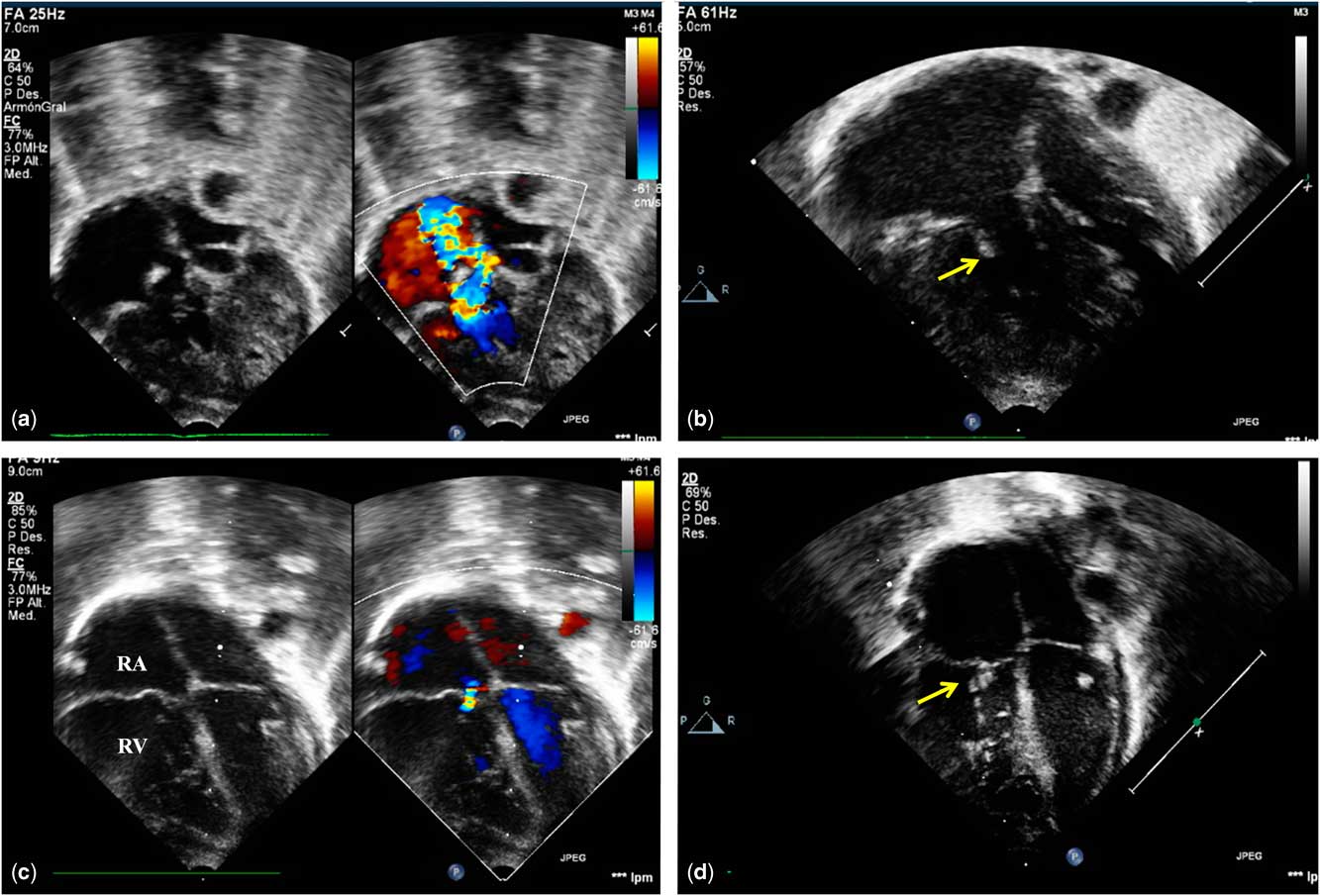
Immediately after birth, he was ventilated and transferred to the ICU. Echocardiography showed a flail tricuspid valve due to anterior papillary muscle rupture and severe tricuspid regurgitation (Fig 1a and b; Supplementary video 1). The tricuspid annulus was 13×10 mm, the tricuspid annular plane systolic excursion was 16 mm, and right ventricle-to-right atrium gradient was 25 mmHg. Paradoxical septal movement (types II–III) was noted. Enlargement of the right atrium (21×21 mm) and signs of mild-to-moderate pulmonary hypertension were present.
Transthoracic echocardiography. Four-chamber view. ( a and b ) Preoperative findings. Severe tricuspid regurgitation. Chordae rupture (anterior leaflet) is noted (yellow arrow). ( c and d ) Findings at 20-month follow-up. Mild tricuspid regurgitation. Polytetrafluoroethylene neochordae is demonstrated (yellow arrow). RA=right atrium; RV=right ventricle.

Treatment with milrinone and dopamine was initiated. Treatment for acute myocarditis was also initiated using hydrocortisone and immunoglobulin. Anti-SSA/Ro, anti-SSB-La, and anti-nuclear antibodies were positive in the neonate. No systemic involvement was detected, thus confirming the diagnosis of neonatal cardiac lupus.
He was extubated on the 5th day after birth. Over subsequent weeks, symptoms of heart failure significantly improved. His left ventricular ejection fraction was 70%, and tricuspid regurgitation remained unchanged. He was discharged home on day 25 after birth on medical treatment with captopril, sildenafil, and furosemide. On subsequent echocardiograms, the absence of hyperechoic foci and improvement in the paradoxical septal movement (type I) were noted. The right ventricle-to-right atrium gradient significantly increased to 32 mmHg.
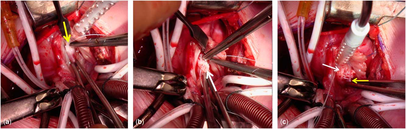
At 2 months of age (weight 3.6 kg), he underwent tricuspid valve repair. A 7/0 polytetrafluoroethylene neochordae was placed in the anterior leaflet with attachment to the papillary muscle, with no annuloplasty associated (Fig 2). A valve orifice of 12 mm (Z score 0) was maintained. The total cardiopulmonary bypass time was 46 minutes, and aortic cross-clamping time was 23 minutes. No postoperative complications were detected, and echocardiographic assessment showed good mobility of the tricuspid valve and mild regurgitation. He was discharged on postoperative day 26.
Surgeon’s view. ( a ) Ventricular surface. Insertion of neochordae (yellow arrow). ( b ) Anterior leaflet of the tricuspid valve. Insertion of neochordae into the fibrosis at the edge of the valve (white arrow). ( c ) Both insertions of the neochordae are shown.

At the 20-month follow-up, he is progressing well, with good biventricular function, normal-sized right ventricle, and mild tricuspid regurgitation (Fig 1c and d; Supplementary video 1).
Discussion
This model of passively acquired autoimmunity is responsible for a proinflammatory and profibrosing process in the fetus that may extend beyond the conduction tissue, involving the myocardium and endocardium. In general terms, cardiac structural lesions have been reported in 10–40% of children with congenital heart block due to lupus, but the occurrence of these lesions has never been studied in the absence of cardiac block.Reference Vinet, Pineau and Scott 5
Although the presence of valvular disease in this setting is exceptional, immunoglobulin and complement deposition in the valvular structure may occur, leading to several forms of lesions, such as valvular thickening.Reference Zuily, Regnault and Selton-Suty 6
On the other hand, myocarditis may also promote any isolated valvular involvement secondary to the subsequent phases of the autoimmune injury, even without conduction disturbances. In an environment of immune reaction within the myocardium extending to the subvalvular apparatus, the inflammatory components may lead to fetal discomfort and hypoxic insult, representing factors of excessive fibre tension, papillary muscle rupture, and critical valve regurgitation.
In general terms, abnormalities of the tricuspid valve – dysplasia or straddling – are rare causes of tricuspid regurgitation in neonates; nevertheless, tricuspid regurgitation caused by flail leaflets in this exceptional autoimmune scenario constitutes a serious disease with high morbidity and mortality.
Following birth, inflammation of the surrounding tissues may make unfeasible an optimal repair. Subsequently, the management of heart failure in the neonatal period is mandatory for establishing optimal timing for surgery. It needs to be considered early in the course of the disease, but only when the myocarditis component has resolved.
Replacement or augmentation of chordae tendinae has proved to be a useful technique of valve repair in infants when the chordal apparatus is involved, with excellent outcomes.Reference Veerbhadran, Pillai and Sasidharan 7 , Reference Boon, Hazekamp and Hoohenkerk 8 In our case, despite the patient’s growth, significant valvular restriction by the artificial chordae has not been observed.
At long-term follow-up, routine cardiac evaluation is needed not only for valvular assessment, but also for detection of other silent cardiac abnormalities related to cardiac lupus.
The spectrum of cardiac manifestations associated with anti-SSA/Ro and SSB/La antibodies is undoubtedly expanding. The recognition of flail tricuspid valve as the final event of an underlying myocardial autoimmune inflammatory process may be added as a manifestation of neonatal lupus. A firm understanding of these exceptional abnormalities is needed to prevent such complications after birth.
Acknowledgements
None.
Financial Support
This research received no specific grant from any funding agency or from commercial or not-for-profit sectors.
Conflicts of Interest
None.
Informed Consent
Informed consent was obtained from the legal guardians of the patient.
Supplementary Material
To view supplementary material for this article, please visit https://doi.org/10.1017/S1047951117000543