I. INTRODUCTION
Copper(II), bis[(2E)-3-methoxy-2-[(2,6-dimethylphenyl)imino]-4-[(2,6-dimethylphenyl) imino-κN]-3-pentanolato-κO] complex (Figure 1) is a reaction intermediate in the synthesis of keto-β − diimine ligands (Yokota et al., Reference Yokota, Tachi and Itoh2002). The complexation of these ligands to nickel leads to the formation of efficient catalysts for olefin polymerization (Azoulay et al., Reference Azoulay, Schneider, Galland and Bazan2009) similarly to more frequently used α − diimine complexes of Ni and Pd (Johnson et al., Reference Johnson, Killian and Brookhart1995). These keto-β-diimine Ni complexes allow the living polymerization of ethylene and higher olefins leading to polyolefins with the controlled molar mass.
Structural formula of the title compound, copper(II), bis[(2E)-3-methoxy-2-[(2,6-dimethylphenyl)imino]-4-[(2,6-dimethylphenyl) imino-κN]-3-pentanolato-κO] complex.

II. EXPERIMENTAL
A. Sample preparation
Preparation of Cu(II) complex is based on oxidative degradation of β-diimine by copper acetate under aerobic conditions reported by Yokota et al. (Reference Yokota, Tachi and Itoh2002).
Starting compounds for the synthesis were a β-diimine ligand, prepared according to Feldman et al. (Reference Feldman, McLain, Parthasarathy, Marshall, Calabrese and Arthur1997), and copper(II) acetate monohydrated yielding a copper(II) β-diimine complex. The copper(II) β-diimine complex was converted to the title complex (copper(II), bis[(2E)-3-methoxy-2-[(2,6-dimethylphenyl)imino]-4-[(2,6-dimethylphenyl)imino-κN]-3-pentanolato-κO]) by a reaction with molecular oxygen.
B. Diffraction data collection and reduction
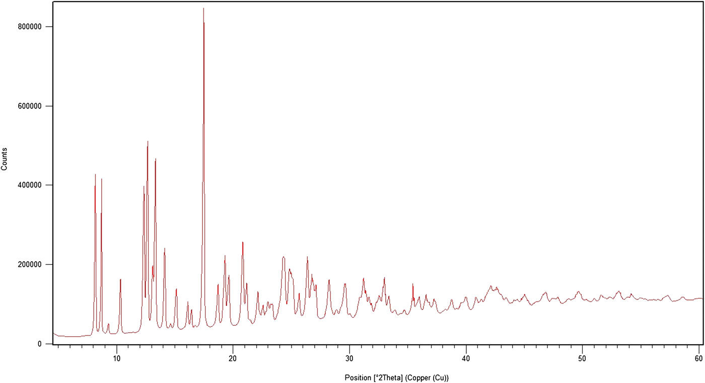
The diffraction pattern for the title compound was collected at room temperature using an X'Pert PRO θ–θ powder diffractometer with parafocusing Bragg–Brentano geometry and CuKα 1 radiation (λ = 1.5406 Å, generator setting: 40 kV, 30 mA). An ultrafast X'Celerator detector was employed to collect XRD data over the angular range from 4 to 60° 2θ with a step size of 0.017° 2θ and a counting time of 20.32 s/step. The experimental powder diffraction pattern is depicted in Figure 2. The software package HighScore PlusV3.0d of PANalytical, Almelo, Netherlands was used to smooth the data, to fit the background, and to eliminate the Kα 2 component. The top of the smoothed peak method was used to determine the peak positions and intensities of the diffraction peaks.
X-ray powder diffraction pattern of the title compound using CuKα 1 radiation (λ = 1.5406 Å).

Automatic indexing of the experimental XRD pattern was done using DICVOL04 (Boultif and Louër, Reference Boultif and Louër2004).
III. RESULTS
The experimental powder diffraction pattern is depicted in Figure 2. Automatic indexing results obtained by DICVOL04 (Table 1) show that the title compound is triclinic with a space group P-1 and unit-cell parameters: a = 8.683(3) Å, b = 11.216(3) Å, c = 11.753(4) Å, α = 66.27(3), β = 84.61(3), γ = 78.85(3), unit-cell volume V = 1027.77 Å3, and Z = 1. The figures of merits are F 20 = 115.3(0.0062, 28) (Smith and Snyder, Reference Smith and Snyder1979) and M 20 = 40.8 (de Wolf, Reference De Wolff1968). All lines were indexed and are consistent with the P-1 space group.
Indexed X-ray powder diffraction data for the title compound, C44H54CuN4O4l.. Only the peaks with I rel of 1 or greater are presented [a = 8.683(3) Å, b = 11.216(3) Å, c = 11.753(4) Å, α = 66.27(3), β = 84.61(3), γ = 78.85(3), unit-cell volume V = 1027.77 Å3, Z = 1, and space group P-1]. All measured lines were indexed and are consistent with the P-1 space group. The d-values were calculated using CuKα 1 radiation (λ = 1.5406 Å).

ACKNOWLEDGEMENT
This work was supported by the grant TA 03011394 of the Technological Agency of the Czech Republic.