Introduction
Schistosomiasis is considered to be a major health problem, as more than 200 million people are currently infected with Schistosoma species, with 600 million at risk (Yorston & Murray McGavin, Reference Yorston, Murray McGavin, Cook and Zumla2009). Schistosomiasis is one of the highly endemic parasitic diseases in 78 countries in Africa, the Americas, the Middle East and South-East Asia (WHO, 2014). There is also an increased number of imported cases of schistosomiasis to Europe due to expanding international travel (Grobusch et al., Reference Grobusch, Muhlberger, Jelinek, Bisoffi, Corachan, Harms, Matteelli, Fry, Hatz, Gjorup, Schmid, Knobloch, Puente, Bronner, Kapaun, Clerinx, Nielsen, Fleischer, Beran, da Cunha, Schulze, Myrvang and Hellgren2003). The pathological effect of Schistosoma mansoni (S. mansoni) is induced directly through egg deposition and embolization, with the characteristic granuloma formation mainly in the liver, colon and lung (Cheever, Reference Cheever1993). The granulomatous formation is mediated by a cellular immune response to soluble egg antigen (SEA) which induces a T-lymphocyte-dependent delayed hypersensitivity response (Jacobs et al., Reference Jacobs, VanDam, Bogers, Deelder and Van Marck1999). Later on, as the infection becomes chronic, the granulomatous response is down-modulated by T- and B-regulatory cells and, alternatively, activated macrophages that limit the extent of immune-mediated pathology (Wilson et al., Reference Wilson, Mentink-Kane, Pesce, Ramalingam, Thompson and Wynn2007). Infection with S. mansoni may affect almost any organ of the body indirectly through deposition of the SEA and the circulating immune complexes, which are considered to be an important mechanism in the pathogenesis of the disease (El Garem, Reference El Garem1998). The presence of S. mansoni antigens in different tissues of the host, such as liver, spleen, kidney, heart, ovaries and testes, as a part of systemic immune complex deposition, has been demonstrated in many studies (El Dosoky et al., Reference El-Dosoky, Van Marck and Deelder1984; Eid, Reference Eid1988; Yassein, Reference Yassien2001; El Nouby, Reference El-Nouby2003). Moreover, S. mansoni is responsible for altering the haemodynamics of the portal circulation, lung arterial and venous systemic systems. Therefore, associated haemodynamic changes in the ocular circulation blood flow are also probable (Delgado de Souza et al., Reference Delgado de Souza, Teixeira Brandt, Ventura and Orefice2002).
Schistosoma mansoni can affect the eye by either or both mechanisms – egg embolization and immune complex deposition – however, most of the literature about ocular schistosomiasis has presented case studies. Schistosomiasis may reach any part of the eye, including the orbit, eyelid, conjunctiva and eye wall layers. Schistosomiasis was reported to cause eyelid granuloma (Kabo & Warter, Reference Kabo and Warter1993), a periocular granuloma that presented as a subconjunctival tumour (Randriamora et al., Reference Randriamora, Rabarijaona, Rabearivony, Bernardin and Rasoavelonoro2004), optical nerve granuloma (Creed, Reference Creed1993), subretinal granuloma and vasculitis, and even an open-angle glaucoma (Bialasiewicz et al., Reference Bialasiewicz, Hassenstein and Schaudig2001).
Schistosomiasis can cause other pathological effects in the eye besides granuloma formation. Schistosomal ocular inflammatory disease was detected in association with ectopic cutaneous schistosomiasis (Milligan & Burns, Reference Milligan and Burns1988). In addition, yellowish-white bilateral nodules of various sizes in the choroid were detected in patients with hepatosplenic schistosomiasis. These lesions proved to be schistosomal in origin (Orefice et al., Reference Orefice, Simal and Pitella1985).
Schistosomiasis can affect the retina, which is the most sensitive layer of the eye. A case of pure macular oedema was diagnosed to be due to schistosomal infection after exclusion of other aetiologies (Vedy et al., Reference Vedy, Carrica, Rivaud, Chanut and Graveline1979). Moreover, schistosomiasis may lead to inflammation of retinal pigment epithelium (Dickinson et al., Reference Dickinson, Rosenthal and Nicholson1990). Using retinal fluorescein contrast arrival time, it was found that S. mansoni infection may be incriminated in the delay of ocular venous blood flow drainage in patients with hepatosplenic schistosomiasis (Delgado de Souza et al., Reference Delgado de Souza, Teixeira Brandt, Ventura and Orefice2002).
The problem in studying schistosomal infection of the human eye is that biopsies are almost impossible to take, and histopathological examination of suspicious lesions can be only undertaken post-mortem or after enucleation. Thus, this experimental study on hamsters gives insight into the possible effects and pathological processes occurring in the eye during S. mansoni infection.
Materials and methods
Experimental infections
This study was carried out on 55 laboratory-bred, parasite-free, golden Syrian hamsters of both sexes (6–8 weeks old and weighing from 120 to 150 g). Hamsters were housed in appropriate cages and fed with a commercial rodent chow and tap water ad libitum, in accordance with institutional and national guidelines. Schistosoma mansoni cercariae shed from laboratory-bred Biomphalaria alexandrina snails were obtained from the Schistosme Biological Supply Program, Theodor Bilharz Research Institute, Giza, according to Frandsen & Christensen (Reference Frandsen and Christensen1984).
Fifty hamsters were infected by S. mansoni cercariae in a dose of 60 cercariae per hamster, injected subcutaneously according to the method described by Peters & Warren (Reference Peters and Warren1969). Five hamsters remained uninfected as negative controls and were sacrificed at the end of the experiment (20 weeks). Ten infected hamsters were sacrificed on weeks 8, 12, 16 and 20 post-infection (pi), and ten hamsters were kept as a reserve to compensate for any dead ones. Eight hamsters died during the course of the experiment. Eyes were removed from each sacrificed hamster, fixed in 10% buffered formalin and processed for histopathological and immunohistochemical studies.
Histopathological and immunohistochemical studies
Formalin-fixed, paraffin-embedded sections (5 μm thickness) were prepared and stained with haematoxylin and eosin (H&E) for histopathological examination (Drury & Wallington, Reference Drury and Wallington1980), and with immunoperoxidase by avidin–biotin complex technique for immunostaining of S. mansoni antigen in the eye tissues (Rogers, Reference Rogers1985) using monoclonal antibodies to repetitive carbohydrate epitopes of S. mansoni soluble egg antigen (114-5B1-A and 114-4D12-A) (Deedler et al., Reference Deedler, De Jonge, Boerman, Fillie, Hilberath, Rotmans, Gerritse and Lexschut1989) and biotinylated goat anti-polyvalent (TP-015-HD, Lab Vision Corporation, California, USA) as a secondary antibody.
Results
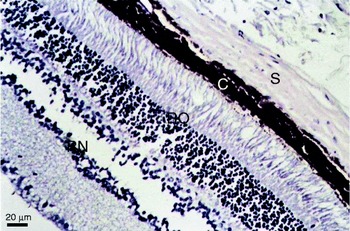
The histopathological examination of eye sections of the control hamsters revealed the normal histological structure of the eye tissue. The sclera consists of dense fibro-elastic tissue arranged in bundles parallel to the surface. The choroid is a layer of loose vascular supporting tissue with numerous large, heavily pigmented melanocytes lying between the sclera externally and the retina internally. The retina is formed typically of ten layers, arranged from the outermost layer as: the pigmented epithelial cells separating the retina from the choroid, rod and cones, outer limiting membrane, outer nuclear layer, outer plexiform layer, inner nuclear layer, inner plexiform layer, ganglion cell layer, afferent fibres and inner limiting membrane that demarcates the innermost aspect of the retina from the vitreous (fig. 1).
Transverse section of the eye of a non-infected hamster to show the sclera (S), choroid (C), outer layers of the retina (RO) and inner layers of the retina (RN).

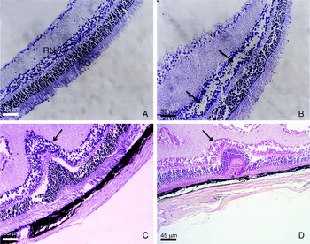
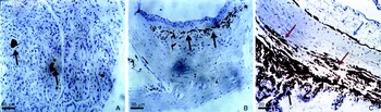
In S. mansoni infected hamsters examined on weeks 8 and 12 pi, no histopathological changes were detected, with preservation of the normal retinal architecture (fig. 2A). Histopathological changes were detected in 12.5% of infected hamsters examined later on weeks 16 and 20 pi. The lesions were in the form of looseness of the innermost layers of the retina, detected on week 16 pi (fig. 2B), whereas, on week 20 pi, there was disruption of the normal retinal architecture with hyperplastic polypoid growth of the retinal layers. In addition, oedematous changes in the innermost layer of the retina were revealed (fig. 2C). Retinal detachment and retinal folds were detected, with partial destruction of the photoreceptor layer (fig. 2D). Neither Schistosoma eggs nor granulomata were detected in eye sections throughout the experimental period. However, immunohistochemical staining of S. mansoni antigen in the eye tissue revealed positive schistosomal antigen deposition in 35% of infected hamsters. The deposition of the antigen started early on week 12 pi in the lacrimal gland (fig. 3A). Later, on weeks 16 and 20 pi, moderate expression of schistosomal antigen was detected at the subepithelial lining of the conjuctival sac (fig. 3B). In addition, marked subchoroidal and scleral antigen deposition was revealed (fig. 3C).
Transverse sections of the retina of S. mansoni infected hamsters to show: (A) on week 12 pi, normal retinal architecture (RO, outer layers and RN, inner layers of the retina); (B) on week 16 pi, loose and oedematous inner retinal layers (arrowed); and on week 20 pi (C) hyperplastic polypoid growth and oedema of the retina (arrowed) and (D) disruption and folding (arrowed) of the normal retinal architecture.

Immunoperoxidase-stained transverse sections of the eyes of S. mansoni infected hamsters to show: (A) on week 12 pi, moderate deposition of schistosomal antigen in the lacrimal gland (arrowed); (B) on week 16 pi, moderate subepithelial conjunctival deposition of schistosomal antigen (arrowed); (C) on week 20 pi, marked deposition of schistosomal antigen in the sclera (blue arrow) and subchoroidal region (red arrows), in addition to dense melanin pigmentation of the choroid and ciliary body (black arrows).

Discussion
The unique structure of the human eye renders it vulnerable to a number of uncommon infectious parasitic diseases. Once anatomical barriers are breached, host defences directed against these parasitic antigens are often insufficient to prevent loss of vision. Therefore, early identification and treatment of the involved patient are paramount (Klotz et al., Reference Klotz, Penn, Negvesky and Butrus2000). Since schistosomiasis is a chronic endemic disease in Egypt and some other parts of the world, ocular involvement is highly expected. Yet, diagnosing ocular schistosomiasis is a challenge and is generally made by exclusion, due to the impossibility of taking biopsies from the internal structures of the eye in humans. This work is an attempt to study the pathology and the possible pathogenesis of S. mansoni infection in the eyes of infected hamsters.
Although humans are rarely infected intraocularly with trematodes, ectopic ocular lesions produced by S. mansoni adults and eggs have been described (Newton et al., Reference Newton, Kanchanaranya and Previte1968). It was reported that in cases with schistosmiasis, almost all organs of the body, including the eye, may contain Schistosoma eggs, as shown by post-mortem tissue digestion (Bell, Reference Bell1995). However, in the present study no S. mansoni eggs or granuloma could be detected in all examined eye sections throughout the experimental period. Stein & Char (Reference Stein and Char1982) examined the possible development of schistosomal granuloma inside the eye tissue through intraocular injection of S. mansoni eggs (500 eggs) or egg extract directly into the eyes of the rabbits. They observed the production of inflammatory reactions involving vitreous, choroid, retina and optic nerve, depending on the tissue involved and the number of eggs injected. They described a granuloma similar to those observed in the livers of infected hosts, characterized by an intense eosinophilia and infiltration with polymorphonuclear leucocytes, plasma cells and fibroblasts. Eosinophilia is reported to mediate destruction of the parasites and to cause most of the pathology in the choroid and retina (Hsu et al., Reference Hsu, Hsu, Mitros, Helms and Solomon1980). However, this is not the case in our study, where hamsters were systemically infected in a trial to imitate the natural route of infection that occurs in humans. A similar study (Remígio et al., Reference Remígio, Brandt, Pontes Filho and Albuquerque2009) detected a retinal granuloma in only one case with otherwise normal architecture and thickness of the retina. They suggested that this may be due to a low level of parasite infection.
In the present study, 12.5% of infected hamsters showed detectable histopathological changes in their eye tissues. The ocular lesions started to appear on week 16 pi in the form of retinal oedema and looseness of the innermost layers of the retina. These findings are in agreement with those of Vedy et al. (Reference Vedy, Carrica, Rivaud, Chanut and Graveline1979) who described pure macular oedema in a patient who was diagnosed to be infected with S. mansoni after ruling out all other aetiologies. On week 20 pi, there were marked histopathological changes in eye sections of infected hamsters. These changes were in the form of retinal folds with retinal detachment and infiltration with inflammatory cells. Similar lesions were detected in the eyes of S. mansoni infected patients, in whom inflammation of the retinal pigment epithelium was reported (Dickinson et al., Reference Dickinson, Rosenthal and Nicholson1990). Also, UL'danov (Reference UL'danov1978) reported a case of schistosomal maculitis that led to a marked decrease in the visual acuity of the affected eye.
Schistosomal antigen and/or immune complex deposition in the eye tissues was detected in 35% of infected hamsters. The detection of schistosomal antigen in the present study began in the lacrimal gland early on week 12 pi. Later, on weeks 16 and 20 pi, moderate subepithelial conjuctival deposits were detected as well as marked subchoroidal and scleral deposition. Schistosomal immune complexes could be formed in the circulation then deposited in the tissues, or they may be formed in situ following the deposition of SEA in the tissues where the anti-schistosomal antibodies become bound to the antigens, forming immune complexes (Eid, Reference Eid1988).
The early detection of S. mansoni antigen in the lacrimal gland could be explained by the early appearance of SEA in the blood on week 8 pi, which was mainly deposited along the blood vessels (El Nouby, Reference El-Nouby2003). Subsequently, the SEA and circulating immune complexes in the blood may be excreted in other body secretions (Khalil et al., Reference Khalil, Makled, El-Sebai and El-Naggar1987). Therefore, in the present study, the antigenic deposition in the lacrimal gland could be followed by excretion of SEA in the lacrimal gland secretion (tears). The deposition of subconjunctival schistosomal antigen with absence of histopathological involvement of the conjunctiva could be explained by the time needed by the antigen to induce an inflammatory reaction. However, Randriamora et al. (Reference Randriamora, Rabarijaona, Rabearivony, Bernardin and Rasoavelonoro2004) detected S. mansoni eggs in the conjunctiva with formation of subconjunctival granuloma in a 12-year-old girl infected with S. mansoni.
Similarly, the subchoroidal and scleral deposition of schistosomal antigen in the present study would be explained by the high vascularity of both the sclera and choroid. The sclera is penetrated by branches of the ophthalmic artery and the choroid contains larger blood vessels in its superficial aspect with a rich capillary plexus in its deeper aspect (Snell & Lemp, Reference Snell and Lemp1998). Similar choroidal lesions were recorded by Pittella & Orefice (Reference Pittella and Orefice1985), who described what they considered ‘The first case of granulomatous choroiditis produced by S. mansoni’ in a patient who had hepatosplenic and cardiopulmonary forms of the disease and presented with cerebral schistosomiasis. In the same year, Orefice et al. (Reference Orefice, Simal and Pitella1985) studied five patients with hepatosplenic schistosomiasis. All cases showed yellowish-white, multiple, bilateral nodules of variable sizes, located in the choroidal plane. They suggested that the detected lesions were of schistosomal origin.
In the absence of detection of Schistosoma eggs or granulomatous reaction, the question of ocular damage in schistosomiasis is a matter of debate. Some investigators suggested that ocular lesions in schistosomiasis may be due to the migrating schistosomula in the small blood vessels of the eye, with the subsequent damage in the eye (Vedy et al., Reference Vedy, Carrica, Rivaud, Chanut and Graveline1979). On the other hand, Stein & Char (Reference Stein and Char1982) explained the retino-choroidal pathological involvement in cases of S. mansoni infection by lytic or toxic damage resulting from eosinophil-released substances.
Our results clearly demonstrated the deposition of schistosomal antigen at different sites of the eye. The antigenic deposition appeared earlier than histopathological changes, but the intensity of the deposition was marked at the time of detection of the histopathological changes in the retina. Therefore, the deposition of schistosomal antigen and/or immune complexes seems to be the key player in the pathogenesis of the ocular lesions. The role of deposition of schistosomal immune complexes in the pathogenesis of lesions in different organs has been proved (Sobh, Reference Sobh1994; Yassein, Reference Yassien2001; El-Nouby, Reference El-Nouby2003). In this context, Stein & Char (Reference Stein and Char1982) showed that the damage of ocular structures in schistosomiasis may result from mediators released by effector cells that lead to autosensitization to retinal tissue. However, Corsi et al. (Reference Corsi, Nucci, Knafelz, Bulgarini, Di Iorio, Polito, De Risi, Ardenti Morini and Paone1998) suggested that the risk of the development of ocular manifestations in parasitic infections does not seem to be related to the severity of infection, its duration or the use of anti-parasitic therapy, but may reflect a genetic predisposition.
In conclusion, the present study provided an insight into the pathology and the possible mechanism of pathogenesis of human ocular schistosomiasis. Although no S. mansoni eggs could be detected, we did detect some striking ocular pathological changes that could be correlated with the deposition of the schistosomal antigen and/or immune complexes in the eye tissues. Therefore, schistosomiasis should be suspected in cases with unexplained ophthalmological findings, especially in endemic areas.
Acknowledgements
Soluble egg antigen (114-5B1-A and 114-4D12-A) was obtained as a generous gift from Professor Dr A.M. Deelder, Leiden University Medical Center, The Netherlands.
Conflict of interest
None.
Ethical standards
The authors assert that all procedures contributing to this work comply with the ethical standards of the relevant national guides on the care and use of laboratory animals and have been approved by the Research Ethics Committee, Quality Assurance Unit, Faculty of Medicine, Tanta University.