Introduction
Ascospores are the most important means of dispersal in lichenized and non-lichenized Ascomycota. As an immediate result of nuclear fusion and subsequent meiotic cell division, they are responsible for establishing thalli with genetic variation, thus enabling natural selection among individuals of a population (Ahmadjian & Hale Reference Ahmadjian and Hale1973; Jennings & Lysek Reference Jennings and Lysek1996; Deacon Reference Deacon2005). Since the advent of light microscopy, ascospores have also become one of the most important character complexes in the taxonomy of lichenized and non-lichenized fungi (Müller Argoviensis Reference Müller Argoviensis1887; Zahlbruckner Reference Zahlbruckner, Engler and Prantl1905; Bellemère & Letroit-Galinou Reference Bellemère, Letrouit-Galinou and Galun1988; Bellemère Reference Bellemère and Hawksworth1994). Ascospores are rich in morpho-anatomical and chemical features, such as wall thickness and the formation of a gelatinous halo, the number and orientation of septa, endospore development, pigmentation, ornamentation, and chemical reactions with agents such as iodine solution (Ahmadjian & Hale Reference Ahmadjian and Hale1973; Kohn & Korf Reference Kohn and Korf1975; Aptroot Reference Aptroot1991; Harris Reference Harris1995; Staiger Reference Staiger2002; Frisch et al. Reference Frisch, Kalb and Grube2006).
For a long time, ascospore septation and pigmentation were among the most important characters used to separate genera especially in crustose lichens, a classification put forward chiefly by Müller Argoviensis (Reference Müller Argoviensis1887) and Zahlbruckner (Reference Zahlbruckner, Engler and Prantl1905). In many lineages of lichenized fungi, however, it has been shown that such a classification is not natural and that species displaying ascospores of different size, septation, and pigmentation, might in fact be closely related (Harris Reference Harris1986, Reference Harris1995; Staiger Reference Staiger2002; Frisch et al. Reference Frisch, Kalb and Grube2006). On the other hand, certain ascospore characters are characteristic for monophyletic lineages. This is especially true for the distinction between eusepta (formed centrifugally starting with a septal plate) and distosepta (formed centripetally and lacking a septal plate): most families or even orders of lichenized fungi have only euseptate ascospores, while others, including the groups studied here, have distosepta in addition to eusepta (Harris Reference Harris1986, Reference Harris1995; Makhija & Patwardhan Reference Makhija and Patwardhan1988, Reference Makhija and Patwardhan1993; Aptroot Reference Aptroot1991; Kirk et al. Reference Kirk, Cannon, Minter and Stalpers2008). Additionally, ascospore pigmentation is often constant in such lineages, and the colour of the pigmentation is specific to certain groups. For example, all Physciaceae have dark brown ascospores, whereas almost all of the related Teloschistaceae (with a few exceptions) have colourless ascospores; likewise, almost all Lecanorales have colourless ascospores. In pyrenocarpous lichens, the situation is comparable: most or all Monoblastiaceae, Porinaceae, Strigulaceae, and Trypetheliaceae have colourless ascospores, whereas in the Pyrenulaceae they are mostly grey-brown (Harris Reference Harris1986, Reference Harris1995; Aptroot Reference Aptroot1991; Aptroot et al. Reference Aptroot, Lücking, Sipman, Umaña and Chaves2008).
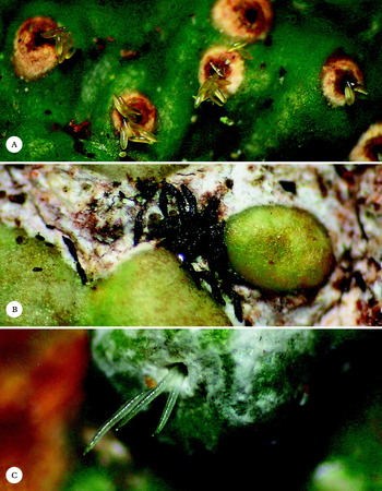
The reasons behind the enormous variation in ascospore features in crustose lichens are largely unknown. Seasonal or diurnal variation in ascospore discharge has been observed in many groups of lichenized fungi (Abbayes Reference Abbayes1951; Pyatt Reference Pyatt1968a, b, Reference Pyatt1969; Ostrofsky & Denison Reference Ostrofsky and Denison1980; Clayden Reference Clayden1997; Yamamoto et al. Reference Yamamoto, Kinoshita, Takahagi, Kroken, Kurokawa and Yoshimura1998; Yamamoto Reference Yamamoto2002). Ascospores are generally believed to be dispersed by abiotic agents (wind, water), either passively (in mazaedia) or after forcible discharge from the asci (Bailey & Garrett Reference Bailey and Garrett1968; Garrett Reference Garrett1971). However, there is evidence that invertebrates play a role in the dispersal of ascospores and other lichen propagules (Pyatt Reference Pyatt1968c; Gerson Reference Gerson1973; Bailey Reference Bailey, Brown, Hawksworth and Bailey1976; Sipman Reference Sipman1983; Lorentsson & Mattsson Reference Lorentsson and Mattsson1999; Leal & Oliveira Reference Leal and Oliveira2000; Dyer & Letourneau Reference Dyer and Letourneau2007). Size and shape of ascospores affect their properties both regarding abiotic and biotic dispersal, and pigmentation appears to be a means of protection against UV radiation (Shields et al. Reference Shields, Durrell and Sparrow1961; Durrell Reference Durrell1964; Bragg Reference Bragg1981; Roper et al. Reference Roper, Pepper, Brenner and Pringle2008). Tropical crustose lichens have a marked tendency to form large and often pigmented ascospores with either thick walls and septa or with muriform cell division (Sipman & Harris Reference Sipman, Harris, Lieth and Werger1989; Aptroot Reference Aptroot1991; Staiger Reference Staiger2002; Frisch et al. Reference Frisch, Kalb and Grube2006). Both factors might play a role in physical stabilization of large ascospores, but might also affect how ascospores germinate. The reasons for evolving such large ascospores are unknown, but it is commonly observed that large ascospores are extruded from the ascomata and remain on the surrounding thallus surface (Fig. 1). Often, hyaline ascospores become dark brown after being expelled from the ascomata. It has been suggested that ants may play a role in the dispersal of ascospores (Lorentsson & Mattsson Reference Lorentsson and Mattsson1999; Leal & Oliveira Reference Leal and Oliveira2000; Dyer & Letourneau Reference Dyer and Letourneau2007; Rivas Plata & Lumbsch Reference Rivas Plata and Lumbsch2011).
Ascospore ‘discharge’ in selected Trypetheliaceae and Graphidaceae. A, Laurera megasperma, hyaline ascospores protruding through the ostiole; B, Architrypethelium nitens, dark brown ascospores deposited on the thallus surface, becoming almost black with time; C, Ocellularia subpraestans, hyaline ascospores protruding through the apothecial pore.

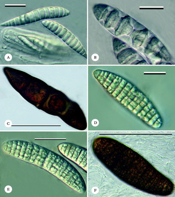
Together with the Graphidaceae and Pyrenulaceae, the Trypetheliaceae are the most important family of tropical crustose lichens (Sipman & Harris Reference Sipman, Harris, Lieth and Werger1989; Komposch & Hafellner Reference Komposch and Hafellner2002; Rivas Plata et al. Reference Rivas Plata, Lücking and Lumbsch2008; Lücking et al. Reference Lücking, Rivas Plata, Chaves, Umaña and Sipman2009) and exhibit a remarkable variation in ascospore characteristics with regard to size, septation, wall and septa morphology and pigmentation and ornamentation (Fig. 2). Besides ascoma morphology, ascospore types have been used for generic delimitation, placing species with transversely septate ascospores in Astrothelium (fused ostioles) and Trypethelium (separate ostioles) and those with muriform ascospores in Campylothelium (lateral ostioles), Cryptothelium (fused ostioles), and Laurera (separate ostioles; Harris Reference Harris1986, Reference Harris1995; Makhija & Patwardhan Reference Makhija and Patwardhan1988, Reference Makhija and Patwardhan1993; Aptroot Reference Aptroot1991; Aptroot et al. Reference Aptroot, Lücking, Sipman, Umaña and Chaves2008). The genus Architrypethelium has been described as having very large, transversely septate ascospores which can either be hyaline or dark brown (Aptroot Reference Aptroot1991). The genus Aptrootia is characterized by a very unusual ecology growing over mosses in montane regions; it has very large, richly muriform, dark brown ascospores that look very different from any other ascospores known in the family (Lücking et al. Reference Lücking, Sipman, Umaña, Chaves and Lumbsch2007). Considering extremes such as the small, transversely septate, hyaline ascospores with diamond-shaped lumina in many species of Astrothelium and Trypethelium versus the large, muriform, dark brown ascospores with rectangular lumina in Aptrootia, one would be tempted to accept ascospore characteristics as an important character complex in delimiting genera within the Trypetheliaceae. However, preliminary molecular analyses suggest that there is limited correlation between phylogenetic relatedness and ascospore type in the family (Del Prado et al. Reference Del Prado, Schmitt, Kautz, Palice and Lumbsch2006; Nelsen et al. Reference Nelsen, Lücking, Grube, Mbatchou, Muggia, Rivas Plata and Lumbsch2009).
Trypetheliaceae, variation of ascospore types. A, Trypethelium eluteriae; B, Astrothelium diplocarpoides; C, Architrypethelium nitens; D, Bathelium tuberculosum; E, Laurera megasperma; F, Aptrootia robusta. Scales: A, B, D = 20 µm; E = 50 µm; C = 100 µm; F = 200 µm.

Besides Graphidaceae, Trypetheliaceae includes species with some of the largest ascospores known among Ascomycota, either with muriform cell division or with few large cells and thick walls and septa. Such megalosporous ascospores, that is ascospores over 50 × 15 µm and thick-walled when non- or sparsely septate and over 100 × 20 µm (up to over 800 × 60 µm in the largest ascospores) when multiseptate or muriform, provide a unique opportunity to study ascospore development and to provide insight into how ascospores of different types are ontogenetically related to each other. In this paper, we studied ascospore development in five species of megalosporous Trypetheliaceae, representing five different ascospore types (septate-astrothelioid, septate-architrypethelioid hyaline and dark brown, and muriform hyaline and dark brown), in order to elucidate the importance of ascospore characteristics in terms of phylogeny and ecology. For comparison, we also documented ascospore discharge and germination in Ocellularia subpraestans (Graphidaceae).
Material and Methods
Specimens were studied and photographically documented using a LEICA MS5 dissecting microscope in combination with a NIKON Coolpix 5400 digital camera and an OLYMPUS SZX12 dissecting microscope with a JENOPTIC ProgRes C3 digital microscope camera. Light microscopic studies and images were photographed with an OLYMPUS BH-2 compound microscope with a JENOPTIC ProgRes C3 digital microscope camera and a ZEISS Axioscop 2 compound microscope with a JENOPTIC ProgRes C5 digital microscope camera. Sections were studied and photographed on material mounted in tap water; sodium hypochloride (C solution) was used to analyze the structure of walls, septa, and endospore deposits in dark brown ascospores of Aptrootia and Architrypethelium (Eriksson Reference Eriksson1989).
The following material was studied:
Trypetheliaceae
Aptrootia terricola (Aptroot) Lücking, Umaña & Chaves: Costa Rica, Lücking 17211 (F).
Aptrootia robusta (P. M. McCarthy & Kantvilas) Aptroot: Australia (Tasmania), Lumbsch 20012 (F).
Architrypethelium nitens (Fée) Aptroot: Panama, Lücking 27038 (F).
Architrypethelium seminudum (Mont.) Aptroot: Costa Rica, Lücking 15212r (F).
Astrothelium diplocarpoides Müll. Arg.: USA (Florida), Lücking 26627 (F).
Bathelium tuberculosum (Makhija & Patwardhan) R. C. Harris: India, Lumbsch 19733j (F).
Laurera gigantospora (Müll. Arg.) Zahlbr.: Philippines, Rivas Plata 2128 (F).
Laurera megasperma (Mont.) Riddle: USA (Florida), Lücking 26710 (F).
Laurera phaeomelodes (Müll. Arg.) Zahlbr.: Panama, Lücking s. n. (F).
Trypethelium eluteriae Spreng.: Colombia, Moncada & Gonzales 3399 (F).
Graphidaceae
Ocellularia subpraestans Hale: Panama, Lücking 33043 (F).
Developmental sequences were determined by using the number of septa and the size of the ascospores in a given hymenium, as well as the development of the cell content (granulose versus clear) and wall pigment. Ascospores with fewer septa, smaller size, granulose cell content, and lack of pigment, were considered younger than ascospores with more numerous septa, larger size, clear content, or pigmentation. For that purpose, a large number of ascospores found in a given hymenium was photographed and the ascospores were put into sequence using the above criteria. Ambiguities or conflicts were resolved as follows: if ascospores showed the same number of septa but variable endospore development, ascospores of the preceding and the following stage in number of septa were used as reference. For example, if 3-septate ascospores occurred with and without an endospore but 1-septate ascospores lacked an endospore and 5–7-septate ascospores consistently produced an endospore, then the 3-septate stage lacking endospore was assumed to be prior to the 3-septate stage with an endospore. If ascospore sequences according to septa and size did not correlate, ascospores of the same septal stage were measured to assess the amount of variation in size, and sequences were rearranged to account for such variation. A particular stage in ascospore development was assumed to be genuine (rather than representing an artefact) if it was observed in at least five ascospores per hymenium and at least five hymenia per specimen. Ascospores that featured certain elements of mature ascospores (such as septa) but did not exhibit all mature characters (such as clear cell content, full number of septa or fully developed pigmentation) are referred to as premature.
Results
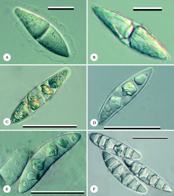
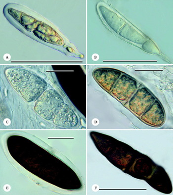
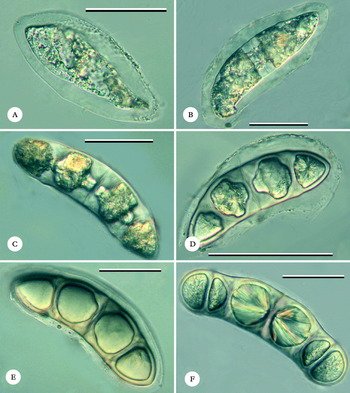
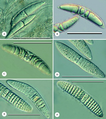
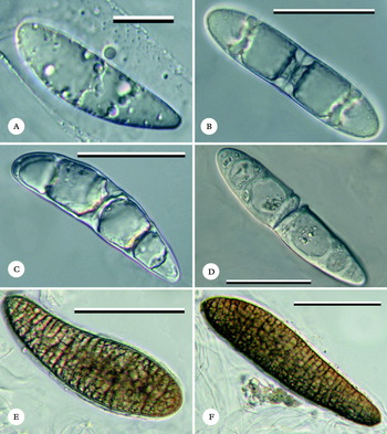
All species of Trypetheliaceae studied show identical ascospore initials, starting out with thin-walled, hyaline ascospores forming a thin euseptum in the centre (Figs 3A, 4A & B, 5A, 6A, 7A). Considering the principal transverse septa only (since the development of the small muriform septa is not readily observed using light microscopy), the first and central septum is the only principal euseptum laid down during the development of the ascospores of the members of the family studied here. All subsequent principal (transverse) septa are distosepta, in which endospore material is chiefly deposited close to the outer wall, resembling the so-called trigons in liverwort leaf cells and eventually producing the characteristic diamond-shaped outline of the septa and lumina (Figs 3B, 5B, 6B, 7B & C). Further septa are laid down from the centre to the periphery in macrocephalic fashion (Figs 3C & D, 4C, 5C & D, 6C, 7B & C), meaning that they divide the remaining cell unequally, but most species produce only three main septa, so no macrocephalic septal sequence can be observed.
Astrothelium diplocarpoides, ascospore development. A, young ascospore forming first median septum; B, young ascospore with endospore development at median septum; C, young ascospore laying down additional septa in macrocephalic fashion; D, premature ascospore with endospore well-developed; E, premature ascospore with thickened, softened endospore (‘amorphous’); F, mature ascospores with diamond-shaped septa and lumina. Scales: A & B = 20 µm; C–F = 50 µm.

Architrypethelium nitens, ascospore development. A, young ascus with ascospore initial; B, young ascospore forming first median septum; C, young ascospore laying down additional septa in macrocephalic fashion, with partial endospore development; D, premature ascospore with endospore well-developed at median septum and with initial pigmentation; E & F, mature, fully pigmented ascospores with endospore at median septum secondarily reduced. Scales: A, B, F = 100 µm; C–E = 50 µm.

Architrypethelium seminudum, ascospore development. A, young ascospore still without septum; B, young ascospore with endospore development at median septum; C, young ascospore laying down additional septa in macrocephalic fashion, with conspicuous endospore development; D, premature ascospore with endospore well-developed; E & F, mature ascospores with endospore secondarily reduced and needle-shaped crystals partially visible in cell lumina. Scales: A–C, E, F = 50 µm; D = 100 µm.

In species with astrothelioid ascospores, such as Astrothelium diplocarpoides, mature ascospores retain the diamond-shaped outline of septa and lumina, often in a very pronounced manner (Fig. 3E & F). This ascospore type is usually called trypethelioid, but species of Trypethelium s. str. actually have a different ascospore type with thin septa and walls (Fig. 2A). In species of Architrypethelium, the stages prior to maturity resemble those of astrothelioid ascospores but in maturity the endospore appears relatively less pronounced, with more rounded to rectangular lumina, due to the substantially larger ascospore size and apparent lack of further endospore production (Figs 4C–F, 5C–F). Architrypethelium nitens develops heavily pigmented, mature ascospores (Fig. 4E & F) which otherwise closely resemble hyaline ascospores of the same type, such as in A. seminudum (Fig. 5E & F).
Ascospores of Laurera gigantospora undergo a remarkable change from the premature to the mature stage (Fig. 6). At first, transverse septa are laid down macrocephalically from centre to periphery and premature ascospores with all transverse septa developed closely resemble the astrothelioid ascospores of Astrothelium diplocarpoides, with pronounced endospore and diamond-shaped septa and lumina (Fig. 6A–C). In the next stage, the endospore is partially resorbed and the ascospores go through a more or less amorphous stage regarding septal structure, during which the longitudinal septa are developed (Fig. 6D & E). Mature spores have distinct horizontal rows of cells with more or less rounded lumina, looking very different from the premature, astrothelioid ascospores (Fig. 6E & F). Ascospore development is often markedly asynchronous in the two halves of the ascospores (Fig. 6E).
Laurera gigantospora, ascospore development. A, ascus with young ascospores forming first median septum; B, young ascospores with endospore development at median septum and forming additional septa; C, young ascospore laying down additional septa in macrocephalic fashion; D, premature ascospore with ‘amorphous' muriform stage. E; premature and mature ascospores showing different stages and asynchronous development in ascospore halves; F, mature ascospore with fully developed muriform septation; the cells are arranged in distinct rows. Scale = 100 µm.

Even more striking are the changes undergone by the ascospores in Aptrootia terricola (Fig. 7). The premature, hyaline ascospores are 3–5-septate and exhibit endospore development very similar to that found in Architrypethelium species (Fig. 7B & C). The next stage is more or less amorphous with regard to septal structure, with the main septa less conspicuous (Fig. 7C), before very rapidly the further transverse and longitudinal septa are developed and the ascospores become dark brown (Fig. 7E & F).
Aptrootia terricola, ascospore development. A, young ascospore forming first median septum; B, young ascospore with endospore development at median septum and forming additional septa; C, young ascospore with secondarily reduced endospore; D, premature ascospore with ‘amorphous' septa; E & F, mature ascospore with fully developed pigmentation and muriform septation. Scales: A = 20 µm; B–D = 50 µm; E & F = 100 µm.

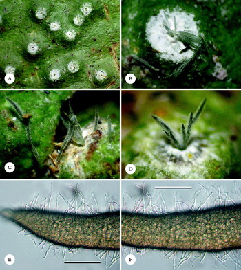
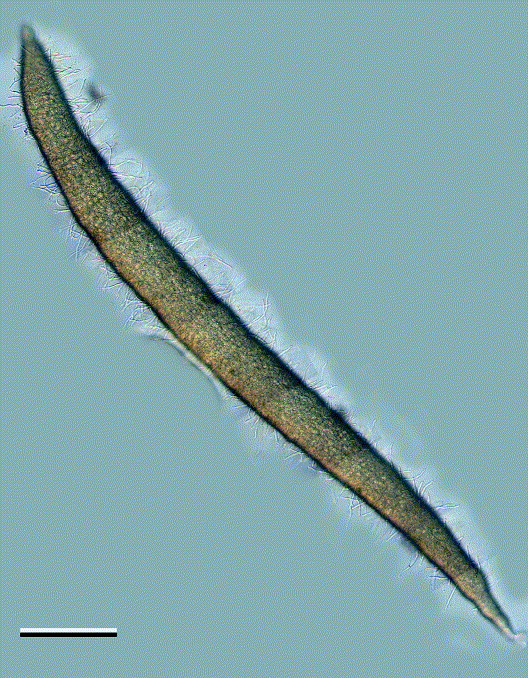
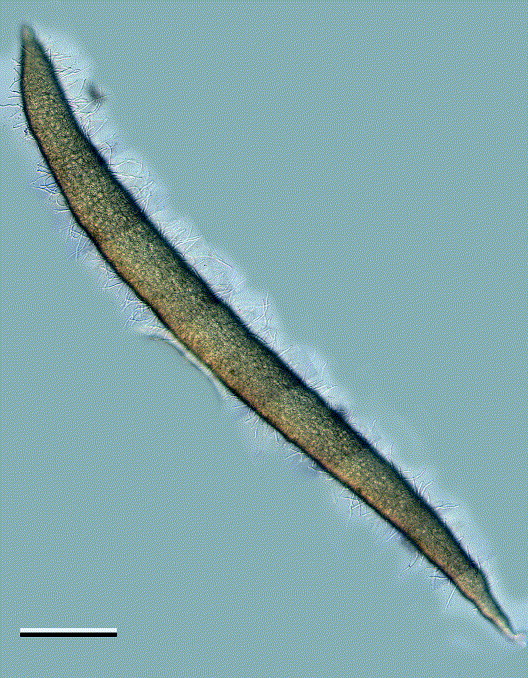
Ascospore discharge and deposition on the thallus surface near the ascomata in Ocellularia subpraestans resembles that of Trypetheliaceae (Figs 1, 8A–D). While still protruding from the ostiole, the ascospores appear as if ‘mouldy’ (Fig. 8C & D), but actually appear to begin germinating from the peripheral cells, in addition to becoming slightly pigmented (Fig. 8E & F). Mature ascospores of this species were 560–880 × 40–65 µm in size (Fig. 9).
Ocellularia subpraestans, ascospore discharge. A, thallus with apothecia; B–D, ascospores protruding through apothecial pore and immediately beginning to germinate; E & F, detail of germinating ascospores; each cell of the richly muriform ascospore produces germinating hyphae. Scale in E–F = 50 µm.

Ocellularia subpraestans, composite image of discharged and germinating ascospore. Scale = 100 µm.
Discussion
Generic delimitation in the lichen family Trypetheliaceae, as well as in other families of crustose lichens, traditionally relied on ascomata and ascospore characteristics (Zahlbruckner Reference Zahlbruckner, Engler and Prantl1905; Harris Reference Harris1986, Reference Harris1995; Makhija & Patwardhan Reference Makhija and Patwardhan1988, Reference Makhija and Patwardhan1993; Aptroot Reference Aptroot1991; Staiger Reference Staiger2002; Frisch et al. Reference Frisch, Kalb and Grube2006). Considering the very different ascospore types found in the family, this concept appeared to be justified to a certain degree. However, preliminary molecular phylogenetic analysis suggests that neither ascoma morphology nor ascospore type are reliable characters for genus delimitation in Trypetheliaceae, and clades include species with both small and large ascospores (Del Prado et al. Reference Del Prado, Schmitt, Kautz, Palice and Lumbsch2006; Nelsen et al. Reference Nelsen, Lücking, Grube, Mbatchou, Muggia, Rivas Plata and Lumbsch2009). Still, it seems difficult to accept that species with such different ascospores as Astrothelium galbineum and Trypethelium nitidiusculum on the one hand, with small astrothelioid ascospores, could be closely related to or even congeneric with species with large, muriform, or dark brown pigmented ascospores typical of Aptrootia terricola or Laurera gigantospora.
Our study shows that, in addition to molecular phylogeny (Del Prado et al. Reference Del Prado, Schmitt, Kautz, Palice and Lumbsch2006; Nelsen et al. Reference Nelsen, Lücking, Grube, Mbatchou, Muggia, Rivas Plata and Lumbsch2009), ascospore ontogeny provides evidence for potentially close relationships between species with very disparate ascospore morphology in the Trypetheliaceae. The different ascospore types share basic developmental features that could be interpreted as synapomorphic for the family as a whole, but as plesiomorphic for individual clades within the family, such as the initial euseptum followed by a variable number of distosepta, as well as the wall and septa thickenings occurring in intermediate stages but subsequently disappearing. Studies of more species are required but our results support initial findings by Makhija & Patwardhan (Reference Makhija and Patwardhan1988: fig. 19) that Trypetheliaceae form only one central euseptum, contrary to other pyrenocarpous families such as Monoblastiaceae, Pyrenulaceae and Strigulaceae, in which the number of eusepta is variable (Aptroot Reference Aptroot1991; Harris Reference Harris1995; Aptroot et al. Reference Aptroot, Lücking, Sipman, Umaña and Chaves2008).
Since basic, plesiomorphic ontogenetic features are repeated in ascospores of different mature types, repeated changes from one ascospore type to another between related lineages are plausible and support the notion that similar mature ascospore types are homoplastic across the family. Thus, species with large, muriform ascospores, such as Laurera gigantospora and Aptrootia terricola, exhibit premature stages that correspond to premature or mature ascospores found in Architrypethelium and Astrothelium, respectively. The latter are identical to those of the small-spored taxa, such as Astrothelium galbineum and Trypethelium nitidiusculum, except that they are much larger. Thus, between the seemingly very different ascospores of Astrothelium galbineum and Laurera gigantospora, there are only three steps: increase in size, partial resorption of the endospore through an amorphous septal stage, and final development of distinct muriform septation. Likewise, between Architrypethelium nitens and Aptrootia terricola, the main differences are two additional steps to partially resorb the endospore and lay down the final transverse and longitudinal septa. Architrypethelium seminudum is connected to Astrothelium diplocarpoides via a similar premature stage.
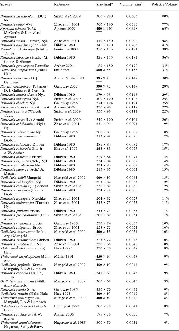
There is a marked tendency to form large ascospores in crustose and especially tropical lichens (Sipman & Harris Reference Sipman, Harris, Lieth and Werger1989). Santesson (Reference Santesson1952) and Pentecost (Reference Pentecost1981) report the ascospores of Varicellaria rhodocarpa and Bacidia marginalis as the longest known among lichens, but by far longer ascospores are documented here for Ocellularia subpraestans, with dimensions up to 880 × 65 µm. Hale (Reference Hale1978a) recorded the ascospores for that species as being up to 300 µm long, but our own investigations of the type revealed ascospores up to 650 × 50 µm. In most specimens studied from Costa Rica and Panama, the ascospores regularly reach 750 × 50 µm in size and exceptionally over 800 × 60 µm. These are the longest spores known among all fungi (Ingold Reference Ingold2001) and this underlines the observation that Ocellularia includes several species with the longest ascospores known (Table 1). However, more voluminous ascospores are formed by several species of Pertusaria s. lat. and by the Trypetheliaceae and Aptrootia robusta (Table 1). For unknown reasons, almost all fungi with very large, voluminous ascospores are found among lichens; the largest ascospores among non-lichenized fungi appear to be those of Podosphaera vertesensis, but these are less than 10% of the volume of the largest ascospores found in Pertusaria (Table 1). Some workers consider certain non-lichenized fungi to have very large ascospores (Raja & Shearer Reference Raja and Shearer2006; Eriksson Reference Eriksson2009), but these are small compared to lichenized fungi and their size range falls far outside that of the 50 species with the largest ascospores (49 of which are lichens; Table 1).
Compilation of lichenized and non-lichenized Ascomycota with the largest ascospores reported, according to various sources (sorted in descending order of ascospore volume)

* the ten longest ascospores are underlined and in bold.
† species currently classified as Thelotrema belonging in Ocellularia s. lat.
Although there is a distinct phylogenetic constraint, as shown by most of the large-spored species representing Pertusaria and related taxa, the tendency to form such large ascospores in crustose lichens suggests a functional role of ascospore size with regard to dispersal and establishment. In species with small ascospores that produce eight per ascus, it has been found that these are discharged in packages rather than individually, thus acting as a single, large propagule (Bailey & Garrett Reference Bailey and Garrett1968; Garty & Delarea Reference Garty and Delarea1988). One possible effect of ascospore size is discharge distance, but Bailey & Garrett (Reference Bailey and Garrett1968) found no significant correlation between maximum discharge distance and ascospore or projectile size, although larger projectiles tended to have a slightly wider discharge range. Pentecost (Reference Pentecost1981) studied the range of ascospore types and sizes in lichens of the British Isles and found correlations with substratum types, but these correlations are in many cases not causal but produced by phylogenetic relationships. For example, Teloschistaceae prefer limestone and at the same time have a very uniform, polarilocular ascospore type, which is not necessarily functionally related to the substratum.
Perhaps most remarkable in the development of muriform ascospores in the Trypetheliaceae, as seen for instance in Laurera gigantospora, is the amorphous premature stage in which the endospore material built with the initial transverse septa is partially resorbed. Such a development appears to be unique for Trypetheliaceae and suggests a vestigial feature of originally transversely septate, astrothelioid ascospores, which are subsequently transformed into muriform ascospores. Formation of muriform ascospores even after substantial investment into transverse distosepta which are then resorbed indicates some sort of constraint (selective pressure) towards muriform ascospores; it also appears that direct formation of muriform ascospores, without first forming and then resorbing transverse distosepta, has not evolved in these species. The alternative explanation of random drift to evolve muriform ascospores seems less likely since the resorption of the transverse distosepta indicates a complex mechanism of transformation from one stage into the other. Since many Trypetheliaceae, such as Architrypethelium spp., Astrothelium ultralucens, and Trypethelium phaeothelium and T. pustulatum, produce very large, robust ascospores with transverse septa only, size alone cannot explain the tendency to form muriform ascospores. The main difference between the two types is the number of cells and nuclei (Zopf Reference Zopf1905a, b). Sanders & Lücking (Reference Sanders and Lücking2002) illustrated the germination of muriform ascospores in Gyalectidium (Gomphillaceae), which suggests that each peripheral cell germinates individually and a single cell might be able to germinate, associate with a compatible photobiont, and establish a new thallus. A similar phenomenon was observed here in Ocellularia subpraestans, in which the ascospores appear to start germinating immediately after protruding through the apothecial pore, and has also been documented for Pertusaria (Pyatt Reference Pyatt1968d; Ahmadjian & Hale Reference Ahmadjian and Hale1973).
It appears that in muriform ascospores with numerous cells, there is a greater likelihood that some cells survive dispersal to eventually germinate. This would be consistent with possible biotic dispersal by invertebrates, because feeding on ascospores would lead to their partial destruction but leave some cells intact. Studies on ascospore germination after being externally or internally dispersed by invertebrates are rare (Lücking & Bernecker-Lücking Reference Lücking and Bernecker-Lücking2000). Pyatt (Reference Pyatt1968c) showed that broken ascospores from the digestive tract of rotifers could germinate, and Meier et al. (Reference Meier, Scherrer and Honegger2002) demonstrated that lichen mycobiont remnants are able to germinate after passing through the intestine of mites. Even with abiotic dispersal only, germination of multiple cells from muriform ascospores and searching for compatible algal photobionts in the surrounding area seems more advantageous than from small, few-celled or non-septate ascospores.
Rivas Plata & Lumbsch (Reference Rivas Plata and Lumbsch2011) suggested that ants might play a role in the dispersal of large ascospores, since these resemble the Beltian and Müllerian bodies in Acacia and Cecropia plants that attract ant symbioses (Rickson Reference Rickson1969, Reference Rickson1976; Eubanks et al. Reference Eubanks, Nesci, Petersen, Liu and Sanchez1997; Herrera & Pellmyr Reference Herrera and Pellmyr2002; Meehan et al. Reference Meehan, Olson, Reudink, Kurt and Curry2009), and ants would then mistakenly carry such ascospores, or parts of them, away when trying to feed on them. This assumption would be consistent with the observation that lichens producing large ascospores deposit them directly on the thallus surface around the ascomata. At first glance, this appears to contradict findings that larger ascospores have a wider discharge range (Bailey & Garrett Reference Bailey and Garrett1968), but that finding seems to apply mostly to ascomata with exposed hymenia (apothecia). In many tropical lichens, a tendency to hide the hymenium within perithecioid or lirellate ascomata can be observed and then the ascospores protrude through the opening rather than being discharged into the air above the thallus (see Figs 1 & 8). The purpose of doing so would either be to grow daughter thalli directly on top or in the vicinity of the mother thallus, or to put the ascospores on ‘display’. Some trees exhibit a strategy of forming large, heavy seeds that disperse directly underneath the mother tree, possibly to guarantee successful growth under the same conditions as the mother tree, whereas others tend to disperse seeds as far as possible to decrease predation and intraspecific competition (Janzen Reference Janzen1970; Connell Reference Connell, Den Boer and Gradwell1971). In crustose lichens, it can often be observed that thalli overgrow old or dead thalli of other lichens; however, this appears to happen randomly and there is no evidence suggesting that thalli regularly overgrow old or dead thalli of the same species. Our findings that the large ascospores of Ocellularia subpraestans germinate immediately after being extruded from the apothecium appear to support the idea of daughter thalli establishing directly on the mother thallus, but anatomical studies of the thalli showed no evidence of layered growth with subsequent generations on top of each other. It also seems unlikely that germinating hyphae from the ascospores are ‘absorbed’ into the mother thallus, as this would make little sense in terms of reproductive biology. The fact that the ‘germ’ hyphae remain rather short and uniform, even in ascospores resting on the thallus surface, suggests that their function has been modified to serve another purpose.
Therefore, the alternative hypothesis that these lichens put their ascospores on ‘display’, albeit being speculative, has some appeal. If the purpose is dispersal by invertebrates such as ants or termites, which usually move up and down the tree trunks, it would indeed make little sense to actively discharge the ascospores over a larger distance, since most ascospores would inevitably fall to the ground between leaf litter where they would stand little chance of surviving or being transported to a viable substratum. That ascospores can develop very specific adaptations towards dispersal and establishment was shown by Cuny et al. (Reference Cuny, van Haluwyn and Caron1997) for Diploschistes muscorum, a juvenile lichen parasite, in which ascospore germination was stimulated by the presence of fumarprotocetraric acid, the main component of the host Cladonia. It is therefore possible that the initial germ tubes of the ascospores in Ocellularia subpraestans serve another purpose: making the ascospores stick to passing invertebrates by the hairy surface. Tibell (Reference Tibell1971) suggested that ascospores with a rough surface produced in mazaedia are possibly removed passively by small animals. Additionally, some insect larvae stick lichen parts (soredia) to their bodies for the purpose of camouflage (Skorepa & Sharp Reference Skorepa and Sharp1971; Wilson & Methven Reference Wilson and Methven1997).
The observation that large ascospores are deposited on or near the mother thallus is in contrast with the observation by Smith (Reference Smith1995) that the lichen biotas of oceanic island groups, such as Galapagos and Hawaii, have a higher proportion of species with large and pigmented ascospores compared to mainland biotas. However, that conclusion is potentially biased by the comparison with extratropical biotas in North America and the British Isles. Tropical mainland lichen biota have a very high proportion of species with large and/or pigmented ascospores (Sipman & Harris Reference Sipman, Harris, Lieth and Werger1989; Lücking et al. Reference Lücking, Rivas Plata, Chaves, Umaña and Sipman2009), and there is no evidence that there would be a bias towards such species in island biotas, suggesting improved properties for long distance dispersal of such ascospores. In contrast, initial comparative analysis of the composition of lichen biotas between the Galapagos Islands and areas in mainland Central and South America shows a decreased proportion of species with large ascospores in the island biota of Trypetheliaceae, Graphidaceae, and Pyrenulaceae. For example, 39% of species of Graphis on the Galapagos Islands have large ascospores, whereas the percentage amounts to 44% in Costa Rica (Lücking et al. Reference Lücking, Chaves, Sipman, Umaña and Aptroot2008; Bungartz et al. Reference Bungartz, Lücking and Aptroot2010).
Material of the species studied was collected in the frame of the NSF-funded projects “Ticolichen” (DEB 0206125 to The Field Museum; PI Robert Lücking) and “Neotropical Epiphytic Microlichens – An Innovative Inventory of a Highly Diverse yet Little Known Group of Symbiotic Organisms” (DEB 715660 to The Field Museum; PI R. Lücking). The Caterpillar® company provided additional funds to study lichens from Panama. The internship of the first author (GS) at the Botany Department of The Field Museum was sponsored by the internship program of the Sustainability Studies @ at Roosevelt University, Chicago [as “Biodiversity” (SUST 330 Biodiversity) course, organized by Julian Kerbis Peterhans]. William Sanders and an additional reviewer provided a thorough review of the manuscript and helped to improve it considerably.