
I. INTRODUCTION
The ability to integrate nanoparticles (NPs) into ordered two- and three-dimensional self-assembly architectures is a highly promising tool for diverse applications like magnetic storage, photocatalysis, magnetic resonance imaging, and drug delivery. Reference Ramasamy and Gupta1–Reference Yoo, Nam, Qi, Lee, Park, Belcher and Hammond3 These hierarchically ordered structures have the advantage of precise control over inter-particle distance and material properties, distinct from their individual components. Reference Bronstein4,Reference Glasgow and Tullman-Ercek5 This opens the scope to engineer “artificial solids” with carefully engineered material properties. While much research has been directed at chemical methods like evaporation induced self-assembly, biohybrid nano-architectures offer an incredible range of structural flexibility and an environment-friendly approach, but remain relatively unexplored. In particular, protein cages are attractive building blocks for hierarchical ordering because they possess a remarkable variety of structural packing and surface charge distribution. Reference Smith, Hawes and Bundy6,Reference Courchesne, Klug, Chen, Kooi, Yun, Hong, Fang, Belcher and Hammond7 Additionally, large quantities of protein cages can be reproducibly synthesized within a narrow size range using cost-effective bio-amplification techniques. Reference Zhou, Bedwell, Li, Prevelige and Gupta8 Among the protein cages, bacteriophage P22 is one of the best-characterized systems in terms of macromolecular self-assembly. This study investigates the assembly of P22 protein cages with rigid inorganic NPs and soft polymer linkers to form large area building blocks for more complex biomimetic structures. The study represents an important step toward engineering advanced bio-inspired materials. So far, the assembly of P22 protein cages with inorganic NPs and polymer has not been reported.
P22 is considered a hallmark model for double-stranded DNA (ds-DNA) viruses. Reference Parent, Khayat, Tu, Suhanovsky, Cortines, Teschke, Johnson and Baker9 The Salmonella typhimuriam bacteriophage P22 assembles via a scaffold protein (SP) into T = 7 60 nm icosahedral symmetry containing a 50 nm central cavity for DNA packaging. Reference Prevelige, Thomas and King10 415 molecules of coat protein (CP), 60–300 SP, and a dodecameric portal protein complex co-assemble to form the P22 procapsid. In vitro co-assembly of CP and SP produces virus-like particles (VLPs) virtually indistinguishable from those assembled in vivo. Reference Prevelige, Thomas and King10 The VLPs are attractive because they contain the capsid proteins without the infectious viral genome. In addition, P22 has a robust architecture with unique surface features like the portal complex incorporated at one of the pentameric vertices to allow for entry and egress of the DNA. P22 shows a range of conformational arrangement. For example, expanded P22 morphologies with popped out pentamers can be readily obtained via heat-treatment. Reference Morris and Prevelige11 Additionally, P22 VLPs contain pockets of local positive and negative charges, while the overall VLP is negatively charged above the isoelectric point. Therefore, both electrostatic interaction and the sophisticated structural features can be exploited to achieve ordered assembly and multidimensional molecular engineering with P22.
Recently, P22 VLPs have been successfully arranged into higher ordered assemblies using the linker protein, Dec. Reference Uchida, LaFrance, Broomell, Prevelige and Douglas12 In this system, Dec served as a soft and site-specific linker material for connecting expanded P22 VLPs into an extended lattice. Other soft materials like dendrimers and diblock copolymers are also known to induce hierarchical ordering of protein cage units like cowpea chlorotic mottle virus (CCMV) and ferritin, but fewer reports are available on P22. For example, well-defined assembly of ferritin cages was driven by electrostatic interaction with cationic diblock co-polymer, poly(N-methyl-2-vinyl pyridinium iodide-block-poly(ethyleneoxide)). Reference Tahka, Laiho and Kostiainen13,Reference Kostiainen, Hiekkataipale, de la Torre, Noltea and Cornelissen14 In contrast, inorganic NPs offer unique possibility of facilitating ordered arrangement of P22 because the NPs are rigid and crystalline as compared to the soft binding protein units. Kostiainen et al. reported a unique AB8 fcc binary superlattice of CCMV and Au NPs for the first time via controlled electrostatic interaction. Reference Kostiainen, Hiekkataipale, Laiho, Lemieux, Seitsonen, Ruokolainen and Ceci15 Using a similar approach, a third NP component could also be integrated into a CCMV-avidin binary superlattice. Reference Liljestrom, Mikkila and Kostiainen16
Despite the immense structural possibilities offered by biohybrid assembly, it is not trivial to control the interaction between the polymers or inorganic NPs and protein cages. In particular, the NPs must be highly stable in the aqueous phase and the length over which the electrostatic interaction of the binary components are active (Debye screening length, see Supplementary Material) must be accurately controlled for different virus systems. Reference Kostiainen, Hiekkataipale, Laiho, Lemieux, Seitsonen, Ruokolainen and Ceci15 The size and surface charge distribution of the VLPs are also known to play a dominant role in the final ordered arrangement. Considering these challenges, the ordered arrangement of P22 VLPs using inorganic NPs has not been attempted thus far. Indeed, the self-assembly of P22 over a large area will be highly useful to integrate semiconductor NPs for bottom-up synthesis of bio-inspired photocatalytic materials. Previously, we have reported the synthesis of semiconductor NPs CdSe, CdS, and ZnS using P22 cages. Reference Zhou, Bedwell, Li, Bao, Prevelige and Gupta2,Reference Zhou, Bedwell, Li, Prevelige and Gupta8 To exploit the full potential of protein cage architectures for designing of new photocatalytic and magnetic materials, the marked difference between the self-assembly of P22 VLPs using rigid NPs and soft dendrimer units should be explored in depth. These electrostatic interactions will help outline the factors inducing large area P22 assembly.
Here we report the assembly of P22 VLPs using two different linker materials, inorganic NPs, and polyamidoamine G3 (PAMAM) dendrimers. In these systems, polyethyleneimine (PEI) coated inorganic NPs (iron oxide, CoFe2O4, and Au) serve as the model for rigid linkers while PAMAM is a model for soft linker material. NaCl solution is used to control the Debye screening length for suitable ordering of P22 VLPs. The effect of PAMAM:P22 number ratio on the assembly of P22 is investigated. The terminal groups of PAMAM dendrimer likely interact with P22 VLPs. In addition, the ability of positively charged iron oxide NPs to induce self-assembled architectures of P22 is reported. In case of the NPs, controlled electrostatic attraction plays the key role in the assembly of P22. To this end, the effect of pH, NP:P22 number ratio, and capping agents on the ordering of P22 is investigated. These environmental-friendly P22-based nano-architectures will be particularly promising building blocks for hierarchical biomimetic structures to tailor key material properties like band gap for photocatalytic applications and saturation magnetization for magnetic resonance imaging and therapeutic applications. Reference Shen, Bao, Prevelige and Gupta17
II. EXPERIMENTAL DETAILS
A. Materials
All reagents including uranyl acetate (Sigma Aldrich, St. Louis, Missouri), iron(III) chloride (Sigma Aldrich), cobalt(II) chloride (Sigma Aldrich), sodium oleate (Spectrum Chemicals, New Brunswick, New Jersey), gold(III) chloride hydrate (HAuCl4·xH2O) (Sigma Aldrich), polyethyleneimine (PEI, Sigma Aldrich), 8-amino-1-octanethiol (Sigma Aldrich), oleic acid (OA, Thermo Fisher Scientific, Waltham, Massachusetts) 1-octadecene (Sigma Aldrich), n-hexane (Thermo Fisher Scientific), ethyl alcohol (Thermo Fisher Scientific) are used as purchased. All water used in the experiments is deionized via a Milli-Q Advantage water purification system (EMD Millipore, Billerica, Massachusetts). PEI-iron oxide NPs are purchased from Ocean NanoTech (San Diego, California). Polyamidoamine G3 (PAMAM) dendrimer has been purchased from Dendritech Inc. (Midland, Michigan) and used without further purification.
B. Preparation and purification of P22 virus-like particles
P22 procapsid is synthesized by co-expression of the coat protein (CP) and scaffold protein (SP) in E. coli, as reported earlier. Reference Lanman, Tuma and Prevelige18,Reference Kale, Bao, Zhou, Prevelige and Gupta19
C. Synthesis of CoFe2O4 and Au nanoparticles
Iron–cobalt oleate precursor for CoFe2O4 NPs is prepared by mixing iron chloride (4 mmol, 0.65 g), cobalt chloride (2 mmol, 0.2 g) with sodium oleate (16 mmol, 4.87 g) in a solvent mixture (hexane, 20 mL, DI water, 10 mL, and ethanol, 10 mL) at 60 °C for 4 h. Subsequent evaporation of ethanol and hexane is conducted at 70 °C and 110 °C. Reference Bao, Shen, An, Padhan, Turner and Gupta20,Reference Bao, Shen, Wang, Ma, Mazumdar and Gupta21 After phase separation, the organic phase containing the iron–cobalt oleate precursor is washed with DI water multiple times and dried overnight in the chemical hood. The entire process is performed in air without inert gas protection. The iron–cobalt oleate paste is subsequently used to synthesize CoFe2O4 NPs. Typically, 5 g of the mixed oleate complex is heated in 1-octadecene (20 mL) at 320 °C for 2 h in the presence of ligand, OA to form CoFe2O4 NPs. To render these organic phase NPs hydrophilic, a second ligand layer of PEI is coated on the NPs via a modified bilayer phase transfer method previously described. Reference Palchoudhury, Hyder, Vanderlick and Geerts22
The Au NPs are synthesized via a “modified Frens” method replacing the traditionally used citrate ligand with the cationic polymer, 8-amino-1-octanethiol. Reference Bastus, Comenge and Puntes23 These aqueous phase Au NPs are subsequently used for self-assembly studies of P22.
D. P22 assembly by PAMAM dendrimer and inorganic nanoparticles
To study the assembly of VLPs in the presence of electrostatic interaction from rigid linkers, the P22 VLPs are mixed with three different NPs (e.g., PEI–CoFe2O4 NPs, PEI–iron oxide NPs, and 8-amino-1-octanethiol-coated Au NPs). The mixing is systematically conducted at different NP:P22 number ratios (e.g., 0.9:1, 2.4:1, 4.3:1, 6.5:1, 7:1, and 11:1). Gentle mixing via pipettes is used to maintain the integrity of the sample. Each experiment is repeated in the pH range of 6–7.5. Different NaCl concentrations (0, 10, 50, and 100 mM) are used for each run to investigate the effect of Debye length.
Similarly, P22 VLPs are gently mixed with PAMAM-G3 at different PAMAM:P22 number ratios (e.g., 2:1, 6:1, 8:1, 10:1, and 100:1), keeping the pH ∼ 7.5. Further, each experiment is repeated with different NaCl concentrations (10 mM, 100 mM, and 1 M).
The detailed calculations for NP:P22 and PAMAM:P22 number ratios are derived in the Supplementary Material.
E. Characterization
The size and morphology of the P22 assembly with both NPs and dendrimers are investigated on a FEI Tecnai F-20 transmission electron microscope (TEM; FEI Company, Hillsboro, Oregon). Prior to imaging, the grid containing the bio-hybrid sample is stained with 1% uranyl acetate solution (2 μL) to render the P22 visible. Reference Uchida, Morris, Kang, Jolley, Lucon, Liepold, LaFrance, Prevelige and Douglas24 The hydrodynamic sizes and zeta potential of the NPs, P22 VLP, and the self-assembled P22 VLP samples are monitored using a dynamic light scattering (DLS) instrument equipped with Zetasizer Nano (Malvern Instruments, Worcestershire, United Kingdom).
III. RESULTS AND DISCUSSION
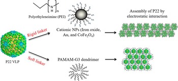
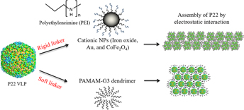
Virus capsids such as P22 VLPs are attractive building blocks for templated synthesis of NPs and bottom-up self-assembly because they can be easily produced through inexpensive bio-amplification techniques, can be genetically modified, and contain multiple functional ligands for potential interaction sites. We have earlier demonstrated the site-specific nucleation and growth of light-harvesting NPs within P22 VLPs, but little work has been reported on the self-assembly of P22 in the presence of NPs and dendrimers. Reference Shen, Bao, Prevelige and Gupta17 Figure 1 shows the schematic representation of the experiments conducted to explore the difference in assembly of P22 VLPs in the presence of rigid and soft cationic linkers. The inorganic NPs serve as the rigid material while the PAMAM dendrimer is considered the soft linker. Reference Uchida, LaFrance, Broomell, Prevelige and Douglas12 We aim to induce large area assembly of P22 VLPs through controlled electrostatic interaction between the negatively charged VLP and the positively-charged polymer or the inorganic NPs (Fig. 1). Due to the nature of the polymer as compared to the NPs, the assembly of P22 varies in the presence of the two components. These protein cage architectures can potentially serve as biomimetic platforms for bottom-up self-assembly of semiconductor NPs to control the band gap for photocatalytic applications.
Schematic drawing of the bio-hybrid synthesis route for assembled P22 VLPs via rigid (inorganic NPs) and soft (PAMAM) cationic linkers.

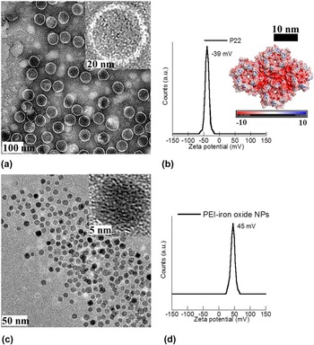
Figure 2(a) shows the TEM image of wild-type P22 VLPs synthesized for this study. The VLPs are monodisperse spheres of ∼60 nm in size, as suggested from the high resolution transmission electron microscope (HRTEM) image [Fig. 2(a), inset]. The DLS hydrodynamic size of the P22 VLP is 68 nm, slightly higher than the dimension measured from TEM image. This difference in size is likely because the hydrodynamic dimension is calculated from the Stokes–Einstein equation, assuming a perfectly spherical morphology [Fig. S1(a)]. The charge distribution on the hexameric units of P22 is an important parameter for large area self-assembly and is visualized in detail with a molecular modeling program, UCSF Chimera [Fig. 2(b), inset]. Reference Pettersen, Goddard, Huang, Couch, Greenblatt, Meng and Ferrin25–Reference Janner27 Negatively charged surface residues are shown in red and the positively-charged regions are shown in blue. The hexameric and pentameric units are marked in the figure for clarity. The overall surface charge of the aqueous phase P22 VLPs, measured on the Malvern Zetasizer Nano series is −39 mV, indicating good colloidal stability [Fig. 2(b)]. Typically, a colloidal system is considered stable if the absolute zeta-potential value is greater than 30 mV. Reference Xu, Qin, Palchoudhury and Bao28 Fig. 2(c) shows the PEI-coated iron oxide NPs used in subsequent experiments as rigid linker model to self-assemble P22. The NPs are 10 nm in size with a narrow size distribution and a hydrodynamic diameter of 55 nm [Fig. S1(b)]. They form stable positively-charged suspension in water, as indicated by the high absolute zeta-potential value [Fig. 2(d)]. Therefore, these iron oxide NPs have the potential for electrostatic interaction with the negatively-charged P22 to facilitate packing of the VLPs over a large area.
P22 and iron oxide NPs. (a) TEM image of wild-type P22 VLPs, inset, HRTEM, (b) zeta-potential data of P22, inset charge distribution on P22, (c) TEM image of PEI–iron oxide NPs, inset HRTEM, and (d) zeta-potential measurements of c.

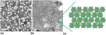
The aqueous PEI–iron oxide NPs are mixed with P22 VLP solution at different NP:P22 number ratio (0.9:1, 2.4:1, 4.3:1, 6.5:1, 7:1, and 11:1; Supplementary Material). The isoelectric point of P22, based on amino acid sequence of the coat protein is ∼4.97, indicating that the VLPs carry an overall negative charge at pH > 5 (Fig. S2). Reference Uchida, LaFrance, Broomell, Prevelige and Douglas12 In contrast, the amine-coated iron oxide NPs show positive zeta-potential values beyond slightly acidic pH (Fig. S3). Therefore, a pH range of 6–7.5 was chosen to investigate the assembly of P22 driven by electrostatics. The Debye screening length, κ−1 is the distance over which the electrostatic interactions are active and is an important parameter in this type of self-assembly. To control the Debye screening lengths in our experiments, different concentrations of NaCl solution (0, 10, 50, and 100 mM) are used. High NaCl concentration acts as a screen for the surface charges on the NPs and P22 while absence of NaCl induces very strong electrostatic interaction between the NP and VLP. It was found that at low NP:P22 number ratio such as 2.4:1, large area 2-dimensional assembly of P22 is possible only with no NaCl in solution and a pH of 6.5. This is expected as the PEI–iron oxide NPs are positively charged at slightly acidic pH. The NPs pack around groups of VLPs, as suggested by the TEM image [Fig. 3(a)]. Figure S4 shows the TEM images at different NP:P22 number ratio to compare the packing of VLPs. A denser monolayer packing of P22 VLPs surrounded with NPs can be obtained with NP:P22 number ratios in the range of 6.5:1 to 7:1 as compared to lower number ratios (e.g., 2.4:1). This is clearly visible from the dark contrast in the bright field TEM image [Fig. S4(b)]. As the NP:P22 number ratio is increased to >10:1, P22 VLPs assemble in small area clusters with the NPs around them, likely due to the very strong electrostatic interactions (Fig. S4). To screen the electrostatic charges, NaCl solution is added to the NP–VLP mixtures at high number ratios. The best suitable result is obtained at a NP:P22 number ratio of 6.5:1 in the presence of 50 mM NaCl and a pH of 6 [Fig. 3(b)]. The Debye length and electrostatic interactions in this case facilitate assembly of wild-type P22 VLPs over large areas (>12 μm). This VLP array shows a dense packing of P22 with the NPs, based on the dark contrast in the TEM image. Figure 3(c) shows the schematic representation of the assembly of P22 with iron oxide NPs. Table 1 (Supplementary Material) summarizes the experimental details and corresponding Debye lengths of the electrostatic VLP complexes formed under the influence of rigid linkers like PEI–iron oxide NPs. At large Debye length ∼3.1 nm, the strong electrostatic interaction between the VLP and NPs induces smaller area assembly of P22 VLPs, as compared to the moderate Debye length (∼1.4 nm). The NP:P22 number ratio and the Debye length are found to be a primary influences on the assembly of P22 VLPs, based on the TEM and DLS observations. This is a critical finding because the few VLP–NP assemblies reported so far were calculated based on the NP:VLP mass ratio rather than the number ratio.
Representative TEM image of PEI–iron oxide NP induced self-assembly of P22 at different NP:P22 number ratio. (a) 2.4:1, no NaCl, pH = 6.5, (b) 6.5:1, 50 mM NaCl, pH = 6, and (c) schematic drawing of VLP–iron oxide NP assembly.

Figure 4 shows the assembly of P22 VLPs in the presence of other NPs of comparable hydrodynamic sizes (∼55–60 nm, Fig. S1c and S1d) like CoFe2O4 and Au. The freshly synthesized cobalt ferrite NPs of size ∼12 nm are coated with PEI and are highly monodisperse. Unlike the PEI–iron oxide NPs, these mixed oxide NPs are sparsely dispersed in P22 and do not fill the edges around the P22 VLPs [Fig. 4(a)]. The ligand, PEI is known to have a strong binding to iron oxide NP surfaces. Reference Xu, Qin, Palchoudhury and Bao28 Therefore, the sparse packing of P22 with CoFe2O4 NPs can be caused by the slightly different binding interaction of the PEI ligand on the CoFe2O4 NP surfaces, as compared to the iron oxide. Reference Palchoudhury, Hyder, Vanderlick and Geerts22 Au NP (size ∼7 nm) is coated with 8-amino-1-octanethiol and induces VLP–NP assembly as shown in Fig. 4(b), distinct from the other NPs, as suggested from the TEM image. Here, the dark regions of size ∼2 µm show NP–VLP three-dimensional assembly. Au NPs appear darker in the bright field TEM image due to their high atomic number. The cationic Au NPs pack on the negatively charged VLP surfaces, as opposed to the iron oxide NPs which is filled around the edges of P22. Notably, all three NPs (iron oxide, CoFe2O4, and Au) are relatively comparable in size, but show different packing with P22 protein cages. This is likely due to the different ligands on the Au and iron oxide NP surfaces. In particular, all these P22 assemblies are vastly different from previous literature reports, where the NPs primarily decorated the inside and outside of the engineered procapsids. Reference Zhou, Bedwell, Li, Bao, Prevelige and Gupta2,Reference Kale, Bao, Zhou, Prevelige and Gupta19 To the best of our knowledge, the packing or interaction of P22 protein cages with inorganic NPs has not been reported, so far. Detailed investigation on the effect of ligand packing on VLP assembly is in process.
TEM image showing P22 assembly induced by different cationic NPs. (a) PEI–CoFe2O4 NPs, NP:P22 number ratio 4:1, pH = 6.5, 100 mM NaCl and (b) 8-amino-1-octanethiol–Au NPs, NP:P22 number ratio 6.5:1, pH = 6.5, 100 mM NaCl.

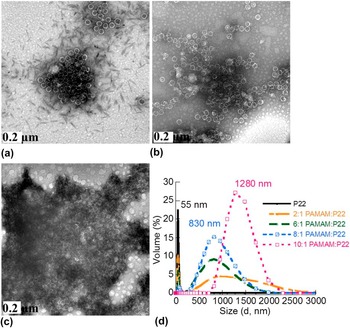
Recently, successful decoration of P22 with soft linkers like Dec protein has been reported. Reference Uchida, LaFrance, Broomell, Prevelige and Douglas12 These soft linkers offer higher flexibility in structural features and surface functionality. However, the possibility to assemble P22 VLPs using the soft linkers has not been explored, so far. Earlier, the cationic PAMAM polymer was successfully used to form electrostatic VLP–polymer complexes with CCMV. Reference Kostiainen, Hiekkataipale, de la Torre, Noltea and Cornelissen14 Therefore, we selected PAMAM-G3 as our model soft linker material. PAMAM-G3 dendrimer (M W 6909) is a soft material of size ∼3.6 nm containing 32 surface groups. Among these surface moieties, multiple amine functional groups are present which can be protonated at physiological pH ∼8. Reference Mikkila, Rosilo, Nummelin, Seitsonen, Ruokolainen and Kostiainen29 The aqueous solutions of PAMAM and P22 are mixed at different PAMAM:P22 number ratios (e.g., 2:1, 6:1, 8:1, 10:1, and 100:1) to investigate the ordering of VLPs in the presence of soft linkers. The pH is kept constant at 7.5. The PAMAM–P22 interactions are systematically monitored via TEM and DLS at high (1 M), moderate (100 mM), and low (10 mM) NaCl concentrations. Reference Kostiainen, Hiekkataipale, de la Torre, Noltea and Cornelissen14 Interestingly, at very high PAMAM:P22 ratios (100:1), whisker-like micelles of the polymer are observed that facilitate small three-dimensional assembly of the VLPs, based on the TEM image [Fig. 5(a)]. This is likely due to the strong VLP–polymer interaction under these conditions. Keeping the same Debye length, the P22 VLPs assemble over a larger area with decreasing PAMAM:P22 number ratio up to 8:1 [Fig. 5(b)]. This is expected as the number of strong electrostatic interactions decrease with the PAMAM:P22 number ratio. In addition, the PAMAM micelles disappear at these lower concentrations, as seen from the TEM image. To screen the strong electrostatic interaction, a high concentration of NaCl (1 M) is used in the subsequent experiment. The charge screening has a significant influence on the P22–PAMAM assembly, as indicated by the TEM image in Fig. 5(c). A densely-packed and larger area assembly of P22 is observed with 1 M NaCl at the same PAMAM:P22 number ratio. Figure 5(d) shows the DLS results for P22 assembly before and after the addition of PAMAM-G3 at different number ratios. The individual P22 VLPs of size 55 nm assemble in larger architectures (D h ∼ 830 nm) in the presence of PAMAM. Reference Kostiainen, Hiekkataipale, de la Torre, Noltea and Cornelissen14 The peak at 830 nm becomes sharper and more prominent as the PAMAM:P22 ratio increases from 2:1 to 8:1, indicating largest area assembly of VLPs at 8:1. The peak corresponding to individual P22 VLP (∼55 nm) decreases with addition of more PAMAM, also suggesting increasing assembly of VLPs at higher PAMAM:P22 number ratios. At 10:1 PAMAM:P22 ratio, large aggregates (D h ∼ 1280 nm) are observed with absence of any individual P22. This is in accordance with our TEM results.
Self-assembly of P22 by PAMAM-G3 dendrimer at different PAMAM:P22 number ratios, pH ∼ 7.5. (a) TEM image at 100:1 PAMAM:P22, 100 mM NaCl, (b) TEM image at 8:1 PAMAM:P22, 100 mM NaCl, (c) 8:1 PAMAM:P22 with 1 M NaCl, and (d) DLS plots showing P22 assembly with addition of different amounts of PAMAM; pDI ∼ 0.15.

IV. CONCLUSION
In conclusion, assembly of P22 VLPs via electrostatic interactions in the presence of rigid NPs and soft PAMAM dendrimers is demonstrated for the first time. The number ratio of the linker: P22 and Debye screening length play dominant roles in the assembly. Different VLP assembly is observed with soft and rigid linker materials. The PEI–iron oxide NPs tend to assemble around the edges of P22 VLPs. Dense assembly of VLPs is induced at 6.5:1 NP:P22 number ratio. The surface ligand, charge, and the ligand packing on the NP surface are found to be important parameters to control P22 assembly, based on the results from three different NPs (e.g., iron oxide, CoFe2O4, and Au). In comparison, the soft PAMAM dendrimer induces assembly of P22 VLPs of hydrodynamic size ∼830 nm through its multiple protonable amine groups. The best suitable P22 assembly is obtained at 8:1 PAMAM:P22 number ratio, based on TEM and DLS data. These results provide insight into the interaction and extended area assembly of individual P22 VLPs with rigid inorganic NPs and soft dendrimers for the first time. Despite the elegant structural assembly of protein cages, they have not been explored to the level of their DNA counterparts for building biomimetic nano-architectures. Reference Yang, Wang, Zhu and Mao30,Reference Glover, Giger, Kim, Naik and Clark31 Our results will be significant in building higher-order structures based on protein cage templates. These nanostructures will be attractive in developing new bio-inspired materials for photocatalytic application, which is currently under investigation. Reference Yang, Wang, Zhu and Mao30
Supplementary Material
To view supplementary material for this article, please visit https://doi.org/10.1557/jmr.2016.439.
Experimental details: table showing Debye length and reaction conditions for interaction of P22 with NPs and PAMAM; DLS plots of P22, PEI–iron oxide NPs, and PAMAM; zeta-potential of P22, PEI-iron oxide NPs, CoFe2O4 NPs and Au NPs at different pH; TEM images of VLP–NP mixture.
ACKNOWLEDGMENT
This work was supported by the US DOE, Office of Basic Energy Sciences, Division of Materials Sciences and Engineering Award DE-FG02-08ER46537. The authors acknowledge UA-MINT Center and thank CAF for use of TEM. The authors also acknowledge Dr. Yuping Bao for use of DLS. SP thanks UT Chattanooga for support.