INTRODUCTION
Characterization of the excretory-secretory (E-S) products of Litomosoides sigmodontis revealed the presence of a molecule of 130–160 kDa with an unusual release pattern. Specifically, this molecule was secreted into culture medium by juvenile but not fully mature female worms (Harnett et al. Reference Harnett, Meghji, Worms and Parkhouse1986). Subsequently called Juv-120 (Hintz et al. Reference Hintz, Schares, Taubert, Geyer, Zahner, Stirm and Conraths1998), the molecule was also found to be present on the surface of the microfilaria sheath having been attached in the terminal part of the uterus following synthesis by maternal cells (Schares et al. Reference Schares, Schutzle, Zahner and Conraths1994). Further work indicated that Juv-p120 was also secreted by L. sigmodontis in vivo (Hintz et al. Reference Hintz, Schares, Taubert, Geyer, Zahner, Stirm and Conraths1998). The secretion of the molecule just before the release of microfilariae, led to the suggestion that its role may be to induce anergy in appropriate lymphocytes thereby preventing recognition of cross-reacting epitopes on the microfilaria surface (Hintz et al. Reference Hintz, Schares, Taubert, Geyer, Zahner, Stirm and Conraths1998).
An unusual structural feature of Juv-p120 is that it contains large amounts of N,N-dimethylethanolamine (DMAE) (28·4 mol%) (Hintz et al. Reference Hintz, Schares, Taubert, Geyer, Zahner, Stirm and Conraths1998). The function of this DMAE is unknown but its similarity in structure to choline (it only differs in lacking 1 of choline's 3 methyl groups) may suggest an immunomodulatory role similar to that of phosphorylcholine (PC) present on other filarial nematode E-S products (reviewed by Harnett and Harnett, Reference Harnett and Harnett2001). The unusual presence of DMAE on Juv-p120 allied to a possible role for it in microfilarial survival dictates that the biochemical processes underlying its attachment should be investigated. Such a process has been undertaken with respect to PC using the E-S product, ES-62, whereupon it was found that PC is transferred to antennary N-acetylglucosamine (GlcNAc) residues of an N-type glycan (reviewed by Houston and Harnett, Reference Houston and Harnett2004). Radio-isotope labelling experiments indicate that the transferred PC is derived from exogenous choline and attachment is inhibited when worms are incubated with the choline kinase inhibitor, hemicholinium-3 (HC-3) (Houston and Harnett, Reference Houston and Harnett1999). The nature of the PC donor has also been investigated (Houston et al. Reference Houston, Lochnit, Geyer and Harnett2002) and it appears that it may be CDP-choline or more likely phosphatidylcholine, as the latter can be used to promote transfer to an artificial substrate in vitro (Cipollo et al. Reference Cipollo, Awad, Costello, Robbins and Hirschberg2004).
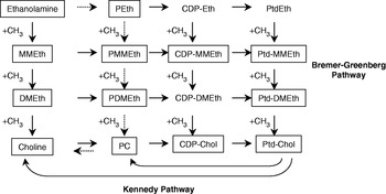
Much less information is available with respect to DMAE. Like PC on ES-62 it appears to be attached via phosphodiester linkages, but it is unknown as to whether it is transferred to carbohydrate as with ES-62 or, as seems more likely, to amino acids (Hintz et al. Reference Hintz, Kasper, Stahl, Geyer, Kalinowski, Kras, Kuhnhardt, Schott, Conraths, Zahner and Stirm1996, Reference Hintz, Schares, Taubert, Geyer, Zahner, Stirm and Conraths1998). Also, it is unknown what the source of DMAE for transfer is. Based on analogy with transfer of PC, it could be that DMAE is transferred from phosphatidyldimethylethanolamine or CDP-dimethylethanolamine. The former compound is a component of the Bremer-Greenberg pathway of phosphatidylcholine biosynthesis (see Fig. 1), which appears to be present in C. elegans (Lochnit and Geyer, Reference Lochnit and Geyer2003). In an attempt to gain evidence in support of this idea, juvenile female L. sigmodontis were pulsed with [3H]-ethanolamine and incorporation of radio-isotope label into Juv-p120 assessed. This work demonstrated that Juv-p120 was only very weakly labelled with [3H]-ethanolamine. However, strong labelling was detected when employing an alternative radio-isotope label for pulsing, [3H]-choline.
A schematic representation of pathways of choline/phosphatidylcholine biosynthesis. Compounds that are boxed are known components of plant pathways of choline synthesis. The feint arrows correspond to the main pathway of choline biosynthesis in glycine betaine-accumulating chenopods (Rhodes and Hanson, Reference Rhodes and Hanson1993) and the ethanolamine to PC stages of this pathway contribute to a third pathway of phosphatidylcholine synthesis in C. elegans (Brendza et al. Reference Brendza, Haakenson, Cahoon, Hicks, Palavilli, Chiapelli, McLaird, McCarter, Williams, Hresko and Jez2007). Eth, ethanolamine; MM, monomethyl; DM, dimethyl; P, phosphoryl; Ptd, phosphatidyl; CDP, cytidine diphosphate.

MATERIALS AND METHODS
L. sigmodontis
Maintenance of L. sigmodontis Chandler, 1931 was undertaken following previously published methods (Diagne et al. Reference Diagne, Petit, Liot, Cabaret and Bain1990; Petit et al. Reference Petit, Diagne, Marechal, Owen, Taylor and Bain1992). Juvenile female worms (5–6 weeks post-infection) were recovered by flushing of the pleural cavity of infected Meriones unguiculatus with 10 ml of PBS as described previously (Babayan et al. Reference Babayan, Ungeheuer, Marin, Attout, Belnoue, Snounou, Renia, Korenaga and Bain2003).
Biosynthetic labelling of L. sigmodontis
Groups of 8–10 juvenile female L. sigmodontis were incubated in RPMI ‘complete’ (Harnett et al. Reference Harnett, Meghji, Worms and Parkhouse1986) lacking choline and serine and 2 MBq of either [methyl-3H]-choline chloride (3·00 TBq mmol−1), [1-3H]-ethanolamine hydrochloride (1·04 Tbq mmol−1) or L-[3-3H]-serine (1·11Tbq mmol−1) (all from Amersham Bioscience UK Ltd, Little Chalfont, UK). After 2·5 h the medium was removed and replaced with RPMI ‘complete’ containing 3% foetal calf serum. The medium was then replaced at 24, 48 and 96 h after initiation of the experiment.
Preparation of parasite-soluble and lipid-phase extracts
On completion of the culture period worms were washed 3 times in PBS, pH 7·4 and then PBS extracts for protein analysis and lipid extracts for phospholipid analysis prepared as described previously for filarial nematodes (Harnett et al. Reference Harnett, Patterson, Copeman and Parkhouse1994; Houston and Harnett, Reference Houston and Harnett1999). The protein content of the PBS extract was estimated using the Bio-Rad protein assay reagent and all extracts adjusted to the same concentration.
Thin layer chromatography
TLC was performed using previously described procedures (Houston and Harnett, Reference Houston and Harnett1999; Houston et al. Reference Houston, Lochnit, Geyer and Harnett2002) unless otherwise indicated. The dried lipid phase was reconstituted in chloroform/methanol (1:1; v/v) and the phospholipid elements separated using chloroform/methanol/water (65/25/4; v/v/v) (Smith et al. Reference Smith, Selkirk and Gounaris1996). Once dried the phospholipids were detected by iodine staining and spots, which co-migrated with phosphatidylethanolamine and phosphatidylcholine standards scraped off and counted for radioactivity using a Beckman LS6500 multi-purpose scintillation counter. The remainder of the track for each isotope label was also scraped off and counted for radioactivity to allow assessment of the amount of total isotope label incorporated into the two phospholipids.
SDS-PAGE/fluorography
Equivalent protein concentrations of PBS extracts of isotope-labelled worms were subjected to SDS-PAGE/fluorography as described previously (Houston and Harnett, Reference Houston and Harnett1999).
Biosynthetic radio-isotope labelling of ES-62
ES-62 of A. viteae was biosynthetically labelled with [3H]-choline as described previously (Houston et al. Reference Houston, Lochnit, Geyer and Harnett2002).
Immunoprecipitation of [ 3H]-choline-labelled extracts
Approximately 600 dpm of [3H]-choline-labelled L. sigmodontis PBS extract or [3H]-choline-labelled ES-62 were added to 10 μl of each of rabbit anti-L. sigmodontis microfilaria sheath protein serum, normal rabbit serum, TEPC 15 (0·1 mg/ml in PBS) (Sigma, Poole, Dorset) or normal mouse serum and the volume made up to 100 μl with PBS, pH 7·4. All samples were vortexed briefly and incubated at 37°C for 1 h. An appropriate amount of goat anti-rabbit IgG or goat anti-mouse IgA was added and samples were incubated overnight at 4°C to allow formation of precipitates. Precipitates were washed 3 times with ice-cold 10 mm-Tris-HCl; 50 mm NaCl; 0·1% (v/v) Nonidet P40, pH 8·3 and then dissolved in formic acid. Measurement of radioactivity was undertaken using a Beckmann LS 6500 multi-purpose scintillation counter. Samples were examined in duplicate and the average value obtained, expressed as a percentage of the total dpm.
Preparation of PC conjugated to BSA (PC-BSA)
PC-BSA and sham-treated BSA were prepared as described previously (Goodridge et al. Reference Goodridge, Marshall, Else, Houston, Eagan, Al-Riyami, Liew, Harnett and Harnett2005).
Dot-blot analysis
Aliquots of PC-BSA and BSA (5 μl of 0·5 mg/ml solutions) were spotted onto a series of nitrocellulose strips (Hybond-C super: Amersham) and allowed to air-dry. The strips were immersed in 4% ovalbumin/Tris-buffered saline (pH 8·5; TBS) for 30 min at room temperature and then washed in TBS (3 times). TEPC 15 (10 μg/ml in 2·5% ovalbumin; TBS, pH 8·5), TEPC 15+10 mm PC-chloride, TEPC 15+10 mm DMAE or TEPC 15+10 mm choline was added, each to a strip and the strips left at room temperature under gentle agitation for 30 min. The strips were washed as before and then immersed in alkaline phosphatase-conjugated anti-mouse IgA under gentle agitation for 30 min. After further washing as before the strips were developed in NBT/BCIP as recommended by Sigma.
RESULTS
Phosphatidylethanolamine is readily biosynthetically labelled in juvenile female Litomosoides sigmodontis
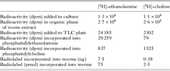
Groups of 8–10 juvenile female L. sigmodontis were pulsed for 2·5 h with 2 Mq [3H]-ethanolamine and then chased for a total of 96 h. Worm extracts were then prepared in the presence of PBS followed by sodium deoxycholate and the latter phase separated as described previously (Houston et al. Reference Houston, Lochnit, Geyer and Harnett2002). The radio-isotope labelled phospholipids in the organic phase were analysed by TLC where it was found that ~84% of the label was incorporated into phosphatidylethanolamine (Table 1). Only 3·5% of the radiolabel was incorporated into phosphatidylcholine. When radio-isotope labelled with [3H]-choline ~66% of the label was incorporated into phosphatidylcholine. However, the total amount of labelled ethanolamine incorporated into the organic phase was ~30 times that of lablelled choline such that the amount of [3H]-choline-labelled phosphatidylcholine generated was actually less (~70%) than that of [3H]-ethanolamine. This suggests that the Bremer-Greenberg pathway and/or the pathway dependent on conversion of phosphorylethanolamine to phosphorylcholine present in C. elegans (Brendza et al. Reference Brendza, Haakenson, Cahoon, Hicks, Palavilli, Chiapelli, McLaird, McCarter, Williams, Hresko and Jez2007) (see Fig. 1) may represent pathways of comparable significance to the Kennedy pathway for phosphatidylcholine biosynthesis in L. sigmodontis. A preference for uptake of [3H]-ethanolamine over [3H]-choline is not unique to L. sigmodontis: a similar ratio of uptake was observed with adult female Acanthocheilonema viteae (results not shown). We also observed the same pattern of radio-isotope labelling in A. viteae with respect to incorporation of the two labels into phospholipids (results not shown).
Incorporation of [3H]-ethanolamine and [3H]-choline into Litomosoides sigmodontis

Juv-p120 is poorly biosynthetically labelled with [ 3H]-ethanolamine but is strongly labelled with [ 3H]-choline
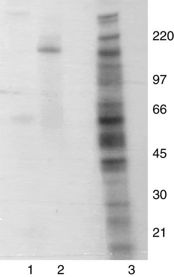
PBS extracts of the isotope-labelled worms and also worms pulsed with [3H]-serine to act as an indicator of the overall protein profile, were examined by SDS-PAGE/fluorography (Fig. 2). The [3H]-serine-labelled extract demonstrated a series of polypeptides of varying intensities, stretching from the top to the bottom of the gel. The ethanolamine-labelled extract revealed a few faint bands each of whose appearance during analysis varied from batch to batch. When labelling with [3H]-choline a band of between 97 and 220 kDa was consistently clearly observed. The corresponding polypeptide could also be detected in extracts of [3H]-ethanolamine labelled worms but only after prolonged exposure (a faint band can be seen in Fig. 2). Save for a suggestion of a smear of lower molecular mass the only labelled polypeptide detected when employing [3H]-choline as the label was the one running between 97 and 220 kDa.
SDS-PAGE/fluorography analysis of (1) [3H]-ethanolamine, (2) [3H]-choline, (3) [3H]-serine-labelled PBS extracts of Litomosoides sigmodontis. The positions reached by [14C]-molecular weight markers (in kDa) during electrophoresis are shown on the right.

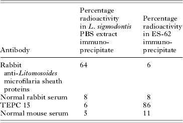
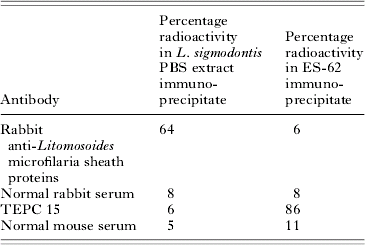
Four pieces of evidence lead us to believe that the polypeptide readily biosynthetically labelled with [3H]-choline is Juv-p120. First of all it corresponds in molecular mass to the only [3H]-ethanolamine-labelled polypeptide that can be (faintly) detected that is of an appropriate molecular mass for Juv-p120. Secondly, Juv-p120 is virtually the only major DMAE-containing protein detected in juvenile female L. sigmodontis extracts (Schares et al. Reference Schares, Schutzle, Zahner and Conraths1994; Hintz et al. Reference Hintz, Schares, Taubert, Geyer, Zahner, Stirm and Conraths1998) and we are only detecting one molecule here, Third, although not definitive, we found the [3H]-choline-labelled molecule to be recognized by an antiserum against Litomosoides microfilaria sheath proteins as determined by simple measurement of radioactivity in immunoprecipitates (Table 2). Fourth, the PC moiety of ES-62 dictates that this E-S product is recognized by the myeloma protein reactive for PC, TEPC 15 (Harnett et al. Reference Harnett, Worms, Grainger, Pyke and Parkhouse1990). We undertook an immunoprecipitation experiment with [3H]-choline-labelled ES-62 and [3H]-choline-labelled L. sigmodontis extract. Although ES-62 was immunoprecipitated, as found previously (Houston et al. Reference Houston, Cushley and Harnett1997), there was no recognition of the L. sigmodontis-labelled molecule (Table 2). Thus, in spite of the labelling with [3H]-choline, the molecule does not appear to contain phosphorylcholine. Juv-p120 is also not recognized by antibodies in serum samples derived from humans infected with filarial nematodes and these antisera contain antibodies against PC (Harnett et al. Reference Harnett, Grainger, Worms and Parkhouse1989). It also fails to bind TEPC 15 (unpublished observation of F. J. Conraths referred to in Hintz et al. Reference Hintz, Schares, Taubert, Geyer, Zahner, Stirm and Conraths1998).
Immunoprecipitation of [3H]-choline-labelled extracts
(Samples were examined in duplicate and the average value obtained expressed as a percentage of the total dpm added (~600).)
DISCUSSION
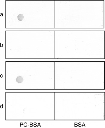
There is very little information on biosynthetic pathways involving DMAE in the literature. In castor bean endosperm, choline synthesis has been demonstrated by sequential methylation of ethanolamine, monomethylethanolamine and DMAE (Rhoads and Hanson, Reference Rhodes and Hanson1993). For this reason we considered that the DMAE that is present on Juv-p120 was most likely to be derived from ethanolamine. However, our data obtained when performing biosynthetic radio-isotope labelling experiments clearly question this in that the use of [3H]-ethanolamine provided poor labelling of Litomosoides proteins whereas [3H]-choline gave strong labelling of a molecule that is almost certainly Juv-p120. The ability to be biosynthetically labelled with [3H]-choline is a property that Juv-p120 shares with another filarial nematode product ES-62 (Houston et al. Reference Houston, Cushley and Harnett1997). In ES-62 the radio-isotope labelling reflects the presence of PC on the protein (Harnett et al. Reference Harnett, Worms, Grainger, Pyke and Parkhouse1990) but previous structural and antigenic characterization indicates that Juv-p120 does not contain PC (Hintz et al. Reference Hintz, Kasper, Stahl, Geyer, Kalinowski, Kras, Kuhnhardt, Schott, Conraths, Zahner and Stirm1996, Reference Hintz, Schares, Taubert, Geyer, Zahner, Stirm and Conraths1998). Consistent with this we found that TEPC 15, a mouse myeloma protein reactive for PC, would not interact with the [3H]-choline-labelled molecule detected in this study. Thus, although PC and DMAE are structurally rather similar they are clearly antigenically distinct. Indeed we found by dot immunoblotting that whereas PC and indeed choline will block binding of TEPC 15 to PC-BSA, DMAE fails to do this (Fig. 3). Thus, we can be certain that the radioactivity we are detecting on Juv-p120 is not due to the presence of PC. This begs the question as to what it is due to and our opinion is that it is most likely due to the presence of radio-isotope labelled DMAE.
Dot-blot analysis of the ability of PC, DMAE and choline to inhibit binding of the anti-PC myeloma protein TEPC 15 to PC-BSA. (a) TEPC 15 alone; (b) TEPC 15+10 mm PC; (c) TEPC 15+10 mm DMAE; (d) TEPC 15+choline.

Labelling of Juv-p120 with [3H]-choline cannot be explained by a process that involves utilization of choline methyl groups for the synthesis of methionine as, if this was the case, it would be expected that a number of polypeptides would be labelled similar to those observed when employing [3H]-serine. Likewise, as it is the methyl groups of the choline that are isotope labelled, a process that involves conversion of the choline into ethanolamine and then methylation (using endogenous groups) of the latter into DMAE cannot explain the labelling. Thus, if the radioactivity we are detecting is due to labelled DMAE and it is derived from ethanolamine it must be from endogenous ethanolamine and the contribution of the isotope-labelled choline is to provide methyl groups. Choline can provide methyl groups via conversion to S-adenosyl methionine (SAM) but culture of worms with S-adenosyl-L-[methyl-3H]methionine did not result in labelling of Juv-p120 (result not shown). Thus it seems unlikely that Juv-p120 is being isotope-labelled as a consequence of donation of radioactive methyl groups to endogenous ethanolamine from [3H]-choline. This proposed failure of ethanolamine to act as a precursor of DMAE on Juv-p120 is clearly consistent with the poor labelling of Juv-p120 obtained when employing [3H]-ethanolamine. In fact we feel the most likely explanation for the (low level) labelling of Juv-p120 by [3H]-ethanolamine that can be eventually detected is that the isotope label is utilized in the biosynthesis of phosphatidylcholine via the pathway dependent on phospho-base methyltranaferases present in C. elegans (Brendza et al. Reference Brendza, Haakenson, Cahoon, Hicks, Palavilli, Chiapelli, McLaird, McCarter, Williams, Hresko and Jez2007) and then the choline is cleaved by phospholipase D and ultilized for DMAE production. Alternatively the choline could be released by cleavage at the PC stage as occurs in plants (Rhodes and Hanson, Reference Rhodes and Hanson1993).
If [3H]-choline is not contributing to labelling of Juv-p120 via the pathways outlined above then we must consider that it is being directly converted to DMAE. There are two ways in which this might happen. First of all, choline residues might be transferred directly to Juv-p120 (from for example, CDP-choline or phosphatidylcholine) and then 1 methyl group removed from each. Second, choline (again perhaps as CDP-choline or phosphatidylcholine) might somehow lose 1 methyl group and then be transferred to Juv-p120. With respect to the former it is known that intact microfilaria sheaths that, as mentioned earlier, contain proteins modified with DMAE, contain 0·2% choline (by weight) but only minute, negligible amounts of ethanolamine and monomethylethanolamine (Hintz et al. Reference Hintz, Kasper, Stahl, Geyer, Kalinowski, Kras, Kuhnhardt, Schott, Conraths, Zahner and Stirm1996). It could be postulated that this reflects the presence of choline residues that were ‘missed’ during the removal of the methyl groups. However, this is simply speculation. The second option is certainly more straightforward but, for either to be correct, the existence of a choline demethylase must be hypothesized. We are not aware of the existence of such an enzyme in nature but it would certainly be interesting to explore any sequence information available on L. sigmodontis (e.g. ESTs) in the search for it by focussing on likely related enzymes. Perhaps one possibility is an enzyme similar to lanosterol 14α-demethylase (CYP51), a member of the cytochrome P450 family involved in cholesterol biosynthesis that is unique amongst family members in being found in both prokaryotes and eukaryotes (Rozman and Waterman, Reference Rozman and Waterman1998).
Finally, when speculating on a role for DMAE on Juv-p120, the idea of immunomodulation is attractive. The appearance of Juv-p120 in the Litomosoides life-cycle, specifically just before the release of DMAE-exposing microfilariae is striking and the structural relationship of DMAE to the known filarial nematode immunomodulator, PC, cannot be ignored. Indeed given the apparent absence of PC-containing molecules in the Litomosoides extract (although PC appears to be present on the surface of the L3 (Al-Qaoud et al. Reference Al-Qaoud, Fleischer and Hoerauf1998)) and the fact that DMAE has not been described in other filarial nematodes to date, there is a strong temptation to ask whether DMAE might play the role of PC in L. sigmodontis.
W. H. would like to thank Dino Rotondo for critical reading of the manuscript.