I. INTRODUCTION
The majority of conventional single-component ceramic or polymer materials cannot satisfy the critical requirements of specific applications such as use as a bone substitute (e.g., bone graft). Hence, other advanced and functionalized biomaterials need to be developed to address this need.Reference NievethithaSethu, Namashivayam, Devendran, Nagarajan, Tsai, Narashiman, Ramachandran and Ambigapathi1,Reference Dziadek, Stodolak-Zych and Cholewa-Kowalska2 Based on the requirements for synthetic bone implants, most of the attention has been focused on developing materials with physiochemical and biological properties similar to those of natural bone.Reference Moncal, Heo, Godzik, Sosnoski, Mrowczynski, Rizk and Ozbolat3
Among the several types of alumina–zirconia composites, zirconia-toughened alumina (ZTA) seems to have the potential to be used as a load-bearing implant because of its high wear resistance, high elasticity, and excellent toughness.Reference Mitic, Stolić, Stojanovic, Najman, Ignjatovic and MiroslavTrajanović4–Reference Kurtz, Arnholt, Huet, Ueno and Walter6 Although ZTA can provide a supportive framework for bone, both alumina and zirconia are chemically inert materials, meaning that they cause no adverse effects or tissue reactions.Reference Gautam, Joyner, Gautam, Rao and Vajtai7,Reference Sequeira, Fernandes, Neves and Almeida8 Known as biomimetic technology, recently nanotechnology has shown advances that have provided unique tools to promote the bioactivity of implants by promoting the bone remodeling and formation process.Reference Ercan and Webster9 Given our experience with such ceramics, modification of the ZTA surface is a practical approach for promoting biomimetic technology.Reference Esfahani, Nemati and Salahi10 Using the combination of polymers and ceramics for creating biomimetic bone implants opens opportunities for new treatment strategies regarding bone fractures, defects, and diseases. Based on the complex characteristics of the bone, the structural diversity of polymers in combination with the bioactivity of calcium phosphate ceramics might offer a potential solution by providing a scaffold for cell adherence and intensifying the proliferation and differentiation of osteoblasts.Reference Nandakumar, Yang, Habibovic and van Blitterswijk11,Reference Sweth, Sahithi, Moorthi, Srinivasan, Ramasamy and Selvamurugan12
The ability of hydroxyapatite (HA) [Ca10(PO4)6(OH)2] to form composites with natural or synthetic polymers promotes the possibility of using such composites in bone regeneration.Reference Gomez-Morales, Iafisco, Delgado-López, Sarda and Drouet13,Reference Shojai, Khorasani and Jamshidi14 Swetha et al.Reference Sweth, Sahithi, Moorthi, Srinivasan, Ramasamy and Selvamurugan12 synthesized a chitosan–gelatin–nano-HA scaffold and found that the adhesion and in vitro proliferation of osteoblasts in simulated body fluid (SBF) were better than those of a pristine polymeric scaffold. Xiong et al.Reference Xiong, Ren, Zhang, Yang, Lang, Min, Zhang, Pei, Yan, Li and Mo15 studied the in vitro behavior of porous nano-HA and polyamide 66 (PA66) composites for use as an implant in the healing of bone defects; they obtained promising results. It has been shown that polyamide 6 (PA6) has the potential to be used in biomedical applications because of its excellent stability in human body fluid, high mechanical strength, and high chemical and thermal stabilityReference Prasad S, Ratha, Adarsh, Anand, Kumar Sinha, Diwan, Annapurna and Biswas16; its combination with ceramic elements produces effective biomaterials for the surface adsorption of proteins.Reference Esfahani, Prabhakaran, Salahi, Tayebifard, Keyanpour-Rad, Rahimipour and Ramakrishna17
Nanostructured HA coatings can establish desirable conditions for bone tissue regeneration as HA mimics the chemistry of nanostructured natural bone.Reference Yang, Xia, Wang, Niu, Song, Xiong, Zheng, Wei and Lu18,Reference Ratha, Anand, Chatterjee, Kundu and Suresh Kumar19 Among the methods of hybrid HA/polymer coating on a ceramic substrate,Reference Latifi, Fathi and Golozar20–Reference Vahabzadeh, Roy, Bandyopadhyay and Bose25 the electrospinning method is a simple procedure of combining ceramic nanoparticles (NPs) with a mechanically robust polymer for the manufacturing of nanofibrous scaffolds for bone tissue engineering applications. High porous coating, thermally stable calcium phases, homogenous HA/polymer matrices, and cost-efficiency are some of the main features of this method.Reference Nandakumar, Yang, Habibovic and van Blitterswijk11 Electrospun nanofibrous meshes have attracted special interests due to their large surface-to-volume ratios, which facilitate the delivery of biological elements for the regeneration of tissue functions.Reference Faridi-Majidi, Nezafati, Pazouki and Hesaraki26,Reference Jing, Mi, Salick, Cordie, McNulty, Peng and Turng27
The purpose of this study was to evaluate the electrospinning method, as a practical coating method, on the bioinert ZTA substrate to prepare a biomimetic and bioactive coating for bone tissue engineering applications. Prior to electrospinning, the HA NPs were synthesized via the wet chemical precipitation method. Water contact angle (WCA) test, in vitro SBF immersion, and cytocompatibility assays using osteosarcoma MG63 cells were performed on ZTA and electrospun PA6/HA-coated ZTA samples.
II. MATERIALS AND METHODS
A. ZTA substrate
The ZTA substrate was prepared by powder composite processing of Al2O3 NPs (mean diameter 27 nm, Nanostructured and Amorphous Materials, Katy, Texas, 99.5% purity, 1015 WW), ZrO2 NPs (mean diameter 30–60 nm, Inframat advanced materials LLC, Manchester, Connecticut, 99.9% purity, 40N-0801), and Y2O3 NPs (mean diameter 20–40 nm, Nanostructured and Amorphous Materials, Inc., Los Alamos, New Mexico, 99.99% purity, 5611RE). The alumina (matrix) and zirconia (toughening phase) particles were mixed at a molar ratio of 85:15, respectively. The mixing was performed homogeneously in acetone, and the mixture was then dried at 80 °C for 24 h. Y2O3 NPs (stabilizer of tetragonal zirconia) (3 mol%) were added to the ZrO2 NPs before mixing with alumina NPs. The ZTA disks (d = 14 mm, h = 2 mm) were prepared by pressing the powder mixture under 250 MPa uniaxial pressure followed by sintering at 1550 °C for 1 h.
B. HA nanopowder synthesis
Aqueous phosphate and calcium salt solutions were separately prepared by dissolving 0.06 mol of diammonium phosphate [(NH4)2HPO4, Merck, Darmstadt, Germany, 99% pure, 1.01207.500] in 50 mL of distilled water and 0.1 mol of calcium acetate [Ca(CH3COO)2·H2O, Merck, 99% pure, 1.09325.0500] in 50 mL of distilled water, respectively. The calcium salt solution was added dropwise to the phosphate solution while stirring to adjust the Ca/P ratio to 1.67. The pH and temperature of the solution were adjusted to 10 and 45 °C, respectively, by means of the addition of 0.1 M ammonia and heating. The precipitated powders were washed thrice and then dried at 80 °C for 10 h.
C. Electrospun coating of PA6/HA fibers
Primarily, HA NPs were dispersed in formic acid (Sigma-Aldrich, 64-18-6), and the N6 pellets (1015b UBE, Mw; 15,000 g/mol) were added to the suspension. To investigate the effect of the amount of HA NPs on the morphology of the final fibers, the PA6/HA ratio in the electrospinning solution was adjusted at three different levels according to Table I. Prior to the electrospinning process, ZTA substrates were cleaned in acetone solution using ultrasound and then attached to aluminum foil. To perform electrospinning, 1 mL of hybrid solution was fed by a syringe pump (IVAC 711) that supplied a constant flow of 0.5 mL/h polymer solution. The applied DC voltage and the distance from needle tip to the aluminum collector were adjusted to obtain an appropriate final fiber shape (see Table I). Finally, the electrospun coated PA6/HA fibers on ZTA were dried at 80 °C for 12 h, and with respect to the P/H ratio, the obtained mats were named M-HR (high ratio), M-MR (medium ratio), and M-LR (low ratio). The coated substrates were then used for characterization and further in vitro studies and evaluations.
Electrospinning conditions for PA6/HA coating on ZTA and their corresponding results.

D. In vitro SBF immersion study
To evaluate the biocompatibility of the PA6/HA-coated ZTA disks, they were immersed inside a container of SBF solution for different periods of time (1, 3, 7, and 21 days). The protocol for the preparation of SBF was adopted from Kokubo et al.Reference Kokubo and Takadama28 After 1 day of immersion, control and coated ZTA samples were removed from SBF, and their surfaces were washed with deionized water. The samples were then dried and studied by scanning electron microscopy (SEM) to characterize the morphological changes on their surface. After 3, 14, and 21 days in SBF, the immersed samples were investigated via the same procedure. Further characterizations were carried out to identify the precipitates from the SBF on the PA6/HA-coated ZTA.
E. In vitro cytocompatibility assay
To study the cytocompatibility of ZTA and PA6/HA-coated ZTA samples, the MG63 osteosarcoma cells were used. The high-glucose Dulbecco’s modified Eagle’s medium (DMEM), supplemented with 10% fetal bovine serum (FBS) and 1% penicillin/streptomycin solution (Thermo Fisher Scientific, Gibco™), was used to culture the MG63 cells. The temperature and humidity of the culture atmosphere were 37 °C and 5% CO2, respectively. The medium was refreshed every 2 days. Prior to the cytocompatibility tests, UV irradiation (2 × 90 min) was applied to all samples for sterilization, and the preparation process was completed by incubation for 24 h at 37 °C. To perform the MTT assay, samples were placed in a nontreated 24-well culture, and MG63 cell seeding was performed at a density of 3.5 × 10⁴ cells per well in 1 mL of complete culture medium. Cell adhesion and proliferation on the ZTA and PA6/HA-coated ZTA samples were evaluated 24 and 72 h after cell seeding. The cell viability percentage was measured in comparison with the positive control by means of UV absorbance spectroscopy. To determine the morphology of osteosarcoma MG63 cells on the ZTA and PA6/HA-coated ZTA samples, the incubated samples (after being cell-cultured for 72 h) were immediately rinsed with phosphate-buffered saline (PBS) thrice and then fixed in 2.5% glutaraldehyde at 4 °C for 24 h. After rinsing with PBS, dehydration occurred through exposure to a gradient of ethanol and drying at 4 °C.
F. Characterization
To characterize the surface of ZTA and coated ZTA, scanning electron microscopy (SEM; model JEOL-JSM 840 A, JEOL Ltd., Tokyo, Japan) was used at an accelerating voltage of 20 kV on gold-sputtered samples. The average fiber diameter and surface porosity percentage of electrospun coatings were determined using ImageJ software (ImageJ 1.38X, NIH, Bethesda, Maryland). The XRD patterns of the ZTA substrate and HA NPs were provided by XRD device (model: Philips PW1730, Cambridge, Massachusetts, Kα Cu, λ = 0.154 nm). Functional molecular groups formed in electrospun PA6/HA mats were evaluated by Fourier transform infrared (FTIR) technique (Perkin Elmer Spectrum 65, Shelton, Connecticut, wavelength range: 4000 to 400 cm−1). The dynamic wetting of the ZTA and electrospun coated ZTA was evaluated by measuring the WCA. For this matter, a droplet of deionized water was automatically dropped onto the surface of the samples, and the variation of the WCA was monitored over thirty seconds. To ensure reliability, measurements were repeated for five different locations of each sample and the average value was reported. To investigate the integrity and adhesion of the coating, the microindentation adhesion test was conducted. For this test, the Vickers indentation test was carried out using a Buehler Illinois-60044 (Lake Bluff, Illinois) micro-hardness tester by loading 1000 grf for 25 s, and then the interface was studied by means of SEM device model JEOL-JSM 840 A (JEOL Ltd.). The cell viability percentage of MG63 cells was calculated by measuring the absorbance at 570 nm using a microplate reader (Synergy HTX, BioTek Instruments, Winooski, Vermont). The morphology of osteosarcoma MG63 cells was studied with SEM microcopy (FEIESEM Quanta 200, FEI, Hillsboro, Oregon) after sputter coating with gold (JEOL-JFC1100E, JEOL Ltd.).
III. RESULTS AND DISCUSSION
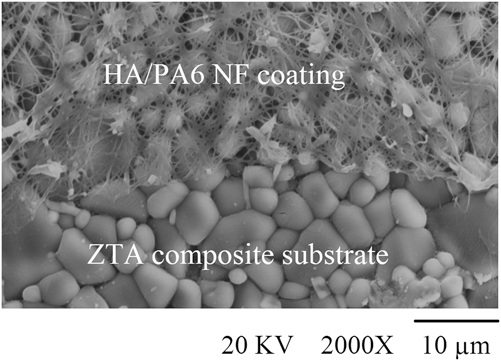
There is an enormous body of literature focusing on delivering new and improved biomimetic materials in the field of bone tissue engineering. As mentioned earlier, this study aimed to improve the bioactivity and biomimetic behavior of ZTA bioceramics via the deposition of hybrid NFs on its surface by electrospinning. Figure 1 shows a SEM micrograph of the edge of the ZTA substrate and NF coating. The zirconia grains have been distributed homogenously inside the alumina grains, having average grain sizes of 0.5 and 1.2 μm, respectively.
SEM top-view micrograph of PA6/HA coating on ZTA composite substrate via electrospinning method.

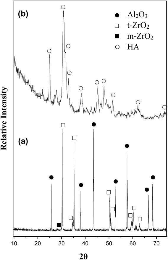
The XRD pattern of the ZTA substrate indicated that tetragonal (t) zirconia had been produced after sintering [see Fig. 2(a)]. The stabilization occurred due to yttria NPs blending with monoclinic zirconia before the formation of composite structure. XRD pattern of the substrate confirms that α-alumina and t-zirconia are the two main phases of the composite matrix. Therefore, it can be said that the ZTA substrate was successfully formed in this study. Incorporation of tetragonal stabilized zirconia can improve the toughness of alumina matrix composites because of martensitic transformation toughening of tetragonal to monoclinic phase. It should be mentioned that zirconia grains act not only as toughening agents in ceramic composites, but also as growth inhibitors for alumina grains (these behaviors have been discussed in detail in the study reported by Esfahani et al.Reference Esfahani, Nemati and Salahi10). Therefore, PA6/HA NFs were coated on a bioinert ZTA surface via electrospinning method, which can improve the bioactivity of ZTA for years following implantation due to the presence of crystalline HA.Reference Allo, Costa, Dixon, Mequanint and Rizkalla29 Furthermore, PA6 was used as the nanofibrous matrix polymer as it contains carboxyl and amine groups (CO(CH3)5NH), which facilitate its interaction with ceramic particles and render it suitable for many biochemical applications such as tissue engineering, drug delivery, and wound dressing.Reference Brydone, Meek and Maclaine30 As shown in Fig. 1, an ultrathin layer of PA6/HA NFs was created on the ZTA surface, and ZTA grains were well masked by bioactive NFs via electrospinning. The average diameter of PA6/HA NFs was calculated to be approximately 100 ± 5 nm. Among different methods for the synthesis of HA NPs,Reference Ben-Arfa, Miranda Salvado, Ferreira and Pullar31 the wet chemical precipitation method was selected because of its high-purity and high-crystallinity products and inexpensive precursors and equipment.Reference Monmaturapoj32 The XRD pattern of NPs synthesized via wet chemical precipitation method is presented in Fig. 2(b). According to the XRD results, the diffraction of X-rays from (211), (112), (300), and (202) planes confirmed the formation of pure HA. Moreover, the crystallinity fraction of 0.71 calculated by Eq. (1)Reference Salehi and Fathi33 explained the crystalline structure of HA NPs. Therefore, it can be assumed that the incorporation of HA NPs inside the PA6 NFs created bioactive inorganic sites that were similar in composition to those of the mineral phase of bone. It is worth emphasizing that the synthetic form of HA is osteoconductive, and its crystalline structure is the same as the crystalline portion of bone.Reference Brydone, Meek and Maclaine30
 $${X_{\rm{C}}} = 1 - \left( {{{{V_{{{112} / {300}}}}} \over {{I_{300}}}}} \right)\quad .$$
XRD patterns of (a) ZTA substrate and (b) HA NPs synthesized via wet chemical precipitation method.

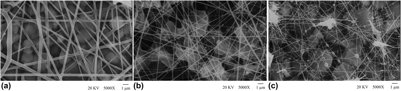
In spite of the existence of bioactive HA elements on the surface of the ZTA substrate, controlling both the morphology and composition of hybrid PA6/HA NFs potentially benefits the biomineralization process. To evaluate these benefits, electrospinning was carried out at three ratios of PA6 to HA (see Table I). SEM micrographs of electrospun mats showed that structures with different morphologies were formed by changing the PA6/HA ratio. When decreasing the PA6/HA ratio from 4 to 0.6, the average diameter of PA6/HA NFs drastically decreased (∼4 times). The average fiber diameter of 310 nm in the sample prepared via M-HR procedure, in addition to the ribbon shape morphology of the fibers, created an undesirable surface on the ZTA substrate [Fig. 3(a)]. Although the finest fibers, with an average diameter of 82 ± 12 nm, were formed by M-LR coating on the ZTA substrate, some defects were detected on the surface of the ZTA such as polymeric beads, broken fibers, and agglomerated HA particles. These defects may affect osteoconductivity heterogeneously [See Fig. 3(c)]. Furthermore, random features attenuate the osteoinductivity to produce a bone’s mineral portion. Hence, a homogenous texture with nanotopography on the surface of an implant is desirable.Reference Shalabi, Gortemaker, Van’t Hof, Jansen and Creugers34 Figure 3(b) demonstrates smooth and perfect fiber formation in the sample prepared via the M-MR coating procedure. Not only a fine fibrous mat is essential to achieve a superactive surface, but also the open surface porosities and consequently high specific surface areas of electrospun PA6/HA coatings can potentially conduct bonelike apatites onto the ZTA surface.Reference Esfahani, Jose and Ramakrishna35 The average surface porosity of M-MR was 64.3%, and in comparison with the M-HR, it made remarkable osteoconductive sites on the bioinert ZTA for bone regeneration. In accordance with the results of this study, it can be elucidated that PA6/HA ratio of 1.15 (M-MR) has the most desirable results in the morphology and arrangement of PA6/HA NFs on ZTA.
SEM images of PA6/HA coating on ZTA via electrospinning method with different P/H ratios: (a) M-HR, (b) M-MR, and (c) M-ML.

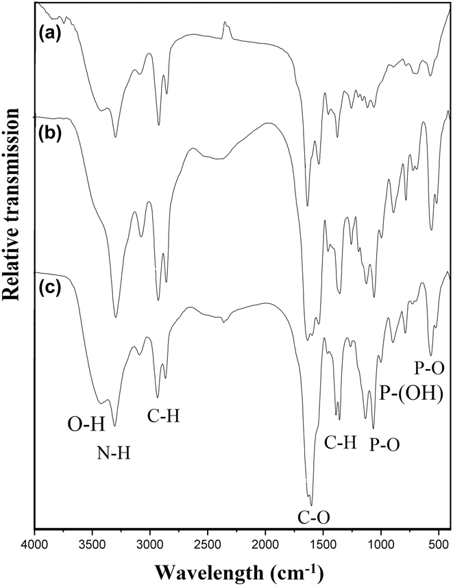
The functional groups in hybrid PA6/HA NFs are essential to be characterized due to the surface characteristics of the electrospun coats, which result in bonelike apatite formation. Hence, the FTIR spectra of electrospun mats were investigated to explore the effect of varying the PA6/HA ratio on the main functional groups of such bioactive coatings (see Fig. 4). The polyamide nature of all of the coated samples was confirmed by detecting prominent peaks at 1541 and 1643 cm−1. The significant band at 3299 cm−1 belongs to the N–H stretching vibration. Other characteristic peaks of PA6 were detected at 1263, 1369, and 2931 cm−1, which refer to the CH2 asymmetric stretching mode. The CH2 stretching vibration band at 2859 cm−1, in addition to the CH2 wagging band at 1170 cm−1, is attributed to the polyamide structure of the electrospun coating.Reference Abdal-hay, Khalil, Al-Jassir and Gamal-Eldeen36 In addition to the representative bonds of polyamide, the characteristic molecular bonds of HA ( ${\rm{PO}}_4^{3 - }$, OH−, and
 ${\rm{HPO}}_4^{2 - }$) were identified in the FTIR spectra of all of the mats. The transmission bands at 580 and 1076 cm−1 are attributed to the bending and stretching vibrations of P–O. A low-intensity band at 928 cm−1 is assigned to the stretching vibrations of P–O(H) in the
 ${\rm{HPO}}_4^{2 - }$ group. The weak band at 3576 cm−1 belongs to the structural OH bond. It is evident from the FTIR spectra that the characteristic bands of HA declined when increasing the PA6/HA ratio. It is worth mentioning that varying the PA6/HA ratio did not influence the atomic structure of polyamide. According to micrographs and FTIR results, it can be said that the M-MR coating had suitable morphology and arrangement on the ZTA substrate, which increased the surface roughness, and it also had bioactive molecular groups inside the scaffold coating. Therefore, subsequent investigations were performed on the M-MR sample.
FTIR spectra of electrospun PA6/HA mats with different P/H ratios: (a) M-HR, (b) M-MR, and (c) M-ML.

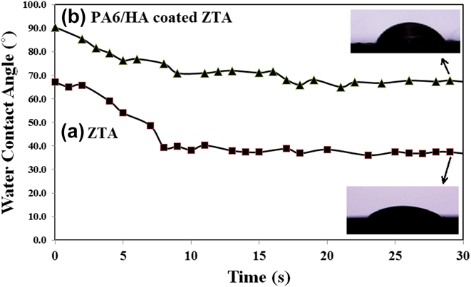
The wetting ability of an implant’s surface is a significant feature in cellular attachment and dispersion during bone regeneration.Reference Ateş, Baran and Yazıcı37 The contact angle of a water droplet represents the wetting ability of the surface, which is also an important surface property of implants. Figure 5 shows the variation of WCA on the ZTA and PA6/HA-coated ZTA during the thirty seconds of contact. The WCA of the ZTA surface was found to be 67 ± 3.5° at the first contact of the water droplet with the surface. On the other hand, the WCA of the PA6/HA-coated ZTA was 90.4 ± 2.8° at the same time. These values explain how the electrospun coating on the ZTA surface changed from being hydrophilic to being hydrophobic. As it can be observed, the WCA decreased to 38.2 ± 0.4° and 70.9 ± 0.7° with respect to the uncoated and coated samples after ten seconds of contact, respectively. Subsequently, the variation of WCA was stable for both surfaces, which can be interpreted as hydrophilic surfaces. The wetting ability is determined by the microstructure and chemistry of the surface.Reference Ateş, Baran and Yazıcı37 The electrospun PA6/HA mats had a different morphology and chemistry on the ZTA surface. It should be mentioned that inorganic materials with high-energy bonds such as ZTA ceramic nanocomposites depict hydrophilic behavior, whereas organic materials have a hydrophobic surface due to the low energy of their bonds on the surface.Reference Redon, Vazquez-Olmos, Mata-Zamora, Ordonez-Medrano, Rivera-Torres and Saniger38 The organic PA6 matrix of the electrospun PA6/HA coating caused the enhancement of the hydrophobicity of the surface, and in contrast, ceramic HA NPs embedded inside the fibers enhanced the wetting ability of the surface. Hence, the WCA decreased after ten seconds of contact. A hydrophilic surface promotes the interaction between artificial implants and collagen and also improves cellular attachment and dispersion.Reference Hughes-Brittain, Qiu, Picot, Wang, Cees and Bastiaansen45 Furthermore, nanopatterning, which is created on the ZTA surface by electrospun PA6/HA mats, modulates the cellular responses due to the typical features of the mechanical environment of the native extracellular matrix (ECM).Reference Santos, Silva, Gomes, Fernandes, Santos and Sencadas39
Variation of WCA on (a) ZTA and (b) PA6/HA-coated ZTA after thirty seconds.

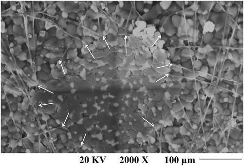
One of the important features of a biocoating is its good adhesion to the substrate.Reference Aly, Mohammed, Al-Meer, Elsaid and Barakat40 The adhesion mechanism of electrospun fibers to ceramic substrates is not clearly known due to the lack of research.Reference Algellai, Tomić, Vuksanović, Dojčinović, Volkov-Husović, Radojević and Jančić Heinemann41–Reference Chlanda, Kijeńska, Rinoldi, Tarnowski, Wierzchoń and Swieszkowski43 Using the microindentation method, an adhesive coating is not expected to deform and delaminate by applying a high force on the coating.Reference Chapman44–46 Figure 6 shows the undeformed PA6/HA nanofibrous coating on the ZTA substrate, even after cutting by the indenter. It seems that the boundary of alumina and zirconia grains at the surface of the ZTA composite was a suitable place for the mechanical interlocking of PA6/HA NFs.
BSE-SEM image of Vickers indent on PA6/HA-coated ZTA. The attached NFs to the edge of pyramid have been marked.

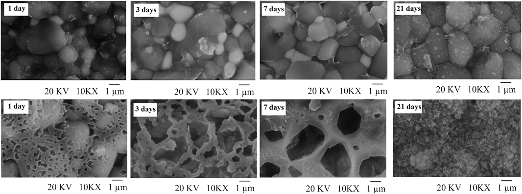
As we know, an essential requirement for a synthetic implant is the ability to rapidly produce a bioactive hydroxycarbonated apatite layer that can bond to the surrounding bone.Reference Prasad S, Ratha, Adarsh, Anand, Kumar Sinha, Diwan, Annapurna and Biswas16 Hence, the ZTA and PA6/HA-coated ZTA samples were immersed in SBF for 1–21 days.Reference Kokubo and Takadama28 Figure 7 shows significant changes on the surface of PA6/HA-coated ZTA, while little change is evidenced on the ZTA baseline control. It seems that the coating formed suitable sites for the precipitation of more bonelike apatite from SBF. The precipitated HA on PA6/HA NFs created more nucleation sites for further bonelike apatite precipitation. Further immersion caused nucleation and growth of more bonelike apatite until a new layer was formed on the PA6/HA-coated ZTA. The creation of open pores on the surface enhances the surface’s response to the bone elements (e.g., biomarkers, bone morphogenetic proteins, and osteoblast cells) present in the surrounding tissue during new bone formation and also improves the mechanical interlocking postimplantation.Reference Burg, Porter and Kellam47 The in vitro study revealed that in contrast to the ZTA substrate, the surface of the PA6/HA-coated ZTA was entirely covered by bonelike apatite after 21 days. The SEM images show that homogenous, smooth precipitations covered the coated surface, which was similar to the biological apatite crystal surface.Reference Gomez-Morales, Iafisco, Delgado-López, Sarda and Drouet13 This modification makes bioinert ZTA an osteoconductive material for supporting bone growth and encouraging the in-growth of new bone.Reference Prasad S, Ratha, Adarsh, Anand, Kumar Sinha, Diwan, Annapurna and Biswas16
SEM micrograph of ZTA (above row) and PA6/HA-coated ZTA (below row) throughout the in vitro study, 1 day to 21 days of immersion in SBF.

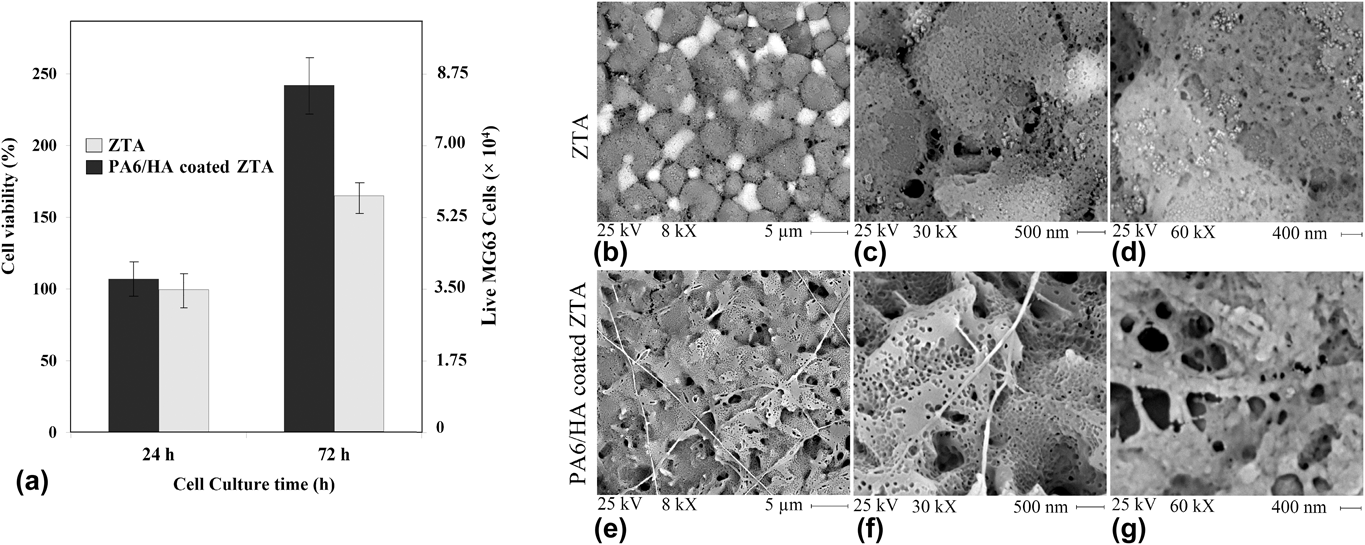
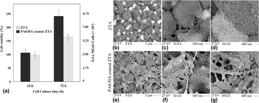
To evaluate the potential of ZTA and PA6/HA-coated ZTA samples in bone regeneration, further in vitro cytocompatibility investigations were carried out. Figure 8(a) displays cell viability on ZTA and PA6/HA-coated ZTA disks after 24 and 72 h. It clearly shows that both ZTA and PA6/HA-coated ZTA samples did not exhibit cytotoxicity for MG63 osteosarcoma cells. This was confirmed by the 101% (3.53 × 10⁴ live cells) and 107% (3.74 × 10⁴ live cells) viabilities of the cells after 24 and 72 h, respectively. According to these results, it can be said that initially, a greater number of cells could adhere to the surface of the coated ZTA and therefore significant cell proliferation, as well as a higher growth rate, was observed. One potential reason behind the promising cell adhesion on PA6/HA might be that the coating provided inductive pathways for cells to attach, proliferate, and differentiate.Reference Wang, Hu, Li, Zhang, Shen, Huang and Shi48 The results revealed that the MG63 osteosarcoma cell viability increased for both ZTA (165%, 5.79 × 10⁴ live cells) and PA6/HA-coated ZTA (243%, 8.47 × 10⁴ live cells) after 72 h. Among several factors that can contribute to this significant difference in cell response, surface properties can play a dominant role.Reference Liu, Li, Cao, Wang, Qu, Wang, Qiu, Di, Wang and Liang49 Although both Al2O3 and ZrO2 ceramics are readily known as biocompatible materials, applying PA6/HA hybrid material promoted the osteogenic differentiation even further due to its osteoinductive and osteoconductive properties.Reference Sequeira, Fernandes, Neves and Almeida8 The nanostructured features of electrospun PA6/HA coating including porosity and pattern could induce the MG63 osteosarcoma cells along the nanofibrous mat to differentiate throughout the open surface porosities and grow along the NFs.Reference Wang, Hu, Li, Zhang, Shen, Huang and Shi48 The roughness created by the nanostructured network also promoted the anchorage and spreading of MG63 cells.Reference Liu, Li, Cao, Wang, Qu, Wang, Qiu, Di, Wang and Liang49,Reference Huang, Hsiao and Chai50 Begam et al.Reference Begam, Kundu, Chanda and Nandi51 showed that rougher surfaces promote better cell adhesion and proliferation.
(a) Viability of MG63 cells on the ZTA and PA6/HA-coated ZTA surfaces after 24 and 72 h assessed by a MTT method. BSE-SEM micrographs of MG63 cells cultured after 3 days on (b, c, d) ZTA and (e, f, g) PA6/HA-coated ZTA in different magnifications (8 kX, 30 kX, and 60 kX).

Osteosarcoma cells are the basic structural and functional building blocks that support bone growth and metabolism.Reference Begam, Kundu, Chanda and Nandi51 Figures 8(b)–8(g) show SEM micrographs of MG63 cells cultured after 72 h on ZTA and PA6/HA-coated ZTA samples at different magnifications. According to the micrographs, the effect of electrospun PA6/HA coating on cell viability was superior to that of ZTA. SEM images of MG63 cells seeded on ZTA revealed that there was no difference between alumina and zirconia grains in terms of the proliferation and differentiation of cells and that the morphology of cultured cells was alike. In contrast, the electrospun PA6/HA coating led to cellular extensions and continuous growth. Furthermore, colonized multilayered cells with numerous cell–cell connections were observed. Moreover, it can be seen that the MG63 cells were well anchored to the NFs. All positive attributes of electrospun PA6/HA mats made this coating favorable for the formation of extracellular matrix (ECM).
According to the obtained results, it can be elucidated that the promising aspects of the electrospinning method can modify the ZTA surface by providing an appropriate texture of osteoconductive PA6/HA. The nanostructured PA6/HA coating might help the development of self-assembled materials with bone characteristics such as high surface area, enhanced chemical reactivity, regeneration, and new bone formation.
IV. CONCLUSION
The present study evaluated the ability of electrospun PA6/HA coating on ZTA to enhance bone regeneration. SEM images showed that the hybrid PA6/HA nanofibrous mats were coated on the ZTA via electrospinning. The results indicated that a PA6/HA ratio of 1.15 (w/w) produced a desirable morphology of the ZTA. The average diameter of NFs and average surface porosity of the coated mat were 120 ± 10 nm and 64.3%, respectively. FTIR spectra revealed that HA NPs could combine with the PA6 matrix. The dynamic measurement of WCA showed that the ZTA surface had hydrophilic behavior, whereas the electrospun PA6/HA coating was hydrophobic upon contact with water, after which the surface became hydrophilic. The microindentation test revealed that PA6/HA coating sufficiently adhered on to the ZTA substrate. The in vitro bioactivity test in SBF showed that a new layer of bonelike apatite homogenously covered the PA6/HA-coated ZTA, while the uncoated ZTA surface was not altered significantly even after 21 days of immersion in SBF. The cytocompatibility of samples revealed that MG63 osteosarcoma cell viability for PA6/HA-coated ZTA (243%) was much higher than for uncoated ZTA (165%) after 72 h. The findings of the current study might modify the practical methods in bone tissue engineering applications.