Introduction
The lingonberry, Vaccinium vitis-idaea var. minus Loddiges, Loddiges, and Loddiges (Ericaceae) is a low-growing, evergreen shrub, which produces edible berries that ripen in mid-September. It is distributed in northern Holarctic regions, typically occurring in rocky and dry peaty soils, tundra regions, wet heaths, barrens, and coastal headlands (Ryan Reference Ryan1978). Larvae of the lingonberry fruitworm, Grapholita libertina Heinrich (Lepidoptera: Tortricidae) feed within developing lingonberry fruit (Torrey Reference Torrey1914; Morris et al. Reference Morris, Penney, Greenslade, Hendrickson and McRae1988). It is known only from North America, reported in Canada from Newfoundland, Nova Scotia, and British Columbia and from the United States of America in California, New Jersey, and Maine (Morris et al. Reference Morris, Penney, Greenslade, Hendrickson and McRae1988; Hillier et al. Reference Hillier, Dixon and Larson2004). Therefore, it is potentially distributed at points between these records and throughout its host plant’s range.
Morris et al. (Reference Morris, Penney, Greenslade, Hendrickson and McRae1988) described the life history of G. libertina in Newfoundland with adults emerging in June through to July and occasionally August and laying eggs on developing berries from July to August. Larvae hatch and bore into the developing berries, where they feed for about four weeks. After reaching maturity, larvae exit berries and overwinter in the prepupal stage in a hibernaculum in debris on the ground. Infestations have been reported as high as 276 larvae per kg of unripe berries, making its presence a significant concern whether for domestic or export markets.
One unidentified species, Phanerotoma Wesmael (Hymenoptera: Braconidae), is a known larval parasitoid of G. libertina (Morris et al. Reference Morris, Penney, Greenslade, Hendrickson and McRae1988). A series of more recent studies (Hillier et al. Reference Hillier, Dixon, Seabrook and Larson2002, Reference Hillier, Dixon and Larson2003, Reference Hillier, Dixon and Larson2004) investigated the potential for monitoring and managing this species using traps with synthetic attractants. During each year of these studies, mass collection of unripe fruit and berries was conducted, and parasitoid numbers and developmental periods were documented during larval counts from fruit.
Material and methods
Methods for fruit and larval collection have been described by Hillier et al. (Reference Hillier, Dixon and Larson2004). Briefly, unripe lingonberries were collected from selected sites in Newfoundland, Canada. In August–September of each year between 1998 and 2000, 1000–10 000 fruits were collected from each site and dissected for the presence of G. libertina larvae. The lingonberry fruitworm is notoriously difficult to rear (Morris et al. Reference Morris, Penney, Greenslade, Hendrickson and McRae1988), particularly from the overwintering prepupal stage to the adult. Larvae were collected from berries at Newfoundland study sites in Pouch Cove, Little Catalina, Freshwater, and Bryant’s Cove (Hillier et al. Reference Hillier, Dixon and Larson2004). Fruit infested with G. libertina were added with vermiculite or screened sand to rearing chambers. The rearing containers (50 mL pill bottles, 500 mL plastic tubs, or 4.2 L Tupperware containers) were held in growth chambers (chamber conditions: 80% relative humidity, 14:10 light:dark hours) at the Atlantic Cool Climate Crop Research Centre, Agriculture and Agri-Food Canada, St. John’s, Newfoundland and Labrador, Canada. Containers were checked every two to three days for emergence.
Mean degree-day accumulations for 10% and 50% emergence of the total population of G. libertina and P. libertinecida were calculated using Arnold’s formula (mean daily temperature−base temperature) (Arnold Reference Arnold1960). A base of 5 °C was used, as recommended by Pruess (Reference Pruess1983).
Following emergence, voucher specimens of G. libertina and P. libertinecida were deposited in the Canadian National Collection at the Eastern Cereal Oilseed Research Centre of Agriculture and Agri-Food Canada (Ottawa, Ontario, Canada). All specimens used for the description of P. libertinecida were from the Canadian National Collection.
Images were made using a DSLR Canon (Melville, New York, United States of America) EOS 60D camera with a 100 mm macro and MPE-65 lens. Multiple images through a range of focal planes from top to bottom were taken of a structure and these combined using Zerene® (Zerene Systems, Richland, Washington, United States of America) to produce a single, focussed image. Specimens were illuminated with a flash Speedlite® 600X (Canon). The final combined image was improved using Adobe Photoshop® CS6 (San Jose, California, United States of America) and plates were assembled using the same software.
Natural history
All specimens documented in the present study were reared from lingonberries or more specifically larvae of the lingonberry fruitworm. Host range and fidelity of Phanerotoma libertinecida is unknown, but it is clear that konobiont endoparasitoid larvae of this species can complete development within G. libertina larvae. Van Achterberg (Reference Van Achterberg1990) reported Phanerotomini as primarily parasitoids of Pyralidae, but to a lesser extent Tortricidae (Olethreutinae), Carposinidae, Gelechiidae, Oecophoridae, and Coleophoridae.
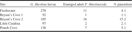
Rearing of G. libertina larvae was largely unsuccessful during 1998–2000, so per cent parasitism rates could not be accurately calculated in these years. Ten P. libertinecida parasitoids emerged from collections in 1998–1999 and 13 P. libertinecida in 1999–2000. In 2000–2001, parasitism as determined by emerged parasitoids, ranged from 1.1% to 15.2% from larvae collected from various sites across Newfoundland (Table 1). Parasitism was also noted by two specimens of an unidentified chalcid wasp. Degree-day accumulations for adult emergence of P. libertinecida was similar to that of G. libertina. Mean emergence (±standard error) for 10% of the populations occurred for each species at 278±43.7 and 278±46.0 for each species, respectively. Fifty per cent emergence occurred at 310±41.0 for P. libertinecida and 323±31.8 degree-days for G. libertina.
Parasitism rates for Phanerotoma libertinecida reared under laboratory conditions from larvae of Grapholita libertina collected in 2000 from field sites in Newfoundland, Canada.

Augmentation of naturally occurring Phanerotoma populations by rearing of parasitoids could be implemented as a control tactic for G. libertina. This would provide pesticide-free biological control of G. libertina populations, and minimises the risks involved with importation of exotic natural enemies to control pest species.
Phanerotoma libertinecida Goulet, new species
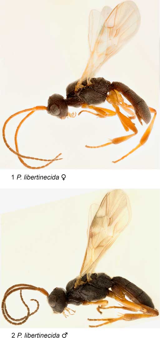
(Figs. 1–5, 8, 10–11, 13, 16, 19, 22–26, 28–29)
Type material. Holotype female (CNC), in perfect condition, labelled [white label] “Freshwater, NL May 13, 1998 ex Lingonberry fruitworm N. K. Hillier”; [red label] “HOLOTYPE Phanerotoma libertinecida ♀ Goulet, 2014”.
Phanerotoma libertinecida, female habitus; 2. Phanerotoma libertinecida, male habitus.

Paratypes. 18 females and 7 males. CANADA. Newfoundland and Labrador: Freshwater, collected 23, 26.viii. 2000, emerged 24, 26. iv. 2001(2 ♀, 2 ♂) [ex Grapholita libertina]; Little Catalina, collected 30. viii. 2000, emerged 22, 24. v. 2001 (2 ♀) [ex Grapholita libertina]; Freshwater 13. v. 1998, ex Lingonberry fruitworm [Grapholita libertina] N. K. Hillier (3 ♂?); Chance Cove, 9. ix, 1996, ex Lingonberry fruitworm [Grapholita libertina] P. L. Dixon (9 ♀); St. John’s 28, 30. vii. 1978, Ray F. Morris (5 ♀) [specimens labelled as “?toreutae” by Zettel, 1987, and reared from ?Grapholita on Lingonberry]; St. John’s 31.vii.1956 Ray F. Morris (1 ♂) [specimen labelled as “?toreutae” by H. Zettel in 1987, and reared from Lingonberry]. Ontario: Simcoe 19.vi.1939, G. E. Shewell (1 ♂) [specimen labelled as “toreutae” by Caltagirone in 1977].
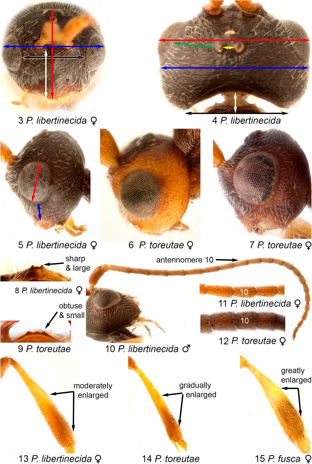
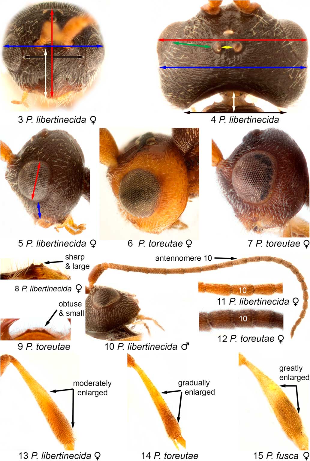
3. Female Phanerotoma libertinecida head capsule in frontal view. Red and blue arrows indicate the capsule is 1.16–1.19 times as wide as long. Face is 1.08–1.50 as wide as median length of the face and clypeus (white and black arrows); 4. Phanerotoma libertinecida head capsule in dorsal view. Head is completely black. Red and blue arrows indicate maximum width of head in dorsal view between outer edges of eyes slightly greater than maximum width between outer edges of genae. White and black arrows indicate the occiput is indented at a depth of 0.2 as long as the width of the indentation. Measurements for the diameter and position of ocelli shown by yellow and green arrows; 5. Female Phanerotoma libertinecida head capsule in lateral view. Malar space in lateral view 0.4–0.5 times as high as eye height (red and blue arrows); 6. Female Phanerotoma toreutae head capsule in lateral view, showing partly reddish-brown colouration; 7. Female Phanerotoma toreutae head capsule in lateral view showing completely reddish-brown colouration; 8. Female Phanerotoma libertinecida clypeus with three sharp and large teeth; 9. Female Phanerotoma toreutae clypeus with three obtuse and small teeth; 10. Male Phanerotoma libertinecida showing full antenna; 11. Female Phanerotoma libertinecida 10th antennomere 1.4–1.9 times as long as wide; 12. Female Phanerotoma toreutae 10th antennomere; 13. Female Phanerotoma libertinecida metatibia showing enlarged in apical 0.5 and dorsal and ventral edges of apical 0.5 subparallel; 14. Female Phanerotoma toreutae metatibia showing narrower width in apical 0.5 than Phanerotoma libertinecida; 15. Female Phanerotoma fusca metatibia showing wider apical 0.5 than Phanerotoma libertinecida.
Diagnostic combination. This species differs from other Nearctic species of Phanerotoma by the completely black body and the light reddish brown base of stigma.
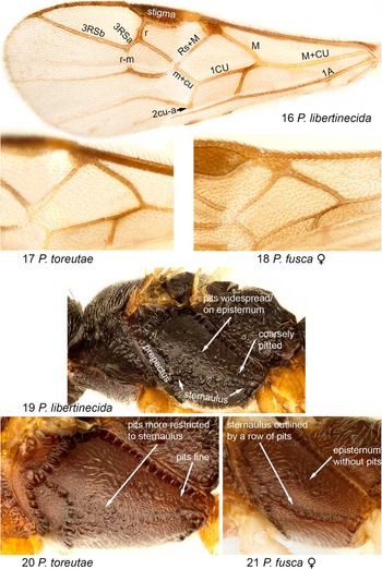
16. Phanerotoma libertinecida forewing; 17. Phanerotoma toreutae forewing showing brown colour in vein M; 18. Phanerotoma fusca forewing vein M, and generally darker venation; 19. Phanerotoma libertinecida thorax in lateral view showing coarse and widespread pits over mesepisternum above sternaulus; 20. Phanerotoma toreutae thorax in lateral view showing finer and less widespread pits than Phanerotoma libertinecida; 21. Phanerotoma fusca thorax in lateral view showing linear pit distribution on sternaulus and absence of pits on episternum.

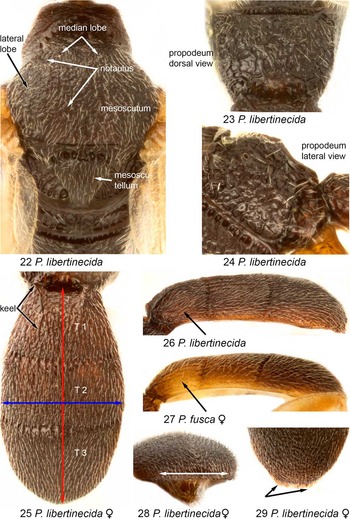
Female description. Colour. Body black; appendages light reddish brown except for brown in apical 0.3–0.4 of metatibia or brown or black tarsomeres 1–5, 3–5, 4 and 5 (metacoxa may be brown or dark brown) and tegula light reddish brown; anterior margin of clypeus brown; scape and pedicel reddish brown to brown, flagellum reddish brown or brown in basal 0.4 shifting to dark brown or black on apical 0.6 (Fig. 1); wings clear, forewing veins brown but veins M+cu, M, part of Rs+M, base of 1 A, m-cu, 2CUa, apical 0.5 of 1CU, and base of stigma light yellow (Fig. 16). Head. Head capsule. Capsule in frontal view 1.16–1.19 times as wide as long (Fig. 3, red and blue arrows); face 1.08–1.50 times as wide as median length of face and clypeus combined, surface coarsely pitted, with beaded sculpticells within pits – thus surface dull (Fig. 3, white and black arrows); maximum width of head in dorsal view between outer edges of eyes slightly greater than maximum width between outer edges of genae (Fig. 4, red and blue arrows); occiput in dorsal view shallowly indented: depth of indentation 0.2 as long as width of indentation (Fig. 4, white and black arrows); ocelli rather small, diameter of lateral ocellus relative to POL=1.00–1.25 and to OOL=3.50–4.75 (Fig. 4, yellow and green arrows); malar space in lateral view 0.4–0.5 times as high as eye height (Fig. 5, red and blue arrows); frons, vertex, and genae with irregular ridges, and surface between ridges with beaded sculpticells – thus surface dull (Figs. 3–5); clypeus very finely pitted and shiny (Fig. 3), anterior edge medially with two (difficult to assess) or usually three teeth (median tooth may be very small) (Fig. 8). Head appendages. Mandible short and wide, its posterior tooth only a little shorter than the frontal tooth. Antenna thickened in central portion, 3rd antennomere 1.1 times as long as the 4th, 10th antennomere 1.4–1.9 times (Fig. 11), 15th 1.4–1.8 times, and 22nd antennomere 1.6–2.8 times longer than wide. Mesosoma. Median lobe of mesoscutum with coarse pits in posterior 0.5 and along notaulus, and with finer pits in anterior 0.5; lateral lobes with fine pits and with beaded sculpticells – thus surface dull; notaulus shallowly impressed, difficult to see though more obvious anteriorly, and outlined by larger pits; mesoscutellum with beaded sculpticells and very fine setigerous punctures (Fig. 2). Mesopleuron and mesoternum mainly with beaded sculpticells – thus surface mainly dull; sternaulus flat, in most specimens not clearly outlined consisting of dense coarse pits forming a wide band (band rarely narrowed centrally) between prepectus and posteroventral area; pits widespread over mesepisternum above sternaulus (Fig. 19). Propodeum with numerous irregular high ridges, and with rounded lateral corners (Figs. 23–24). Appendages. Forewing with vein R at anterior edge 1.1 times as long as the stigma; relative length of r1, 3RSa, and 3RSb as follows 0.9–1.6: 1.0: 5.0–8.1; length of 3RSa 1.0–1.4 as long as r-m (Fig. 16). Mesotibia thin, with no protrusion, longer spur 0.55 times as long as mesotarsomere 1; metafemur 3.9 times longer than wide; metatibia enlarged in apical 0.5 and dorsal and ventral edges of apical 0.5 subparallel (Fig. 13). Metasoma. Carapace (Terga 1–3) in dorsal view 1.02 times as long as mesosoma, and 1.70–1.84 times as long as wide (Fig. 25, red and blue arrows); in lateral view slightly arcuate; relative median length of mediotergites T1, T2, and T3 as follows=0.8–1.2: 1.0: 1.4–1.7 (Fig. 25), apical margin of T3 in posterior view not indented (Fig. 28) and in dorsal view margin narrowly translucent and lateral edge slightly protruding (Fig. 29). Tergum 1 with moderately convergent keels (major ridge originating from anterolateral corner of T1) and at least 0.4 times as long as T1, and surface with prominent and irregular longitudinal ridges (Fig. 25); tergum 2 with prominent and irregular longitudinal ridges becoming less prominent in apical 0.2–0.3 (Fig. 25); tergum 3 in some specimens with little prominent irregular and longitudinal ridges and with a reticulate pattern elsewhere, or with ridges forming a reticulate pattern over most of surface (Fig. 25). Sheath of ovipositor short (three to four times as long as high) and not protruding beyond apex of T3 (Fig. 1).
Male description. As in female except the following. Antenna somewhat more slender centrally, 10th antennomere 2.1–2.6 times (Fig. 10), 15th antennomere 1.7–2.0 times, and 22nd antennomere 1.8–2.0 times as long as wide. Body length: 3.3–4.4 mm.
22. Phanerotoma libertinecida thorax in dorsal view; 23. Phanerotoma libertinecida propodeum in dorsal view; 24. Phanerotoma libertinecida propodeum in lateral view; 25. Phanerotoma libertinecida carapace in dorsal view. Red and blue arrows indicate measurements across carapace=1.02 times as long as mesosoma, and 1.70–1.84 times as long as wide; 26. Phanerotoma libertinecida carapace in lateral view; 27. Phanerotoma fusca carapace in lateral view; 28. Phanerotoma libertinecida female mediotergite T3 in posterior view; 29. Phanerotoma libertinecida female mediotergite T3 in dorsal view.

Taxonomic notes. Following a study of almost all Nearctic species present in the Canadian National Collection and identified by Zettel (Reference Zettel1992) for his monograph of the North American species, specimens of P. libertinecida match only six specimens from St. John’s, Newfoundland and Labrador. Zettel studied these specimens and identified them as “P. ?toreutae”. Using Zettel’s key, most specimens of this species end up at P. toreutae Caltagirone and some at P. fusca Zettel depending on the presence of two or three teeth on the median edge of the clypeus (the median tooth varies from apparently absent to clearly outlined). This is an important but subtle character in Zettel’s key.
If we look at specimens keyed out to P. toreutae, we recorded the following differences.
-
∙ The central portion of the antenna is much less compressed in females of P. libertinecida than in those of P. toreutae (the difference between males and females of P. libertinecida are not marked – the clearest difference is on antennomere 10 (Figs. 11–12)).
-
∙ Pits are coarser along the sternaulus and more widespread on the mesepisternum above the sternaulus in P. libertinecida than in P. toreutae (Figs. 19–20).
-
∙ The lateral teeth along the median edge of clypeus are sharp and clearly more prominent than the obtuse median tooth in P. libertinecida, but obtuse, low and about as large as the obtuse median tooth in P. toreutae (Figs. 8–9).
-
∙ Forewing vein M light yellow in P. libertinecida and brown in P. toreutae (Figs. 16–17)
-
∙ The metatibia of P. libertinecida is wider in apical 0.5 than in P. toreutae (Figs. 13–14).
-
∙ Sheath of ovipositor much shorter (three to four times as long as high, as in Fig. 1) in females of P. libertinecida than in those of P. toreutae (8–10 times as long as high).
-
∙ The head of P. libertinecida is completely black while partly to completely reddish brown in P. toreutae (Figs. 5–7).
-
∙ The host of P. libertinecida, Grapholita libertina Heinrich, is consistently different from the recorded host of P. toreutae, Cydia toreuta (Grote) – both are Lepidoptera: Tortricidae: Olethreutinae. This may be an artefact of hosts studied associated with specific crops.
-
∙ The body length of P. libertinecida is generally shorter (3.3–4.4 mm) than that of P. toreutae (4.0–4.8 mm).
If we look at specimens keyed out to P. fusca, we recorded the following differences.
-
∙ Pits along the sternaulus are more widespread in P. libertinecida and linearly distributed in P. fusca; pits are widespread on the mesepisternum above the sternaulus in P. libertinecida and absent in P. fusca (Figs. 19 and 21).
-
∙ The base of stigma is white in P. libertinecida, but black in P. fusca (wing veins are generally darker in this species) (Figs. 16 and 18).
-
∙ The metatibia of P. libertinecida is narrower than in P. fusca (Figs. 13 and 15).
-
∙ The base of the carapace is black in P. libertinecida and clearly yellowish brown in P. fusca (Figs. 26–27).
-
∙ The body length of P. libertinecida is longer (3.3–4.4 mm) than in P. fusca (2.5–3.2 mm).
We studied two specimens from central northern Yukon (Old Crow) and Northwest Territories (Tuktoyaktuk), Canada. These specimens shared all character states mentioned in the comparison between P. libertinecida and P. toreutae. However, the two specimens from these localities were excluded based on following character states. The specimen from Old Crow, a female, lacks a translucent margin at the posterior end of the carapace and in posterior view the posterior edge of the carapace is deeply indented. The specimen from Tuktoyaktuk, a male, is near the Arctic Ocean. It resembles the Old Crow specimen in having no translucent margin at the posterior end of the carapace. This male has mainly black legs. It is not unusual to find darker specimens at Tuktoyaktuk (based on our personal experience with ground beetles). Both have much darker wing veins: forewing veins M+cu, M, Rs+M, m-cu, 2CUa, apical 0.5 of 1CU are brown. Therefore, we believe that they probably represent an unnamed species close to but distinct from P. libertinecida.
Because the exceptionally dark body colour and the differences between species P. libertinecida keys to, we are confident that we are dealing with a distinct species and worth naming as a significant biocontrol species of a lepidopteran pest species of an unusual agricultural crop, the lingonberry.
Origin of the specific epithet. We all agree that the name of this new species should be derived from the caterpillar host specific name, Grapholita libertina. The ending of the name “-cida” means “killer”. Thus, the name, libertinecida, means the killer of G. libertina.
Host. Grapholita libertina Heinrich (Lepidoptera: Tortricidae).
Distribution. Ontario and Newfoundland and Labrador, Canada.
Acknowledgements
The authors thank J.T. Huber for comments on early drafts of this manuscript. We also acknowledge funding support from the Canada-NL Agri-Food Innovation Program, a Memorial University Special Scholarship for Natural Resource Development to NKH and the Natural Science and Engineering Research Council through a Discovery grant (356109) to NKH.