Introduction
Preeclampsia (PE) is characterized by blood pressure higher than 140/90 mmHg after 20 weeks gestation that seriously affects the perinatal survival rates of mothers and children (Tangeras et al., Reference Tangeras, Austdal, Skrastad, Salvesen, Austqulen, Bathen and Iversen2015). Inadequate trophoblastic invasion was believed to play a role in the pathogenesis of PE (Wu et al., Reference Wu, Yang, Xu, Wang, Zhang, Sagnelli, Hu, Huang and Sun2019).
Extravillous trophoblasts (EVTs) are involved in the establishment of maternal and fetal circulation (Ashton et al., Reference Ashton, Whitley, Dash, Wareling, Crocker, Baker and Cartwright2005; Harris et al., Reference Harris, Keogh, Wareing, Baker, Cartwright, Aplin and Whitley2006). EVTs migrating and invading the decidua, and then entering the maternal spiral arteries of the myometrium, could initiate vascular remodelling process in early pregnancy. This process is crucial for decreased blood flow resistance and increased uteroplacental perfusion (Brosens and Renaer, Reference Brosens and Renaer1972). However, defects in this process would lead to serious pregnancy syndromes, such as PE (Meekins et al., Reference Meekins, Pijnenborg, Hanssens, McFadyen and van Asshe1994). Changes in the microenvironment in vivo could affect the vascular remodelling process. Matrix metalloproteinases (MMPs) could degrade extracellular matrix and help tissue remodelling during pregnancy (Soundararajan and Rao, Reference Soundararajan and Rao2004), among which MMP2 and MMP9 have been regarded as the key enzymes during implantation (Rahat et al., Reference Rahat, Sharma, Bagga, Hamid and Kaur2016). A review demonstrated that reduction in MMP-2 and MMP-9 levels in early gestation reduced the remodelling of spiral arteries (Apicella et al., Reference Apicella, Ruano, Mehats, Miralles and Vaiman2019).
The mechanisms related to the progression of cell invasion and migration in PE remain unclear. Some studies have shown that genetic events could play vital roles in the development and improvement of the PE. lncRNAs are RNAs of more than 200 nucleotides in length, involving multiple physiological activities such as cell growth, differentiation and apoptosis (Zhao et al., Reference Zhao, Zhao, He, Li and Mau2018). Some researchers have demonstrated that lncRNA levels were reduced in PE placentas (Xu et al., Reference Xu, Lian, Zhang, Huang, Zuo, Yang, Chen, Wu and Sun2018a; Wu et al., Reference Wu, Yang, Xu, Wang, Zhang, Sagnelli, Hu, Huang and Sun2019) and implied that lncRNA plays a vital role in the progression of PE. For example, lncRNA HOXA11-AS1 could affect trophoblast cell proliferation by modulating HOXA7 expression via sponged miR-15b-5p (Xu et al., Reference Xu, Wu, Liu, Huang, Zuo, Xia, Jiang, Wang, Chen, Wang and Sun2018b). Search of the Ensembl database showed that lncRNA FOXD2-AS1, FOXD2 adjacent opposite strand RNA 1, had one non-coding exon. It was reported that FOXD2-AS1 was involved in tumour and OA progression. FOXD2-AS1 could reverse resistance to sorafenib in hepatocellular carcinoma mediated by miR-150-5p (Sui et al., Reference Sui, Dong, Yang, Zhang, Dai, Geng, Lu, Yang and Xu2019). Furthermore, the effects of FOXD2-AS1 were reversed via miR-206 inhibitors on chondrocytes viability in osteoarthritis (Cao et al., Reference Cao, Wang, Wang and Huang2018). The review showed that some miRNAs are thought to be involved in placental development by suppressing genes associated with regulation of trophoblast fate, invasion and proliferation (Apicella et al., Reference Apicella, Ruano, Mehats, Miralles and Vaiman2019). However, the role of lncRNA FOXD2-AS1 in trophoblast cells remains unclear. In this study we determined the role of lncRNA FOXD2-AS1 in trophoblast cell proliferation, invasion and migration and elucidated the underlying mechanism.
Materials and methods
Tissue samples and patients
Patients diagnosed with PE in Huai’an First People’s Hospital from 2018 to 2019 were chosen for this study. The plasma from patients with PE or unaffected pregnancies were collected from the relevant research studies and stored at −80°C. In total, 52 PE patients and 52 healthy pregnancies were included in the study. Each patient in the study submitted informed written consents. The study was authorized by the Ethic Committee of Huai’an First People’s Hospital of Nanjing Medical University, China.
Cell culture
HTR-8/SVneo cells were cultured in RPMI 1640 medium (Gibco, USA) containing 10% fetal bovine serum (Thermo Scientific, USA), 100 IU/ml penicillin and 100 μg/ml streptomycin (Invitrogen, USA). Cells were cultured in a 5% CO2 in air atmosphere at 37°C.
Plasmids, microRNAs and cell transfection
shRNA plasmids vector against FOXD2-AS1-1 were obtained from Shanghai GEMAR Pharmaceutical Technology (Shanghai, China), which were respectively shown as shRNA-FOXD2-AS1-1 and shRNA-FOXD2-AS1-2. The plasmid overexpressing lnc-FOXD2-AS1 was provided by the same company. miRs including miR-3127 mimics and corresponding negative control (miR-NC), as well as miR-3127 inhibitors and corresponding negative control were purchased from Guangzhou Rubio Biotechnology (Guangzhou, China). The plasmid and oligonucleotide were transfected into cells using Lipofectamine 2000 reagent (Invitrogen, USA). Cells were then cultured continuously for 36 h.
RNA isolation and qRT-PCR
Total RNA extraction kit was used to extract total RNA (Beyotime, Shanghai, China) following the manufacturer’s instructions. RNA concentration was measured using an ultraviolet light spectrophotometer; FOXD2-AS1 and miRNA-3127 were reverse transcribed into cDNA using the cDNA Reverse Transcription Kit and the miRNA and Reverse Transcription Kit (Beyotime, Shanghai, China). The qPCR reactive system was prepared using the SYBR Select Master Mix (Thermo Scientific, USA). Finally, relative expression levels of FOXD2-AS1 and miR-3127 were detected using a RT-PCR system.
Cell proliferation assay
Cell proliferation was determined using the CCK-8 assay (KeyGen, Nanjing, China). HTR-8/SVneo cells post-transfection were seeded into 96-well plates. Here, 10 μl of CCK-8 solution was added to each well, which was incubated for 24, 48 or 72 h. Later, the cells were incubated at 37°C for 4 h. Absorbance at 450 nm was measured using a microplate reader (Bio-Rad, Hercules, CA, USA).
Cell migration and invasion assays
Cell migration was detected using the wound healing assay. HTR-8/SVneo cells were cultured in 6-well plates for 24 h, then a sterile plastic tip was used to create a wound by the scratching cells. The remaining cells were continuously cultured for 24 h. ImageJ software was used to analyze the photographs taken at 0 and 24 h under an inverted microscope. BD Matrial matrix was put on the upper surface of a transwell chamber. HTR-8/SVneo cells suspended in serum-free medium after 36 h of transfection, were added into the upper chamber. The lower chamber contained 600 μl of medium with 10% FBS. The cells on the upper chamber were cleaned out a cotton swab. The cells that invaded the low chamber through the BD Matrial matrix were fixed with paraformaldehyde and stained with crystal violet. A inverted microscope was used to take photographs for the cells and for cell counting.
Dual-luciferase reporter gene assay
The wild-type FOXD2-AS1 luciferase plasmid (FOXD2-AS1–3′UTR-WT) in which was included the DNA sequence of FOXD2-AS1-1 3′ untranslated region (UTR) containing the miR-3127 binding site was purchased from Promega (Madison, WI, USA). Mutant-type FOXD2-AS1 luciferase plasmid (FOXD2-AS1–3′UTR-MUT) containing the mutant region of miR-3127 binding site was also purchased. The constructed luciferase reporter vectors, FOXD2-AS1–3′UTR-WT/MUT and the miR-3127 mimic or miR-NC were cotransfected into HTR-8/SVneo cells using Lipofectamine 2000. After 48 h, the fluorinase activity of each group of cells was detected using a Dual-Luciferase Reporter Assay (Promega).
Western blot assays
Total proteins from each group were extracted using RIPA protein cell lysate buffer. The BCA method was used to detect protein concentration. Target proteins were separated using 10% SDS-PAGE and transferred into PVDF membranes by electrophoretic transfer. Then, the corresponding primary antibodies (rabbit anti-MMP2, 1:1000; rabbit anti-MMP9, 1:1000; mouse anti-GAPDH, 1:1500; abcam, Cambridge, USA) were added and membranes were incubated overnight at 4°C after sealing. After washing, the blots were incubated at 37°C for 1 h with the corresponding secondary antibody (anti-rabbit, 1:2500; anti-mouse, 1:10,000) coupled to horseradish peroxidase (HPR). Electrochemiluminescence (ECL) was used to develop the colour, and the blot was scanned and analyzed using a gel imager. The intensities of the grey levels of the target band were analyzed using ImageJ software.
Statistical analysis
Data analysis was carried out using GraphPad Prism software. Data from the groups were analyzed using one-way analysis of variance (ANOVA) followed by post hoc tests. A P-value <0.05 was considered to be statistically significant.
Results
FOXD2-AS1 expression was decreased in PE patients
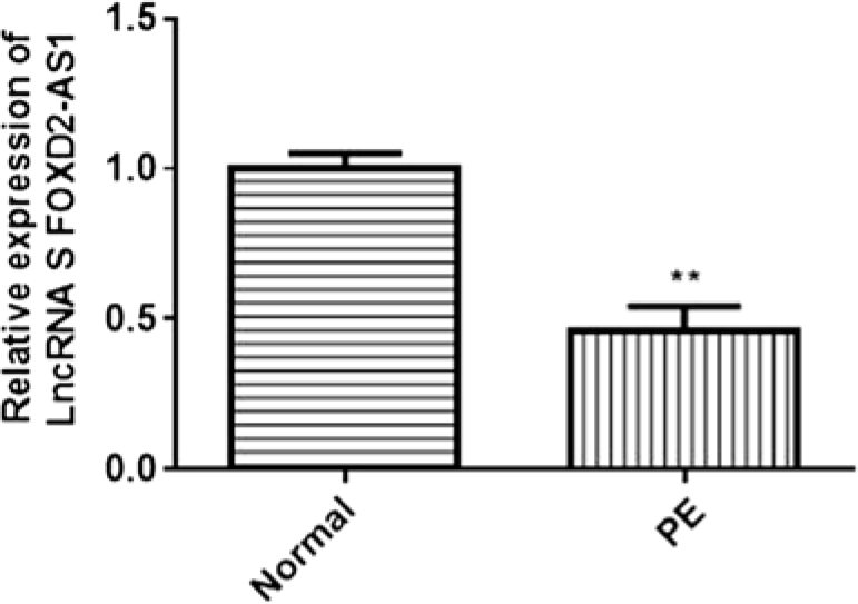
In the present study, we explored FOXD2-AS1 expression in plasma using qRT-PCR. The results showed that FOXD2-AS1 expression was significantly decreased in PE patients and implied that FOXD2-AS1 might play a pivotal role in PE progression (Fig. 1).
FOXD2-AS1 expression was upregulated in pre-eclampsia patients compared with patients with unaffected pregnancies. **P < 0.01 compared with the control group.

FOXD2-AS1 silencing inhibits cell proliferation, invasion and migration in the trophoblast cell
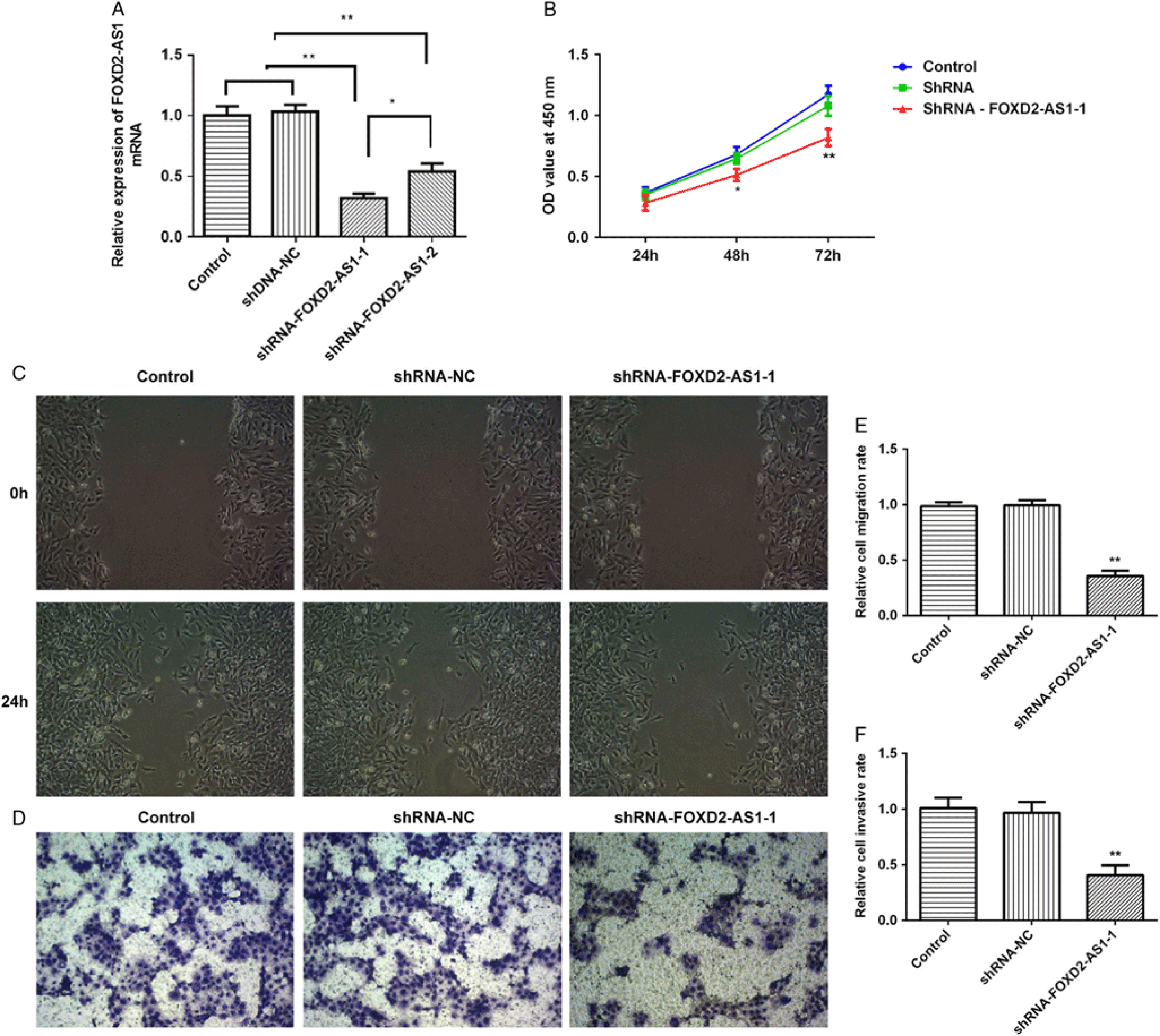
Cell proliferation, migration and invasion are important features of PE progression. FOXD2-AS1 was silenced in HTR-8/SVneo cells by transfection with shRNA-FOXD2-AS1-1 and shRNA-FOXD2-AS1-2. We found that HTR-8/SVneo cells transfected with shRNA-FOXD2-AS1-1 and shRNA-FOXD2-AS1-2 significantly decreased FOXD2-AS1 expression compared with cells transfected with shRNA-NC. shRNA-FOXD2-AS1-1 exhibited greater effects on expression of FOXD2-AS1-1 in HTR-8/SVneo cells as confirmed at transcriptional level (Fig. 2A). Therefore, shRNA-FOXD2-AS1-1 was used as a candidate shRNA for all subsequently studies. CCK-8 assay showed that FOXD2-AS1 silencing in HTR-8/SVneo cells significantly reduced cell proliferation (Fig. 2B). The wound healing assay showed that FOXD2-AS1 silencing in HTR-8/SVneo cells attenuated cell migration abilities (Fig. 2C, E). Furthermore, the transwell invasion assay demonstrated that downregulation of FOXD2-AS1 in HTR-8/SVneo cells caused a significant decrease in the number of invaded cells (Fig. 2D, F).
Downregulation of FOXD2-AS1 suppressed trophoblast cell proliferation, migration and invasion. (A) qRT-PCR analyzed the level of FOXD2-AS1 in HTR-8/SVneo cells transfected with ShRNA-FOXD2-AS1-1, ShRNA-FOXD2-AS1-2 or shRNA-NC. (B) CCK8 assay was used to evaluate the effect of FOXD2-AS1 silencing on HTR-8/SVneo cell proliferation. (C–E) Wound healing assay was used to investigate the effect of FOXD2-AS1 silencing on HTR-8/SVneo cell migratory capacity. (D–F) Transwell invasion assay was used to evaluate the effect of FOXD2-AS1 silencing on HTR-8/SVneo cell invasive capacity. **P < 0.01 compared with the shRNA-NC group.

FOXD2-AS1 overexpression promotes cell proliferation, invasion and migration in the trophoblast cell
FOXD2-AS1 was upregulated in HTR-8/SVneo cells (Fig. 3A). CCK-8, wound healing and transwell assays showed that FOXD2-AS1 overexpression significantly promoted the proliferation, migration and invasion of HTR-8/SVneo cells (Fig. 3B–F).
FOXD2-AS1 promoted cell proliferation, invasion and migration in the trophoblast cell. (A) FOXD2-AS1 expression in HTR-8/SVneo cells transfected with FOXD2-AS1 overexpression plasmids (PcDNA-FOXD2-AS1). (B–F) FOXD2-AS1 overexpression promoted HTR-8/SVneo cells proliferation, invasion and migration in vitro. **P < 0.01 compared with the pcDNA-NC group.

FOXD2-AS1 significantly affects the MMP2 and MMP9 levels
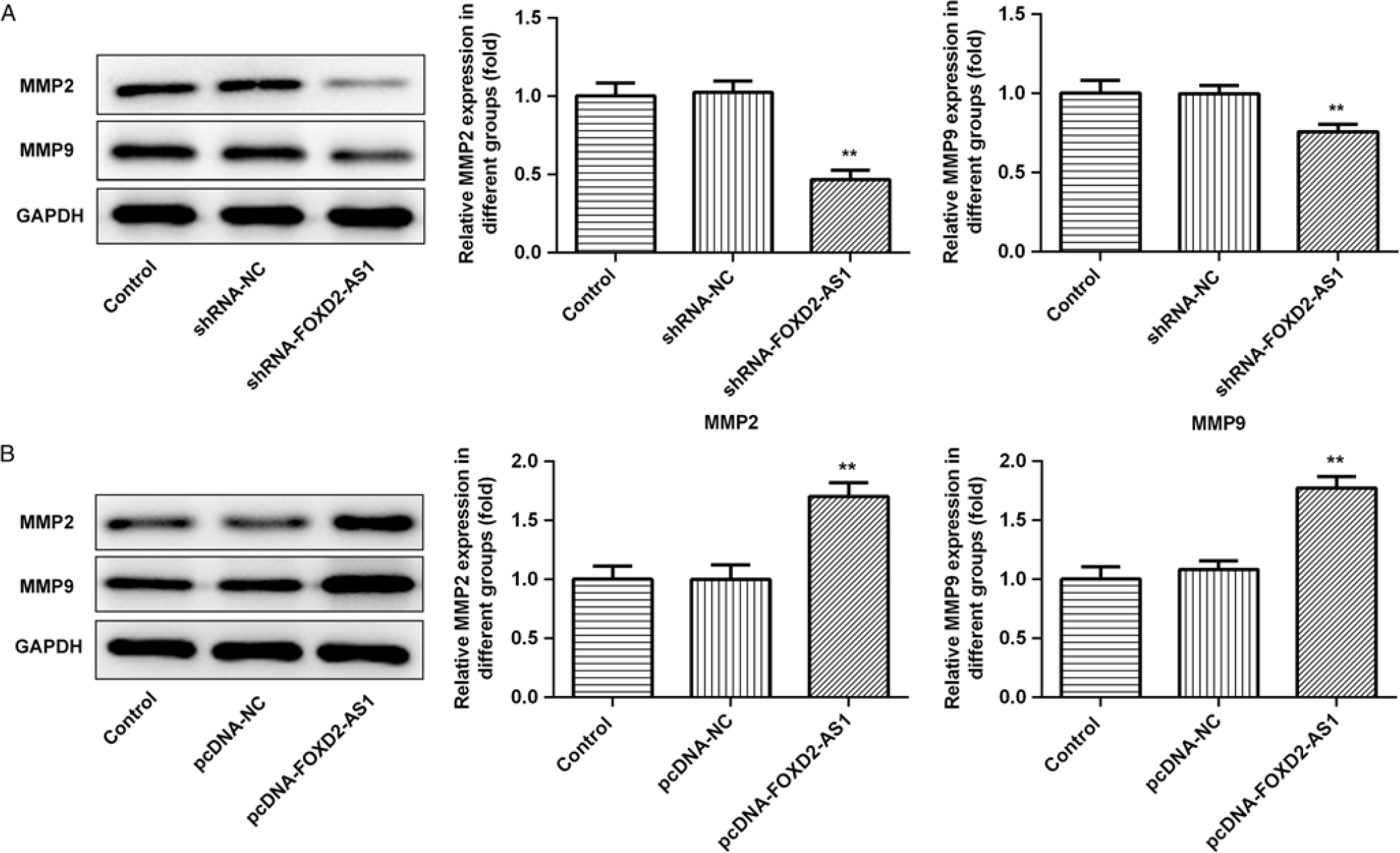
Elevations of MMP-2 and MMP-9 levels could help to maintain normal pregnancy (Eleuterio et al., Reference Eleuterio, Palei, Rangel Machado, Tanus-Santos, Cavalli and Sandrim2015). Western blot analysis indicated that FOXD2-AS1 overexpression or knockdown significantly increased or decreased levels of MMP2 and MMP9 (Fig. 4A, B).
FOXD2-AS1 affected the MMP2 and MMP9 levels in trophoblast cells. (A) FOXD2-AS1 overexpression increased MMP2 and MMP9 levels. (B) FOXD2-AS1 knockdown decreased MMP2 and MMP9 levels. **P < 0.01 compared with pcDNA-NC.

miR-3127 is a target of FOXD2-AS1 in trophoblast cells
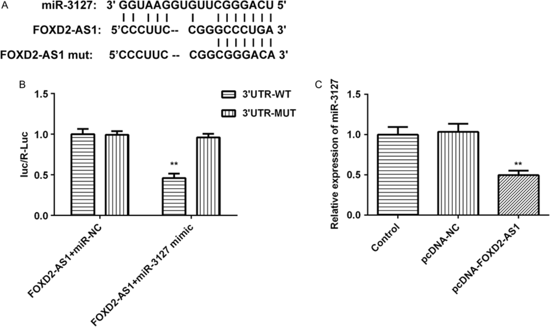
To determine whether FOXD2-AS1 bound miRNAs, the potential binding force of miRNA-3127 and FOXD2-AS1 was predicted using StarBase v.2.0 software (Li et al., Reference Li, Liu, Zhou, Qu and Yang2014) (Fig. 5A). Dual-luciferase assay showed that cotransfection of miR-3127 mimicked the FOXD2-AS1 WT, rather than the FOXD2-AS1 mutant, and significantly suppressed luciferase activity (Fig. 5B). Moreover, qRT-PCR data revealed that FOXD2-AS1 overexpression significantly decreased miR-3127 expression in HTR-8/SVneo cells (Fig. 5C). Taken together, we believe that miR-3127 is a target of FOXD2-AS1 in HTR-8/SVneo cells.
miR-3127 was a target of FOXD2-AS1 in trophoblast cells. (A) Binding and mutant sites between miR-3127 and FOXD2-AS1. (B) Luciferase activity in HTR-8/SVneo cells after cotransfection with miR-3127 mimic/miR-NC and FOXD2-AS1 WT or FOXD2-AS1 Mut reported plasmid. (C) Expression of miR-3127 in HTR-8/SVneo cells transfected with pcDNA-FOXD2-AS1 or pcDNA-NC. **P < 0.01 compared with FOXD2-AS1 3′UTR-MUT + miR-3127 mimic in (B). **P < 0.01 compared with in pcDNA-NC group in (C).

FOXD2-AS1 silencing inhibits cell progression in trophoblast cells by upregulating miR-3127
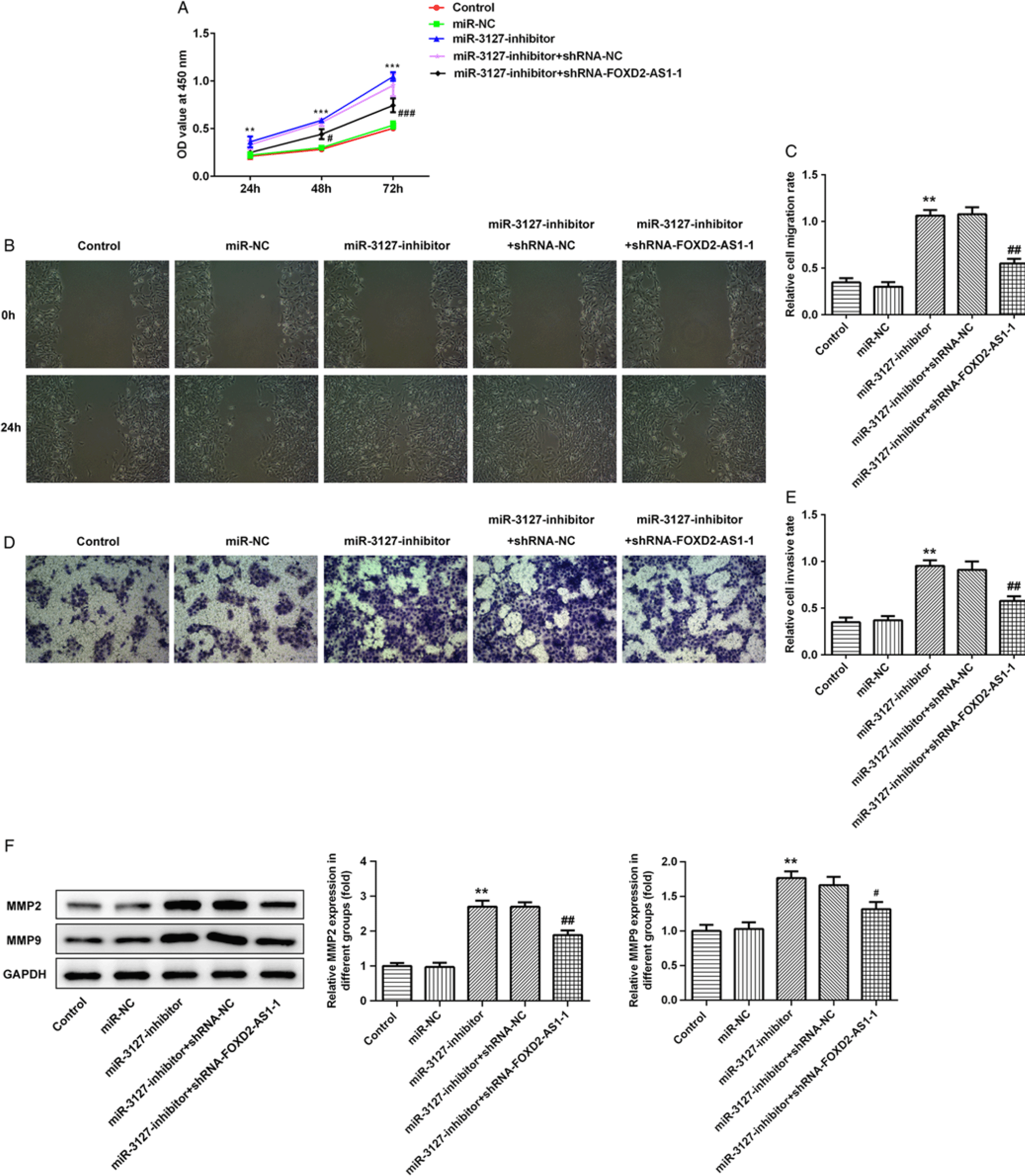
Effects of miR-3127 on proliferation, migration and invasion of HTR-8/SVneo cells were investigated. CCK-8, wound healing and transwell assays showed that the miR-3127 inhibitor significantly increased trophoblast cell proliferation, migration and invasion. Furthermore, downregulation of FOXD2-AS1 notably reversed these effects (Fig. 6A–E). Western blot results indicated that FOXD2-AS1-1 silencing could reduce the levels of MMP2 and MMP9 increased by miR-3127 inhibitors (Fig. 6F).
miR-3127 inhibitor rescued the inhibitory effect on proliferation, migration and invasion of trophoblast cells induced by FOXD2-AS1 silencing. (A–E) Cell proliferation, migration and invasion were detected in FOXD2-AS1 cells transfected with miR-NC, miR-3127 inhibitor, MiR-3127-inhibitor + shRNA-NC, MiR-3127-inhibitor + shRNA-FOXD2-AS1-1. (F) miR inhibitor increased the MMP2 and MMP9 levels. **P < 0.01 compared with miR-NC. ##P < 0.01 compared with miR-3127-inhibitor + shRNA-NC.

Discussion
Preeclampsia is caused by the interaction of both genetic and environmental factors (Janani and Changaee, Reference Janani and Changaee2017). Impaired migration and invasion of trophoblastic cells have been proposed as possible causes of pathogenesis of PE pathogenesis. Abnormal expression of lncRNAs and miRNAs has been found in the placentas of patients with PE. Therefore, exploration and identification of PE-associated lncRNAs and their functions may provide a new route for understanding PE pathogenesis and defining novel therapeutic targets, as well as provide diagnostic and prognostic markers for PE (Wu et al., Reference Wu, Yang, Xu, Wang, Zhang, Sagnelli, Hu, Huang and Sun2019).
In this study, we explored the potential molecular mechanism of lncRNA FOXD2-AS1 using trophoblastic cell lines. FOXD2-AS1 has been mainly researched for its functions in osteoarthritis and cancer. Our study found that expression of FOXD2-AS1 was decreased in the plasma of women with PE compared with women with unaffected pregnancies. In vitro, FOXD2-AS1 overexpression could effectively promote the progression of trophoblast cells, whereas silencing of FOXD2-AS1 expression caused the opposite results. These findings implied that FOXD2-AS1 could be used as a vital regulatory molecule involved in influencing pathologic and physiologic processes of PE and suggested that FOXD2-AS1 may be a promising biomarker for the diagnosis of PE.
Long non-coding RNAs were reported to exert their biological roles by functioning as competing endogenous RNAs (ceRNAs) to negatively regulate miRNA expression (Wang and Chang, Reference Wang and Chang2011). To investigate the underlying mechanisms by which FOXD2-AS1 exerts its biological effects on trophoblast cell proliferation, migration and invasion, it was essential to identify its binding miRNAs.
In the present study, by luciferase reporter assay, miR-3127 was identified as a downstream target of FOXD2-AS1. miR-3127 inhibitor significantly promoted the proliferation, invasion and migration in HTR-8/SVneo cells. This study found that miR-3127 could modulate cell proliferation in hepatocellular carcinoma (Jiang et al., Reference Jiang, Zhang, Guo, Yu, Chen, Li, Tian and Sun2015). FOXD2-AS1 knockdown markedly altered the effect by which miR-3127 inhibitor increased trophoblast cell viability. Furthermore, miR-3127 inhibitor increased MMP2 and MMP9 levels in trophoblast cells, and upregulated MMP2 and MMP9 levels reduced by FOXD2-AS1 knockdown. A previous study showed that MMP2 and MMP9 levels were reduced in PE (Rahat et al., Reference Rahat, Sharma, Bagga, Hamid and Kaur2016). These results suggested that miR-3127 plays a vital role in affecting the progression of PE and that miR-3127 plays a vital role in affecting the progression of PE. Previous studies have shown that miR-3127 could affect cell proliferation and invasion via signalling pathways such as PI3K/AKT, c-Abl/Ras/ERK or Wnt/β-catenin signalling (Sun et al., Reference Sun, Chen, Zhang, Xie, Hou, Hui, Xu, Du, Zhou, Su and Gao2014; Jiang et al., Reference Jiang, Zhang, Guo, Yu, Chen, Li, Tian and Sun2015; Yang et al., Reference Yang, Sun, Wu, Tang, Ding, Xu, Su and Gao2018). However, further study is needed on how miR-3127 produces the effects in preeclampsia.
MMPs could regulate cellular migration, invasion, and angiogenesis during embryonic implantation under normal physiological conditions (Corthorn et al., Reference Corthorn, Rey, Chacon and Valdés2007). A previous study has indicated that overexpression of MMP2 and MMP9 could recover the invasion of HTR-8/SVneo cells decreased by BMAL1 siRNA treatment (Li et al., Reference Li, Zhai, Liu, Hong, Zhao, Zhao, Sun, Du and Chen2017). In the present study, FOXD2-AS1 overexpression or knockdown significantly increased or decreased MMP2 and MMP9 levels in trophoblast cells. These results might imply that FOXD2-AS1 could modulate trophoblast cellular migration and invasion, partly mediated by influencing MMP2 and MMP9 levels.
In conclusion, our study showed that FOXD2-AS1 expression was lessened in placenta in the PE patients. Furthermore, FOXD2-AS1 significantly affected proliferation, invasion and migration of trophoblast cells in vitro by regulating miR-3127. FOXD2-AS1 regulated MMP2 and MMP9 levels by affecting miRNA-3127 levels. Further studies are required to investigate underlying mechanism for developing FOXD2-AS1 as a potential therapeutic target for PE.
Conflicts of interest
The authors declare no conflicts of interest.