Introduction
Pelecaniformes (cormorants, pelicans, gannets and related species) tend to establish a close relationship with an aquatic environment and feed on copepods and fish in that environment. Species of this group of birds are highly important epidemiologically in the spread of several pathogens in different geographical areas, owing to the migratory habit of these birds (Corn et al., Reference Corn, Barre, Thiebot, Creekmore, Garris and Nettles1993; Sick, Reference Sick2001).
Nematodes of the genera Contracaecum (Railliet & Henry, 1912) and Eustrongylides (Jagerskiold, 1909) may constitute a large problem, with several reports describing human infections caused by ingestion of raw or improperly cooked fish. Fish represent an intermediate and/or paratenic host during the life cycle of these nematodes, the pathogenicity of which can be different and more evident in mammals than in birds (Buendia, Reference Buendia1997; Pinto et al., Reference Pinto, Barros, Tortelly, Teixeira and Gomes2004). Studies carried out in the Katy Rybackie breeding colony and in selected Masurian lakes revealed that all of the black cormorants examined in northern Poland were infected with Contracaecum rudolphii (Kanarek et al., Reference Kanarek, Rolbiecki, Sitko, Barus and Rokichi2002; Kanarek & Rokichi, Reference Kanarek and Rokichi2005). Moreover, it has been found that the infection rate of Contracaecum spp. in the stomach of black cormorants is age-dependent (Kuiken et al., Reference Kuiken, Leighton, Wobeser and Wagner1999). Transmission of Eustrongylides tubifex requires two intermediate hosts, the first being aquatic oligochaetes (Measures, Reference Measures1988b) and the second being fish (Measures, Reference Measures1988c). The importance of these nematodes in the aetiology of disease (inducing nodular lesions in the wall of the proventriculus) and mortality in fish-eating birds is well documented (National Wildlife Health Center, 1987).
Reports of accidental human infections associated with distinct clinical signs caused by these helminths emphasize the necessity of an adequate evaluation of the pathogenicity of helminths infecting aquatic birds, by inducing experimental infections in laboratory animals using nematodal larvae recovered from infected fish (Armas de Conroy, 1986; Cancrini et al., Reference Cancrini, Magro and Giannone1997).
The aim of the present study was to conduct a survey of parasitic nematodes in the stomach of great cormorants (Phalacrocorax carbo) in Lake Biwa in Japan and to describe histopathological changes caused by these parasites in the gastric walls of infected birds.
Materials and methods
Study site
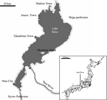
Lake Biwa is the largest freshwater lake in Japan (about 670 km2), and is located in Shiga Prefecture (west-central Honshu Island), to the north of Kyoto City. It is considered to be a breeding ground for several freshwater fish (fig. 1).
Map of Japan illustrating the area of Lake Biwa.

Collection and examination of cormorants for parasites
Thirty adult black great cormorants (P. carbo) of both sexes were shot at Tikubu Island in Lake Biwa with permission from the Shiga Prefectural Government as a part of an animal pest control programme. Dead cormorants were then collected and sent to the Laboratory of Veterinary Pathology at Gifu University to undergo investigation.
All cormorants were necropsied through longitudinal incision, and gonadal examination was done to detect the sex of each bird. This was followed by double ligation of the stomach, anterior and posterior, to preserve all stomach contents. The stomach was incised longitudinally for viewing, and then all free, unattached nematodes were gently extracted by means of a needle and placed in separate Petri dishes. Gastric contents were thoroughly examined to obtain all mixed nematodes, and all recovered helminths were thoroughly washed with normal saline (0.9% NaCl). Nematodes were first observed under a dissecting microscope, and then intact live specimens were relaxed using drops of glacial acetic acid, cleared in lactophenol (one part phenol crystals, lactic acid and distilled water and two parts glycerine) and finally mounted on glass slides using glycerol jelly (Abou-Znada, Reference Abou Znada1993). Prepared slides were examined carefully under a light microscope using different magnifications, and recovered helminths were identified according to Yamaguti (Reference Yamaguti1961), Measures (Reference Measures1988b, Reference Measuresc) and Hartwich (Reference Hartwich1964, Reference Hartwich, Chaubaud, Anderson and Willmott1974).
Histopathology
After removal of nematodes, the mucosal surface of the proventriculus was removed for histological analysis. Pieces of the stomach were fixed in 10% neutral buffered formalin, processed routinely for paraffin embedding, sectioned at 5 μm and stained with haematoxylin and eosin (HE) for examination under the microscope (Bancroft & Stevens, Reference Bancroft and Stevens1996).
Results
Of the 30 adult great cormorants (P. carbo) examined, 26 birds had gastric nematodes, a prevalence rate of 86.7%. Among these, ten were males (83.3%, n = 12) and 16 were females (88.9%, n = 18).
Parasitological investigations
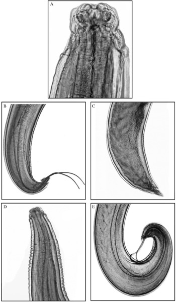
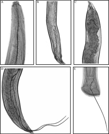
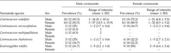
Adenophorean and ascaroid nematodes were removed from the stomach of infected cormorants. Large numbers of Contracaecum spp. were observed during examination of the stomach obtained from cormorants. There were four species of this genus: Contracaecum rudolphii Hartwich, Reference Hartwich1964 (fig. 2A, B and C), Contracaecum multipapillatum Drasche, 1882 (fig. 2D and E), Contracaecum chubutensis Garbin, 2008 (fig. 3A, B and C) and Contracaecum microcephalum Yamaguti, Reference Yamaguti1961 (fig. 3D). Of the four species, the most prevalent was C. rudolphii; both male and female nematodes were found in 10 of the 12 male cormorants, while 13 of the 18 adult females had male C. rudolphii and 16 of the 18 had female parasites. The prevalence rate of C. chubutensis was lower than that of C. rudolphii; both sexes of this nematode were seen in 4 of 18 adult female cormorants, while adult male C. chubutensis was seen in 3 of 12 male birds and 2 of 12 adult female birds. For C. microcephalum, prevalence rates in adult male cormorants were 3 of 12 and 1 of 12 for male and female adult helminths, respectively, while adult male C. microcephalum was seen in 3 of 18 female birds. Finally, C. multipapillatum showed the lowest prevalence of nematodes, with only two live adult male specimens in 1 of 12 male cormorants (table 1). The present investigation reported that C. rudolphii was the most prevalent species and was found in all cases of Contracaecum spp. infection. Moreover, the species co-existed with C. microcephalum, C. multipapillatum and C. chubutensis, causing double infection in these cases. Furthermore, cases of infection with E. tubifex showed mixed infection with C. rudolphii, and gastric walls of these birds exhibited both nodular lesions and haemorrhagic patches that included both types.
(A) C. rudolphii anterior end ( × 100); (B) C. rudolphii male posterior end ( × 20); (C) C. rudolphii female posterior end ( × 20); (D) C. multipapillatum anterior end ( × 40); (E) C. multipapillatum male posterior end ( × 20).

(A) C. chubutensis anterior end ( × 40); (B) C. chubutensis male posterior end ( × 20); (C) C. chubutensis female posterior end ( × 20); (D) C. microcephalum male posterior end ( × 20); (E) Eustrongylides tubifex male posterior end ( × 20).

The prevalence and intensity of infection of cormorants with species of Contracaecum and Eustrongylides, relative to host sex.

The adenophorean nematode Eustrongylides tubifex (Nitzsch in Rudolphi 1819) (Nematoda: Dioctophymatidae) was found in nodular lesions in the wall of the proventriculus. All E. tubifex nematodes were males (fig. 3E), and some were very firmly attached to the stomach wall and could not be extracted. Moreover, the prevalence of infection was higher in adult female cormorants (9 of 18) than in males (5 of 12) (table 1). No other nematodes were detected in necropsied birds.
Pathological features
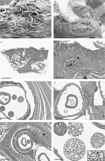
Gross examination showed that nematodes were in association with the surface of the stomach mucosa, and some had penetrated the mucosa and extended deep into the submucosa, muscle layer and serosa, particularly in cases of Contracaecum spp. (fig. 4A). In addition, some nematodes had deeply invaded muscular layers, while others appeared to have induced whitish nodules or tumour-like masses on serosal surfaces, where coiled parasites, denoting the presence of Eustrongylides sp., were frequently observed. These nodules were around 2.0 cm in diameter and protruded into the peritoneal cavity (fig. 4B). In these birds, adult Eustrongylides sp. were associated with the lumen of the proventriculus through small apertures in the mucosa. Either anterior or posterior ends of nematodes had protruded through apertures into the lumen of the stomach, extending as far as 2 cm and then retracting back into the nodule. In infected stomachs, there was marked ulcerative inflammation as well as hyperaemia, especially at sites where parasites attached themselves to the mucosa. In one case, a number of adult nematodes had penetrated through the muscle layer and up into the serosal surface of the stomach, causing peritonitis.
(A) A longitudinal incision in the glandular stomach showed numerous nematodes unattached to the mucosa. Scale bar = 10 mm. (B) The outer serosal surface of the glandular stomach showing whitish nodules or tumour-like masses containing E. tubifex. Scale bar = 10 mm. (C) The wall of the proventriculus showing nematodes in different layers (arrows). Scale bar = 5 mm. (D) A cross-section of nematodes in the lamina propria (arrow); haematoxylin and eosin (HE). Scale bar = 30 μm. (E) A cross-section of nematodes in the muscular layer; HE. Scale bar = 30 μm. (F) Granulomatous reaction consisting of granulocytes, especially eosinophils (arrow), lymphocytes, macrophages and multinucleated giant cells (arrow) surrounding nematodes; HE. Scale bar = 30 μm. (G) Fibrous connective tissue proliferating around a nematode (arrow); HE. Scale bar = 30 μm. (H) A cross-section of an adult female E. tubifex showing the uterus containing eggs (arrow); HE. Scale bar = 30 μm.

Examination of proventricular walls of the infected stomach revealed the presence of numerous nematodes that had penetrated the thickened wall (fig. 4C). Some of these parasites were found in the lamina propria of the glandular stomach (fig. 4D), while others were detected in the muscular layer (fig. 4E). Moreover, some nematodes were seen to have penetrated walls deeply to reach serosal membranes. There were moderate to severe granulomatous inflammatory reactions which consisted of eosinophils, lymphocytes, macrophages and multinucleated giant cells surrounding nematodes (fig. 4F). The mucosal epithelium exhibited severe destruction and infiltration by granulocytes, especially eosinophils, lymphocytes and macrophages. In one case, there was severe peritonitis associated with nematodes that had penetrated through serosal surfaces to the abdominal cavity. In more chronic and severe cases, zones of organizing fibrous connective tissues had formed around nematodes and were encircling parasites (fig. 4G). Occasionally, mineralized foci were seen surrounding necrotic debris in the wall of the proventriculus, especially in muscular and sub-serosal layers, in close association with degenerating or dead nematodes. Pathological alterations in the proventricular wall may be due to both Contracaecum spp. and Eustrongylides sp., as the first might cause some mucosal damage during their penetration (although many of them feed on gastric contents) and the second was already present in nodular lesions, thus coming into contact with the stomach wall and inducing a pathological lesion.
Discussion
During the past few decades, several studies have been conducted that were aimed at establishing the identity of, development of and pathological features induced by both the common anisakid nematode Contracaecum spp. and the adenophorean nematode Eustrongylides sp., in fish-eating birds, primarily cormorants. Environmental changes resulting from anthropogenic actions intensify the exposure of wildlife hosts that act as bioindicators of these changes (Friend, Reference Friend1981; Pinto et al., Reference Pinto, Barros, Tortelly, Teixeira and Gomes2004). Coprological screening is usually insufficient for detecting the presence of avian helminths, as in some nematodal infections e.g. C. multipapillatum, adult nematodes are trapped by tubular fibrous lesions, which are induced by keeping the parasite in contact with the gastric lumen, thus interfering with oviposition, and this can alter the appearance of eggs during faecal examination (Pinto et al., Reference Pinto, Barros, Tortelly, Teixeira and Gomes2004). Although Spalding (Reference Spalding1990) proposed a technique called abdominal palpation for detecting fibrous lesions caused by the migration of larval Eustrongylides sp. in newborn birds, this technique was not suitable for examination of adult birds, as they possess well-developed abdominal musculature in addition to a large amount of fatty tissue that can interfere with the efficacy of this diagnostic method. Therefore, post-mortem examination is the only confirmed method for detecting adult Eustrongylides sp.
The present study revealed a high prevalence of Contracaecum spp., especially C. rudolphii, with infection rates of 83.3% (n = 12) for both nematodal sexes in adult male cormorants and 72.2% and 88.9% (n = 18), respectively, for male and female adult nematodes in female cormorants in Lake Biwa. Similar results were obtained by Kanarek & Bohdanowicz (Reference Kanarek and Bohdanowicz2009), who found that infection rates of the great cormorant P. carbo with C. rudolphii were high for both fourth-stage larvae (preadult) and adult nematodes. Dziekonska-Rynko & Rokicki (Reference Dziekonska-Rynko and Rokicki2008) found that the infection rate among black cormorants from a colony in Lake Selment Wielki was very high, reaching 100%. This high prevalence could possibly be attributed to the frequent occurrence of the life cycle, as a result of several factors; first the outer membrane of the C. rudolphii egg is smooth, so nematode eggs expelled from the uterus do not clump together, and this probably enhances the dispersal of the parasite in aquatic environments (Dziekonska-Rynko & Rokicki, Reference Dziekonska-Rynko and Rokicki2007). Second, the water temperature and the availability of intermediate hosts (fish or copepods) ensure persistence of the parasite in the environment, which in turn may be the underlying cause of the very high prevalence of infection among cormorants.
Some researchers, among them Mozgovoy et al. (Reference Mozgovoy, Shakhmatova and Semenova1965, Reference Mozgovoy, Shakhmatova and Semenova1968), have pointed out that copepods are the obligate intermediate host and larvae of dragonflies serve as the paratenic host for C. rudolphii development. Thomas (Reference Thomas1937a, Reference Thomasb), Huizinga (Reference Huizinga1966) and Bartlett (Reference Bartlett1996), on the other hand, concluded that the role of paratenic host is played by freshwater or marine copepods, while fish serve as an intermediate host by eating free-living larvae or by ingesting infected copepods, and they considered this to be the most common source of infection for cormorants. The present study supported this opinion in that most of the necropsied cormorants showed consumed fish in the stomach on post-mortem examination. This supports the possibility that fish serve as an intermediate host and play a major role in perpetuating the life cycle of C. rudolphii. This appears to contradict the opinion that copepods act as an intermediate host and fish serve only as a transport host, while the strong presence of the parasite appears to indicate a mass occurrence of the nematode in fish.
The present investigation revealed high intensity and infection rates. Since all of the birds examined were adults, this supports a previous report that the prevalence of C. rudolphii and its intensity in great cormorants are dependent on the age of the bird (Kuiken et al., Reference Kuiken, Leighton, Wobeser and Wagner1999); in that study, infection rates were 50% in 1-week-old chicks, 71% in 2-week-old chicks, 83% in 3-week-old chicks and 100% in adult birds. Thus, aquaculturists are advised to control aquatic birds in fish ponds as an effective means of reducing Contracaecum spp. infections, as parasite larvae cause significant problems in terms of hygiene when fish products are consumed or used fresh as feed in fish farms, where the transfer of larvae to cultured fish occurs frequently (Dick et al., Reference Dick, Papst and Paul1987; Fagerholm, Reference Fagerholm1988).
In general, high prevalence and intensity indicate that further studies should focus on avian conservation in this important ecosystem, together with the control of population sources, and it should be realized that the parasite is fairly common in the ichthyofauna. The pathogenicity of the parasite in fish, birds and mammals is still under investigation, but has been described by numerous researchers, among them Abollo et al. (Reference Abollo, Gestal and Pascual2001) and Yang et al. (Reference Yang, Liao and Zeng2000, Reference Yang, Miao, Liao and Wang2001). Furthermore, this threat casts doubt on the safety of fish consumption, particularly prior to thermal treatment. In conclusion, the high prevalence of gastric nematodes, especially C. rudolphii, in great cormorants living in Lake Biwa possibly indicates that these nematodes are able to complete their life cycle.
Contracaecum microcephalum is found worldwide in great cormorants, but is not as common as C. rudolphii (Yamaguti, Reference Yamaguti1961; Anderson, Reference Anderson1992). The present investigation revealed rates of infection by C. microcephalum in adult male cormorants to be 25% and 8.3% (n = 12) for male and female nematodes, respectively. Moreover, in adult female cormorants the infection rate was 16.7% (n = 18) for male parasites. This relatively low rate of infection is possibly due to the number of birds that were examined, in addition to their nature of distribution (Barson & Marshall, Reference Barson and Marshall2004). Similar results were obtained for both C. multipapillatum and C. chubutensis in this study. The infection rate for the former was 8.3% (n = 12) (only male nematodes) in adult male cormorants, while no infection was seen in adult female birds; infection rates for the latter were 25% and 16.7% (n = 12) for both parasite sexes in adult male cormorants, and 22.2% (n = 18) for both nematode sexes in female birds. Malacalza et al. (Reference Malacalza, Poretti and Bertellotti1994) attributed the low mean intensity of Contracaecum spp. in cormorants to the regurgitation habit of birds and considered this phenomenon to be a depuration mechanism, since a high nematode intensity was found in regurgitated pellets.
Males of all nematode species were obtained, but females of some species were not found. However, several parasites were deeply embedded in tumour-like lesions in the wall of the proventriculus and were very difficult to extract. These parasites may have included female nematodes, so percentages cited here may not necessarily have been actual percentages. These results are consistent with those obtained by Karmanova (Reference Karmanova1968), Mace & Anderson (Reference Mace and Anderson1975) and Asakawa et al. (Reference Asakawa, Kimoto and Murata1997), who based their findings on E. excisus, Dioctophyma renale and Eustrongylides sp., respectively, infecting Japanese little grebes.
More than one species of Eustrongylides sp. has been reported from the same host, and this lack of host specificity could act as an important factor in increasing the rate of infection by this parasite (Howard & Moore, Reference Howard and Moore1984; Measures, Reference Measures1988a). Accordingly, the present study suggests that the high intensity of E. tubifex could be related to the water temperature in Lake Biwa, which is ideal for the development of parasite eggs. The presence of large populations of oligochaetes and fish hosts possibly suggests that the infection rate in Lake Biwa is quite high, so these prerequisites, as well as the creation of reservoirs, where large numbers of fish-eating birds congregate during breeding, constitute a favourable environment for an increasing number of tubificid intermediate hosts. All of these encourage the epizootiological status of E. tubifex.
The present investigation revealed that most of the recovered Contracaecum spp. came from fish intermediate hosts ingested by birds: however, others were found to have attached themselves to the gastric wall. Huizinga (Reference Huizinga1971), Fagerholm et al. (Reference Fagerholm, Overstreet and Humphrey-Smith1996) and Abollo et al. (Reference Abollo, Gestal and Pascual2001) suggested that the majority of Contracaecum spp. are found lying unattached within the lumen of the proventriculus and they feed on gut contents rather than on host tissue; however, those that were attached were often observed to have penetrated into the proventricular wall as well as into the flesh of ingested fish, and were detrimental in that the proventricular epithelial lining was destroyed as a result of adult and larval parasites burrowing into it. Other detrimental effects included petechial haemorrhages, ecchymoses and erosive ulcerations. The purpose of parasite attachment is to anchor nematodes against the abrasive movement of bone materials passing from the proventriculus into the intestine. Huizinga (Reference Huizinga1971) reported that, in the absence of food, parasites might feed on the epithelial lining of the host's alimentary tract. In most of the cases examined, lesions were found in the stomach that were caused by parasite invasion of the proventricular mucosa, suggesting that mucosal penetration is a common behaviour of Contracaecum spp., which shows a wide range of host specificity in fish-eating birds belonging to Pelicaniformes. However, it has not yet been established that Contracaecum spp. cause mortality in birds.
Concerning E. tubifex, acute inflammatory response and extensive connective tissue proliferation around the parasite have been described in all cases. Similar results were obtained by Wiese et al. (Reference Wiese, Davidson and Nettles1977) and Measures (Reference Measures1988d), who reported acute to resolved processes of inflammation in various ardeids infected with large numbers of Eustrongylides ignotus. These inflammatory reactions were distinct around lesions containing nematodes, and they continued to increase in size, eventually producing raised tumour-like masses in infected birds. Eustrongylidosis more commonly causes death in nestlings than in juvenile and adult birds. However, aged birds showed distinct lesions that usually proved to be chronic, and parasites appeared to contribute to, rather than cause, deaths of birds (Spalding & Forrester, Reference Spalding and Forrester1993). Acute peritonitis may occur as a result of penetration of the serosal surface by nematodes, in the process introducing some bacterial colonies into the gut of birds.
Acknowledgements
This study was supported partially by a Grant-in-Aid (emerging infection) for scientific research from the Ministry of Health, Labour and Welfare of Japan. Also, the authors thank Miss Cate Swift for conscientious proofreading of this manuscript.