Introduction
The diamondback moth (DBM), Plutella xylostella (L.), is considered a major pest of Brassicaceae in the world (Talekar and Shelton, Reference Talekar and Shelton1993). A number of DBM populations resistant to chemical and biological pesticides have been recorded (Tabashnik et al., Reference Tabashnik, Cushing and Marshall1987; Talekar and Shelton, Reference Talekar and Shelton1993; Baker and Kovaliski, Reference Baker and Kovaliski1999; Zhao et al., Reference Zhao, Li, Collins, Gusukuma-Minuto, Mau, Thompson and Shelton2002). Sustainable strategies for the management of this destructive Brassica pest rely on reduced application of chemical pesticides and the use of biological control agents in integrated pest management (IPM) programs (Talekar and Shelton, Reference Talekar and Shelton1993). Usage of parasitoids to biologically regulate the density of pest insects is attractive in integrated pest management. The endoparasitoid, Diadegma semiclausum (Hellén) (Hymenoptera: Ichneumonidae), is an important natural enemy of DBM in many regions of the world. The introduction of the parasitoid can reduce pest populations in subsequent generations and cause immediate damage reduction in crops (Monnerat et al., Reference Monnerat, Kirk and Bordat2002). Therefore, the exact rate of parasitism is of great importance in making decisions about whether or when to apply insecticides. In practice, determination of economic thresholds of crop damage requires quick and efficient monitoring of parasitism rate in the field. However, there is no rapid and simple method available for producers to conduct these assessments in the field. The most common method for detecting parasitoids is rearing the hosts in the laboratory to observe the emergence of parasitoid. Another technique is through manual dissection of larvae under the microscope, which is time-consuming and requires specific skills. Therefore, development of a rapid and easy method to monitor parasitism levels would provide significant benefits.
Parasitism of lepidopteran larvae by parastic wasps is usually associated with suppression of the host's cellular and humoral immunity (Lavine and Beckage, Reference Lavine and Beckage1995; Schmidt et al., Reference Schmidt, Theopold and Strand2001). One manifestation of impaired host immunity in parasitized insects, a reduced level of plasma-derived melanization, is described to be based on reduced activity of phenoloxidase (PO) (Lavine and Beckage, Reference Lavine and Beckage1995; Carton and Nappi, Reference Carton and Nappi1997; Beck et al., Reference Beck, Theopold and Schmidt2000; Shelby et al., Reference Shelby, Adeyeye, Okot-Kotber and Webb2000; Schmidt et al., Reference Schmidt, Theopold and Strand2001). Active PO is produced when foreign material (e.g. β-1,3-glucans or lipopolysaccharide (LPS)) is detected in the hemocoel, leading to processing of the copper-containing zymogen prophenoloxidase (PPO) into PO via a cascade of serine proteases (Sugumaran et al., Reference Sugumaran, Nellaiappan and Valivittan2000). PO catalyzes the sequential oxidation of tyrosine to L-3, 4-dihydroxyphenylalanine (L-DOPA), quinone and then to melanin. Melanins are a diverse group of natural pigments that are biosynthesized by oxidative coupling of phenolic and/or catecholic precursors (Sugumaran, Reference Sugumaran2002). The study of the ichneumonid, Campoletis sonorensis, parasitising Heliothis virescens (Shelby et al., Reference Shelby, Adeyeye, Okot-Kotber and Webb2000) demonstrated that the C. sonorensis ichnovirus (CsIV)-mediated inhibition of melanization is associated with reduction of enzymatic activity and protein titres of key enzymes in the melanization pathway. Thus, in CsIV-infected larvae, enzymatic deficiencies in the melanization pathway cause inhibition of melanization reactions in parasitized larvae (Shelby et al., Reference Shelby, Adeyeye, Okot-Kotber and Webb2000). Similar reduction of melanization in parasitized larvae has been observed in parasitoids that lack polydnaviruses, such as Venturia canescens, due to the presence of putative protease inhibitors (serpins) in the calyx fluid of the female parasitoid (Beck et al., Reference Beck, Theopold and Schmidt2000). Since suppression of melanization reactions appears to be a universal component of parasitism, we explored this reaction as a diagnostic tool to produce quick and simple methods to detect parasitized larvae in the field.
We also examined a monoclonal antibody (MAb 9A5) against endoparasitoids, Microplitis croceipes (Cresson) and Cotesia marginiventris (Cresson), that has been described (Stuart and Greenstone, Reference Stuart and Greenstone1997; Stuart, Reference Stuart1998) for possible diagnostic properties in the DBM/Diadegma system. Since the antibody showed a broad reactivity to parasitism-specific components for various hymenopteran species (Stuart and Greenstone, Reference Stuart and Greenstone1997), it may provide another potential method for the detection of DBM endoparasitoids.
Material and methods
Insect culture
A laboratory population of DBM (P. xylostella) was established from a field collection in Adelaide and maintained on potted cabbage plants in the insectary (24°C, 14L:10D, 50–70% RH). Diadegma semiclasum was collected from parasitized DBM larvae in Brassica vegetable fields in Adelaide and reared in the insectary on DBM larvae under the same conditions as DBM. For parasitization, 2nd and 3rd instar P. xylostella larvae were placed with D. semiclausum females and were removed immediately following oviposition.
Phenoloxidase activity
Hemolymph samples from P. xylostella larvae was collected in PBS (1.47 mM KH2PO4, 7.3 mM NaH2PO4, 138 mM NaCl, 2.7 mM KCl; pH 7.5) by piercing an abdominal proleg with a pair of forceps. Hemolymph samples (10 μl) from an individual larva was mixed with 10 μl of 18 mM DOPA, and then mixed with 20 μl Micrococcus lysodeikticus (Sigma) solution (in 0.1 M KPO4, pH 7.0). Colour change was observed at 1 h intervals for 3 h.
To test whether the larval body weight plays a role in the melanization reactions, the weight of an individual larva was measured before bleeding, and then the hemolymph was mixed with DOPA and M. lysodeikticus solutions. The hemolymph mixture, which turned to black within 3 h, was regarded as positive; the percentages of the larval hemolymph blacking were calculated against the total number tested. The data were further analysed statistically using the web-based χ2-test (http://schnoodles.com/cgi-bin/web_chi_form.cgi). In the experiment, about 60 non-parasitized and parasitized larvae each were tested.
To test whether the melanization of the epidermis was inhibited after parasitization, larvae were dissected and the epidermis was placed into DOPA solutions to observe the colour change.
Inhibition of coagulation
To test whether there are differences in proteins trapped on nitrocellulose membrane filters, parasitized and non-parasitized larvae were squashed onto membranes and let to air dry. The membranes were then washed with TBST (10 mM Tris-HCl pH 8, 150 mM NaCl, 0.05% Tween 20) for 30 min and overnight to remove non-attached proteins.
About 100 each of non-parasitized and 7-days parasitized larvae were individually squashed onto Nitrocellulose membrane. Then the membrane was washed with TBS (10 mM Tris-HCl pH 8, 150 mM NaCl) for 30 min and the green dots (cross-linking) were recorded. The percentages of the cross-linking were calculated and their difference was tested by web-based χ2-test.
Hemolymph protein collection
Hemolymph from a single 4th instar larva was collected from an abdominal proleg into PBS buffer. Care was taken to avoid rupturing the gut. Hemolymph was centrifuged at 5000 g for 5 min to pellet hemocytes. Cell-free hemolymph was collected and diluted 1:2 with sodium dodecyl sulfate-polyacrylamide gel electrophoresis (SDS-PAGE) loading buffer containing 50 mM Tris-HCl pH 6.8, 5% (v/v) β-Mercaptoethanol, 2% SDS and 0.2% bromophenol blue, heated at 65°C for 5 min and stored at −20°C until use.
Hemolymph proteins were collected from single parasitized and non-parasitized individuals, and the protein extracts were tested individually or pooled together. For an individual larva, 10 μl buffer containing hemolymph was centrifuged and cell-free hemolymph was then mixed with 10 μl 2× loading buffer. For the pooled samples, ten individual larvae were bled in 100 μl buffer with equal volume of 2× loading buffer added after removing hemocytes.
Electrophoresis of hemolymph proteins
Hemolymph proteins were analyzed by SDS-PAGE using the buffer system of Laemmli (Reference Laemmli1970). Frozen samples were thawed and reheated to 65°C for 5 min and 10 μl loaded onto 1.5 mm thick 10% and 15% discontinuous polyacrylamide gels (Laemmli, Reference Laemmli1970). Electrophoresis was performed at constant voltage (100 V) at room temperature using the Bio-Rad Minigel system. The protein molecular weight marker was used according to the supplier's instructions (Sigma).
Coomassie staining and destaining of polyacrylamide gel
Gels were stained by immersion in Coomassie staining solution (0.25 g Coomassie Brilliant Blue R-250 (Sigma) in 50% methanol and 10% glacial acetic acid) and incubated at room temperature for approximately 30 min with gentle agitation. Gels were destained by overnight incubation in destaining solution (10% glacial acetic acid, 30% methanol), with tissue paper to absorb free dye, before being placed on wet Whatman filter paper and wrapped in plastic wrap for image capture.
Western blots
After being separated by SDS-PAGE, proteins were transferred onto a nitrocellulose membrane (Amersham) in a Mini Trans-Blot cell (Bio-Rad) using 220 mA/100 V for about 45 min in a transfer buffer containing 192 mM glycine, 25 mM Tris-base and 20% (v/v) methanol (Towbin et al., Reference Towbin, Staehelin and Gordon1979). Membranes were washed in PBS for 10 min then pre-incubated in the first blocking solution (8% w/v non-fat milk powder and 0.02% sodium azide in PBS) for 1 h at RT with gentle agitation. The primary antiserum MAb 9A5 was added to a final concentration (10 μg ml−1), and the incubation was continued for an additional 2 h or overnight. Then the filters were washed four times, each for 15 min with moderate shaking in TBST. The secondary anti-mouse IgM antibodies (Sigma), which were either alkaline phosphatase-conjugated (diluted at 1:10,000) or peroxidase-conjugated (diluted at 1:1000), were added to membranes in the second blocking solution (5% w/v non-fat milk powder, 150 mM NaCl, 50 mM Tris-HCl; pH 7.5) and membranes were incubated for 1–2 h at RT. The membranes were washed four times, each for 15 min in TBST with moderate shaking. Finally, for the visualisation of the protein bands, the filters were stained in developing solution with Nitro-Blue Tetrazolium (NBT) and 5-Bromo-4-Chloro-3′-Indolyphosphate (BCIP) (sigma) for alkaline phosphatase-conjugated or 4-chloronaphthol (4-CN), diaminobenzidine and hydrogen peroxide (sigma) for peroxidase-conjugated antibodies, as described by (Sambrook et al., Reference Sambrook, Fritsch and Maniatis1989). For dot blot, 5 μl of hemolymph was loaded directly onto the nitrocellulose membrane, and the membranes were then treated as for the gel blots above.
Results and discussion
Inhibition of the melanization reaction
Reduced levels of hemolymph-derived melanization in parasitized insects has been described as a precondition for successful parasitism (Shelby and Webb, Reference Shelby and Webb1999; Beck et al., Reference Beck, Theopold and Schmidt2000; Shelby et al., Reference Shelby, Adeyeye, Okot-Kotber and Webb2000). The inhibition of hemolyph melanization in DBM after parasitization by D. semiclausum was observed in non-parasitized larvae after 3 h but was not observed in the parasitized larvae. The hemolymph of non-parasitized larvae was completely black when left overnight, while hemolymph from parasitized larvae was virtually unchanged. These results indicate that melanization of hemolymph in DBM larvae was inhibited after parasitized by D. semiclausum (data not shown).
Reduced melanization in the parasitized larvae might be due to the inhibition of PO activity and of PO-activating enzymes in the melanization cascade. When H. virescens was parasitized by C. sonorensis, the key enzymes and precursors in the melanization pathway were dramatically reduced (Shelby et al., Reference Shelby, Adeyeye, Okot-Kotber and Webb2000). For example, PO and dopachrome isomerase (DI) were absent or inactivated in the plasma of parasitized larvae. While addition of M. lysodeikticus cells quickly induced melanization in plasma of non-parasitized larvae, it had no effect on plasma from parasitized larvae (Shelby et al., Reference Shelby, Adeyeye, Okot-Kotber and Webb2000). Our results also showed that a quicker colour change, due to melanisation, was indeed observed when DOPA and a M. lysodeikticus cell suspension were mixed together with hemolymph from non-parasitized larvae compared to hemolymph from parasitized larvae. This indicates that the bacteria induce the melanization reaction of non-parasitized larvae in the presence of DOPA, while induction is precluded in the parasitized larvae.
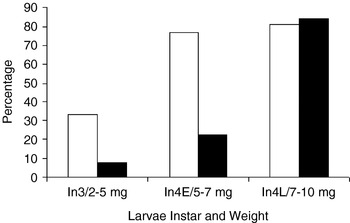
The inhibition of the hemolymph melanisation of the parasitized DBM larvae from non-parasitized ones was further tested. The difference between the non-parasitized and parasitized was compared according to their body weight (fig. 1). The results showed that differences in melanization between parasitized and non-parasitized larvae were significant in 3rd instar with an average body weight of 2–5 mg (In3, χ2=4.55, p⩽0.005; fig. 1) and early 4th instar with an average body weight of 5–7 mg (In4E, χ2=9.81, p⩽0.001; fig. 1). In contrast, the differences in colour change in the later 4th instar larvae with a body weight of 7–10 mg were not significant (In4L, χ2=0.03, p⩽1; fig. 1). There may be several reasons for this; melanization in later instars may not be suppressed by some parasitoids or may be suppressed but not to the low levels that preclude initiation of melanization in the presence of oxygen. Our results indicate that hemolymph melanization in the presence of DOPA and M. lysodeikticus could be used to distinguish parasitized 3rd and early 4th instar DBM larvae from non-parasitized ones.
Inhibition of melanization after parasitization. Hemolymph melanization of parasitized and non-parasitized DBM were tested in presence of DOPA and M. lysodeikticus solutions. Third to 4th instar DBM larvae (about 20 in each group) were used, the body weight of each larva was measured and melanization was tested. The percentage of melanization and its relationship with body weight was compared. Results showed that 3rd and early 4th instar larvae showed significant differences between parasitized and non-parasitized larvae. NP, non-parasitized larvae; P, parasitized larvae; In3/2–5 mg, 3rd instar larvae with a body weight of 2–5 mg; In4E/5–7 mg, 4th instar larvae with a body weight of 5–7 mg; In4L/7–10 mg, later 4th instar larvae with a body weight of 7–10 mg (□, % NP; ■, % P).

A blackening was observed in both parasitized and non-parasitized larvae within 30–60 min of when the epidermis was placed into DOPA solutions (data not shown). This suggests that melanization pathways in the hemolymph were inhibited, whereas activity in the cuticle was not affected, which is consistent with the observation of Shelby et al. (Reference Shelby, Adeyeye, Okot-Kotber and Webb2000). These authors showed that the activities of dopamine N-acetyltransferase (DAT) and plasma quinone methide isomerase, involved in cuticular sclerotin formation (Saul and Sugumaran, Reference Saul and Sugumaran1990), were not affected by parasitism (Shelby et al., Reference Shelby, Adeyeye, Okot-Kotber and Webb2000).
Inhibition of coagulation reaction
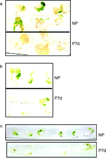
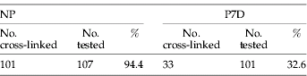
PO is also involved in coagulation reactions together with other abundant storage proteins, such as the lipid carriers lipophorin (Ma et al., Reference Ma, Hay, Li, Asgari and Schmidt2006; Rahman et al., Reference Rahman, Ma, Roberts and Schmidt2006) and hexamerin (Li et al., Reference Li, Scherfer, Korayem, Zhao, Schmidt and Theopold2002; Theopold et al., Reference Theopold, Li, Fabbri, Scherfer and Schmidt2002; Scherfer et al., Reference Scherfer, Karlsson, Loseva, Bidla, Goto, Havemann, Dushay and Theopold2004; Theopold et al., Reference Theopold, Schmidt, Soderhall and Dushay2004). We observed a green signal on the membrane from the remains of non-parasitized DBM larvae (fig. 2a; NP), but not from larvae 7-days post-parasitization when the membranes were washed with TBST overnight (fig. 2a; P7d). It appears that the coagulation reactions include debris from the gut contents that are trapped in the coagulum and, thus, become attached to the membrane. Further tests showed that a difference between the parasitized and non-parasitized larvae could be observed after 30 min washing with TBST (fig. 2b; NP vs P7d) and TBS buffers (fig. 2c; NP vs P7d). The percentage of coagulum-membrane binding in the non-parasitized larvae and parasitized larvae were calculated by using approximately 100 parasitized and naïve larvae each. Results showed that approximately 94% of non-parasitized larvae produced a food-containing coagulum, while 30% of larvae at 7-days post parasitization produced low levels of food-containing coagulation products (table 1). χ2-test showed that the difference is significant (χ2=86.35, p⩽0.001).
Inhibition of coagulation after parasitization. Whole larvae, both parasitized and non-parasitized, were squashed onto nitrocellulose membranes, and the membranes were incubated with the MAb 9A5 antibody. No specific reaction was detected in proteins from parasitized or non-parasitized larvae (data not shown). However, strong green signals (due to chlorophyll-containing coalgulum being bound to the membrane) were mainly observed in the non-parasitized larvae (a). Therefore, to confirm the difference between the non-parasitized and parasitized larvae, individual larva was squashed onto membrane, and washed with different buffers at different times (data not shown). It turned out that membranes washed with TBST (b) and TBS (c) buffer for 30 min produced a clear difference between the parasitized and non-parasitized larvae. NP, non-parasitized larvae; P7d, 7 days post parasitization.

Percentage of cross linking in non-parasitized and parasitized DBM larvae
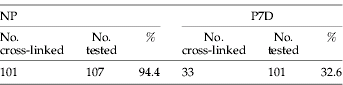
107 NP and 101 P7d larvae were squashed individually onto nitrocellulose membrane and let to air dry. Then, the membrane was washed in TBS buffer for 30 min. The green dots left on the membrane were counted, and the percentage in NP and P7d were calculated. The difference between the NP and P was tested statistically. The χ2 is equal to 86.35 and p is ⩽0.001; therefore, the difference between the NP and P is significant. NP, non-parasitized; P7d, parasitized for 7 days.
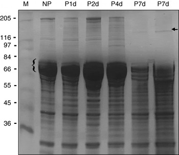
Parasitoids that develop in the hemocoel of their hosts are able to suppress the host's immune system, inducing a decline in the responsiveness to microbial elicitors of a variety of cellular and humoral components (Carton and Nappi, Reference Carton and Nappi1997; Shelby and Webb, Reference Shelby and Webb1999; Bae and Kim, Reference Bae and Kim2004). The situation in the D. semiclausum/P. xylostella system was explored. Hemolymph proteins from the non-parasitized and parasitized (day 1, 3, 4 and 7 of post-parasitization) larvae were subjected to SDS-PAGE and the electrophoresed proteins were visualized by Coomassie Brilliant Blue. This analysis showed that proteins about the 70–80 kDa range are abundant and a series of high molecular weight proteins ranging from 100–250 kDa were present in the non-parasitized and parasitized (1–4 days) larvae (fig. 3). In hemolymph samples of larvae 7 days after parasitization (P7d), proteins ranging from 70–80 kDa were dramatically reduced, and a series of high molecular weight proteins disappeared leaving only weak bands (fig. 3; arrow). Several groups demonstrated that parasitism induced hemolymph protein production in various parasitoid/host systems, including the Cortesia congregata/Pierís brassicae systems (Beckage et al., Reference Beckage, Templeton, Nielsen, Cook and Stolta1987; Rolle & Lawrence, Reference Rolle and Lawrence1994; Ockroy et al., Reference Ockroy, Cole Webb, Trenczek and Dorn2002). Bae and Kim (Reference Bae and Kim2004) reported the reduction of total hemolymph protein, especially an 80 kDa protein in P. xylostella larvae after parasitized by Cotesia plutellae. Our results also showed that the 70–80 kDa proteins, which are involved in coagulation (Li et al., Reference Li, Scherfer, Korayem, Zhao, Schmidt and Theopold2002; Theopold et al., Reference Theopold, Li, Fabbri, Scherfer and Schmidt2002; Scherfer et al., Reference Scherfer, Karlsson, Loseva, Bidla, Goto, Havemann, Dushay and Theopold2004; Theopold et al., Reference Theopold, Schmidt, Soderhall and Dushay2004) are reduced in parasitized larvae. The disappearance of these proteins in P7d larvae might suggest that their coagulation capacity was inhibited, which provides further support to our previous results (fig. 2; P7d) and the paper of Bae and Kim (Reference Bae and Kim2004).
Effect of parasitisation on insect protein components. 10% SDS-PAGE analysis of hemolymph proteins in 4th instar non-parasitized (NP) and DBM larvae at different stages of post-parasitisation (P1d–P7d) was conducted. The hemolymph proteins from individual larva were electrophoresed and the different pattern of each larva was visualized by Coomassie Blue staining. Subsequently, pooled samples of ten larvae were subjected to gel electrophoresis to confirm the different protein patterns. Abundant proteins at molecular weight ranged from 70–80 kDa are present in the non-parasitized (NP) and larvae 1–4 days post-parasitisation (P1d–P4d), but were reduced in the 7 day post-parasitized larvae (P7d). The molecular weight marker is showed in kDa (M), which contained 205 kDa Myosin, 116 kDa β-Galactosidase, 97 kDa phosphorylase, 84 kDa Fructose-6-phosphate kinase, 66 kDa Albumin, 55 kDa Glutamic Dehydrogenase, 45 kDa Ovalbumin, 36 kDa Glyceraldehyde-3-phosphate Dehydrogenase.

Based on the differences in food-containing coagulum bound to membranes and the protein pattern in the PAGE gel, we assume that the green signal produced by non-parasitized larvae is due to the bound coagulum proteins from the hemolymph of non-parasitized larvae, which have trapped chlorophyll-containing gut debris. The coagulation reaction was inhibited after larvae were parasitized for 7 days with less protein complexes formed in these larvae, and less chlorophyll-containing coagulum bound on membranes. Therefore, we can use the signal produced by bound coagulum to identify the parasitism status of larvae in the field.
Reaction of antibody (MAb 9A5) with Diadegma and Plutella proteins
A monoclonal antibody (MAb 9A5) that recognizes the larval and adult stages of M. croceipes and C. marginiventris (Hymenoptera: Braconidae), which are endoparasitoids of the polyphagous pests H. virescens (F.) and Helicoverpa zea (Boddie) (Lepidoptera: Noctuidae), was utilized (Stuart and Greenstone, Reference Stuart and Greenstone1997) for distinguishing parasitized from non-parasitized larvae. The antibody was specific against the two parasitoids in both ELISA and Western blot analysis, but did not react with proteins from the respective lepidopteran hosts (Stuart and Greenstone, Reference Stuart and Greenstone1997). The broad reactivity of MAb 9A5 for Hymenoptera suggested the potential of detecting other parasitoids.
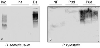
Parasitoid larvae were dissected from DBM host larvae and Western blots were probed with 10 μg ml−1 MAb 9A5 antibody (gift from M.K. Stuart, Kirksville College of Osteopathic Medicine, Kirksville, Missouri) and secondary anti-mouse IgM antibody (Sigma). Our results showed MAb 9A5 reacted with several proteins from 2nd instar (fig. 4a; In2) and adult D. semiclausum (fig. 4a; Ds) but not with protein extracts from the 1st instar larva (fig. 4a; In1).
Probing of parasitoid and host proteins with anti-parasitoid antibody (MAb 9A5). 15% SDS-PAGE gels were used to separate total proteins from 10 μl samples containing 50% of the 1st, 2nd instar and adult D. semiclausum homogenates (a) and 2% of individual non-parasitized (NP) and 3-days (P3d) or 6-days (P6d) post-parasitized DBM larval homogenates (b). One 2nd instar, four 1st instar larvae and one adult D. semiclausum were used (a) while three each DBM larvae of non-parasitized, P3d and P7d were tested (b), respectively. All proteins were probed with MAb 9A5 antibody, which bound to proteins in the 2nd instar, adult D. semiclausum (a: In2, Ds) and P6d host larvae (b: P6d). (a) In2, 2nd instar of D. semiclausum; In1, 1st instar of D. semiclausum; and Ds, adult of D. semiclausum. (b) NP, non-parasitized DBM larvae; P3d, 3-days post-parasitization; and P6d, 6-days post-parasitization.

Although MAb 9A5 reacts with multiple proteins (or glycoproteins), this does not exclude the possibility that the antibody reacts with a single antigen on multigene or post-transcriptionally modified gene products. Since the antibody reacts exclusively with proteins from parasitoids and not with proteins from noctuids examined (Stuart and Greenstone, Reference Stuart and Greenstone1997; Stuart, Reference Stuart1998), it may target a hymenopteran-specific antigen. The specificity of the antibody in the Diadegma/Plutella system was tested by homogenizing a single parasitized and non-parasitized DBM larva, separated in SDS-PAGE and probed with MAb 9A5. Multiple bands were detected in the DBM larvae 6 days after parasitization (fig. 4b; P6d) but not with the DBM larvae 3 days after parasitization (fig. 4b; P3d) and non-parasitized larvae (fig. 4b; NP).
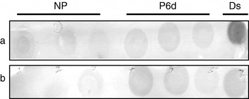
Trowell et al. (Reference Trowell, Forrester, Garsia, Lang, Bird, Hill, Skerritt and Joanne2000) reported that intractable endogenous phosphatases may confound the detection when using phosphatase-conjugated secondary antibody. To avoid the cross-interaction of the endogenous enzyme, phosphatase- and peroxidase-conjugated secondary anti-mouse IgM were used in dot blots. A substrate, for developing the membrane in peroxidase-conjugated secondary antibody, was used at pH 7.2 in which endogenous peroxidase, particularly in larvae, displayed little or no activity at neutral pH. In the dot blot, using the phosphatase-conjugated secondary antibody (fig. 5a), a very strong signal was observed in the Diadegma adult and slightly stronger signals were observed in the P6d than in the NP. In contrast, the dot blot with the peroxidise-conjugated IgM secondary antibody (fig. 5b) showed nearly nothing in the NP and moderate signals in the P6d and Diadegma. These results indicated that the peroxidise-conjugated IgM should be used in the dot blot, since it gave more distinguishable signals between the NP and P6d larvae in the dot blot tests.
Western dot blots of DBM homogenate using anti-parasitoid antibody (MAb 9A5) and two different conjugated secondary antibodies. Individual DBM larva was homogenized into 400 μl SDS-PAGE loading buffer and 5 μl of each was dotted into nitrocellulose membrane. The membrane was incubated with MAb 9A5 and then with alkaline phosphatase- (a) and peroxidase- (b) conjugated secondary antibodies, respectively. Proteins from three non-parasitized (NP), three 6-days post-parasitized larvae (P6d) and one D. semiclausum adult (Ds) were used in the dot blots. The strongest signal was detected in the D. semiclausum adult with phosphate-conjugated secondary antibody (a: Ds). Stronger signals were also detected in the parasitized larvae (P6d) compared with the non-parasitized larvae (NP).

Attempts were made to make the tests easier; dot blots containing proteins from single squashed parasitized and non-parasitized larvae were conducted. Unfortunately, under these conditions, signals were detected in both the parasitized and non-parasitized larvae (data not shown), which may be due to the high protein concentration on the blot that non-specific binding from MAb 9A5 produced in the non-parasitized larvae. Therefore, the MAb 9A5 antibody can only be used to detect parasitism of DBM in dot blots of protein extracts, but not when the whole larva was squashed onto the membrane.
Here we demonstrate that the inhibition of the melanization and coagulation pathways in parasitized DBM larvae by D. semiclasum provides a basis for simple and relatively reliable determination of parasitism rates. There are several advantages in the methods: (i) quick and easy to perform; (ii) no specific skills needed; and (iii) no expensive equipments required. In contrast, the method of rearing DBM larvae until the emergence of parasitoids is a slow process and needs at least a number of days to obtain the results; and the microscopic dissection needs microscope and specific skills to perform. Thus, the melanization of hemolymph and protein cross-linking in DBM can be used to distinguish parasitized from non-parasitized larvae in the field.
In conclusion, several methods have been developed to detect the parasitism of DBM. The inhibition of the hemolymph melanization and coagulation provides easy and rapid diagnosis which can be used in the field. The monoclonal antibody is more specific, but takes longer and has to be performed in the laboratory.
Acknowledgements
We thank Dr Melissa K. Stuart (Kirksville College of Osteopathic Medicine, Kirksville, Missouri) for providing us with MAb 9A5 antibody, Dr Richard Glatz for reading the manuscript and some valuable suggestions, and Dr Garry Hannan (CSIRO Molecular and Health technologies) in helping with the statistical analysis of the data. We would like to thank Ausveg and Horticulture Australia Limited for its funding and the two reviewers for their valuable suggestions.