Introduction
Chronic otitis media with recurrent cholesteatoma is common in otological surgery. Recurrence after initial surgery may be localised in the tympanic cavity only, but can also spread to the mastoid, or involve other highly sensitive structures in the temporal bone.Reference Yung, Tono, Olszewska, Yamamoto, Sudhoff and Sakagami1,Reference Kuo, Shiao, Yung, Sakagami, Sudhoff and Wang2 Otoscopy in the setting of recurrent disease may yield useful information regarding recurrence extent and localisation, but cannot accurately address deeper areas of retraction pockets or perifacial or mastoid disease.Reference Schwarz, Gostian, Shabli, Wolber, Hüttenbrink and Anagiotos3 High-resolution computed tomography (CT) of the temporal bone could be vital in assessing disease and key anatomical characteristics pre-operatively.Reference Swartz4,Reference Stasolla, Magliulo, Cortese, Roncacci and Marini5
Revision surgery is concerned with removing recurrent disease while dealing with other non-affected structures as sparingly as possible.Reference Nadol6 Published literature has focused on the outcomes of two types of surgical procedures most commonly used in revision surgery: canal wall down and canal wall up tympanomastoidectomy. Factors leading to post-operative failure after initial surgery are: inadequate meatoplasty, leaving a high facial ridge in canal wall down procedures; failure to address the mastoid apex cells; post-operative granulation; and mismanagement of high-risk areas, such as the facial recess, posterior wall of the ear canal and sinus tympani.Reference Nadol6,Reference Veldman and Braunius7
Identifying and removing recurrent disease is central in achieving long-term disease control. To date, very few papers have analysed possible correlations between intra-operative findings in revision surgery and pre-operative imaging as predictors of cholesteatoma recurrence or poor surgical outcome, with most of the literature focusing on post-operative imaging.Reference Stasolla, Magliulo, Cortese, Roncacci and Marini5,Reference Yu, See, Ng, Low, Tan and Yuen8–Reference Coralles and Blevins10 In planning for revision surgery, the decision-making process is often based on prior surgical experience, and often depends entirely on the findings encountered upon executing the revision procedure.Reference Veldman and Braunius7,Reference Kasenõmm11–Reference Faramarzi, Motasaddi-Zarandy and Khorsandi13 There is clear clinical value in improving pre-operative planning to address possible disease recurrence patterns, especially to gain information about bone erosion, the integrity of the lateral semi-circular canal and tegmen, and sigmoid plate if the mastoid was drilled previously.
This study analysed the correlations between pre-operative CT imaging, intra-operative details of revision tympanomastoidectomy procedures and surgical outcomes. Describing disease recurrence patterns and determining the reliability of such findings on pre-operative scans could be of great assistance when making decisions regarding the timing, type and extent of revision surgery.
Materials and methods
A retrospective comparative cohort study was conducted comprising 42 patients surgically treated for recurrent chronic otitis media. The study was approved by the University Hospital Center Bioethical Board, adhering to the Helsinki Declaration Revision of 1989.
Inclusion criteria were met if: the patients underwent revision tympanomastoidectomy (either canal wall up or canal wall down), from 1 January 2010 to 1 May 2019, in our tertiary referral centre; the procedure was performed by the same otological team of three surgeons; and complete intra-operative data and pre-operative CT scans were available for analysis. The radiologist was blinded to the data regarding previous surgery. The CT scans were performed in all patients within the two weeks before surgery, and were performed as a routine surgical investigation in patients with active chronic otitis media, even if cholesteatoma recurrence was not immediately suspected. Recurrent cholesteatoma was diagnosed if the pre-operative CT scan showed the presence of a soft tissue mass with bony erosion in the middle-ear space, especially if auditory ossicle erosion was observed. The indication for surgery was continuing otorrhoea and/or evident disease recurrence after previous surgery. The patients’ demographic data, type and instances of previous surgery, intra-operative findings, and CT findings were entered into a comprehensive database. Informed consent was obtained from all patients.
Exclusion criteria were: insufficient intra-operative details, where the analysis of localisations could not be assessed; and patients without pre-operative CT scans available for analysis.
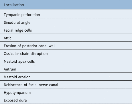
In the 42 patients included in the study, 12 disease recurrence localisations were identified in the revision surgery notes, and these were then correlated with the corresponding CT sites (Table 1).
Localisations of disease recurrence*

* Identified on pre-operative computed tomography scans and during revision surgery
The CT scans were multi-slice, with collimation and 0.5 mm thickness. The scans were analysed by the same neuroradiologist, a member of the otology multidisciplinary team, who was tasked with commenting on the same 12 intra-operative sites noted during surgery.
Two binary outcomes relating to revision surgery were identified: the presence or absence of cholesteatoma, and whether a canal wall up or canal wall down procedure had been performed.
In order to extrapolate the impact of performing CT prior to revision surgery on secondary post-operative outcomes (cholesteatoma recurrence, type of surgery, number of previous surgical procedures and time elapsed from previous surgery), a matched pair analysis was performed. Each patient in the revision surgery group who had pre-operative CT was matched with a patient who had revision surgery performed by the same otologist but without a pre-operative CT examination. The patients were paired to have identical or similar intra-operative findings. This created a subset of 84 participants (42 matched pairs), who were included in the secondary analysis. The presence of pre-operative CT findings was designated as the dependent variable.
Statistical analysis was performed using SPSS Statistics for Windows software, version 22.0 (IBM, Armonk, New York, USA), using standard descriptive statistics and frequency tabulation as indicated. Associations between variables were assessed using a binary logistic regression model with odds ratios, the Kruskal–Wallis test for non-parametric independent samples, Spearman's correlation co-efficient ρ, and principal component analysis using direct oblimin rotation. All tests were performed using a two-sided 5 per cent type I error rate.
Results
The average age of patients was 34.1 years, ranging from 7 to 80 years. There were 21 males and 21 females. Of the total 42 patients, 8 had undergone only one previous surgery for chronic otitis media. Thirty-three patients had undergone multiple surgical procedures, with an average number of 2.9 surgical procedures per patient, with an average time since the previous surgery of nine years. All patients were initially treated for cholesteatoma. Of the 42 patients, 10 had been previously surgically treated in our institution.
Revision procedures encompassed 28 canal wall up and 14 canal wall down tympanomastoidectomy procedures. In the canal wall down revision procedure group, seven patients had previously undergone a canal wall up procedure. Seven patients had extensive disease and had already been treated with a canal wall down procedure prior to revision surgery.
Pre-operative CT identified 25 cholesteatoma recurrences out of 31 cases observed during revision surgery. Thus, the sensitivity rate was 80.65 per cent, with a specificity rate of 81.82 per cent, a positive predictive value of 92.59 per cent and a negative predictive value of 60 per cent (Table 2).
Distribution of revision tympanomastoidectomy patients with cholesteatoma recurrence

CT = computed tomography; CWU = canal wall up; CWD = canal wall down
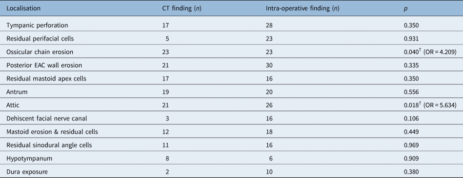
The binary logistic regression model identified a high level of correlation between pre-operative CT diagnosis of likely cholesteatoma and intra-operative findings (p = 0.001, odds ratio = 13.25). Among 12 localisations analysed on pre-operative CT scans, disease presence in the attic (p = 0.018, odds ratio = 5.634) and in the ossicular chain (p = 0.040, odds ratio = 4.209) were identified as significant positive predictors of cholesteatoma. Male patients also had a significantly higher risk for cholesteatoma recurrence (p = 0.018, odds ratio 5.634) (Table 3).
Association between pre-operative CT findings and intra-operative recurrent cholesteatoma findings*

* Analysis includes 12 intra-operatively noted localisations identified as significant in the binary logistic regression model (p = 0.001, odds ratio = 13.25). †Statistically significant association. CT = computed tomography; OR = odds ratio; EAC = external auditory canal
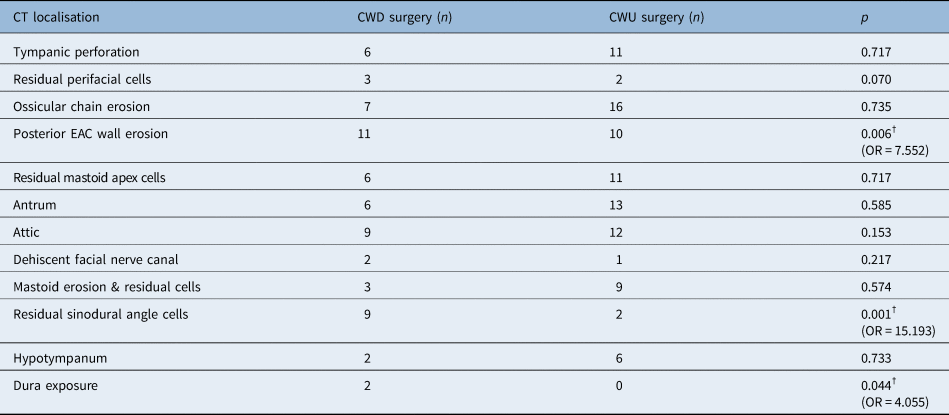
Cholesteatoma identified on CT scans was associated with a significantly higher likelihood of performing canal wall down tympanomastoidectomy (p = 0.033, odds ratio = 4.557). Positive predictors for performing canal wall down surgery were: disease in the sinodural angle (p = 0.001, odds ratio = 15.193), erosion of the posterior canal wall (p = 0.006, odds ratio 7.552) and exposed dura (p = 0.044, odds ratio = 4.055) (Table 4). The likelihood of performing canal wall down surgery was also higher in male patients (p = 0.037, odds ratio = 4.364).
Association between pre-operative CT findings and extent of revision surgery*

* Analysis includes 12 intra-operatively noted localisations identified as significant in the binary logistic regression model (p = 0.033, odds ratio = 4.557). †Statistically significant association. CT = computed tomography; CWD = canal wall down; CWU = canal wall up; EAC = external auditory canal; OR = odds ratio
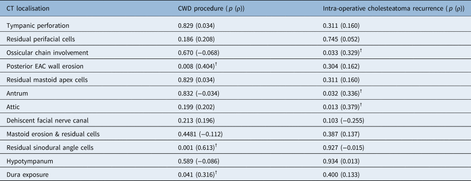
Spearman's correlation identified a significant positive correlation between CT scans and intra-operative findings of cholesteatoma recurrence (ρ = 0.573, p = 0.001). A likely cholesteatoma on pre-operative CT scans correlated well with disease located in the attic (ρ = 0.379, p = 0.013), in the antrum (ρ = 0.336, p = 0.032) and affecting the ossicular chain (ρ = 0.329, p = 0.033). In addition, pre-operative CT scans correlated well with the likelihood of performing canal wall down surgery (ρ = 0.316, p = 0.041) when there was: disease found in the sinodural angle (ρ = 0.613, p = 0.001), posterior canal wall erosion (ρ = 0.404, p = 0.008) and exposed dura (ρ = 0.316, p = 0.041) (Table 5).
Spearman's correlation co-efficient values*

* Demonstrates positive correlations between computed tomography scans and: the likelihood of performing a canal wall down tympanomastoidectomy procedure (ρ = 0.316, p = 0.041) and cholesteatoma recurrence (ρ = 0.573, p = 0.001). †Statistically significant correlation. CT = computed tomography; CWD = canal wall down; EAC = external auditory canal
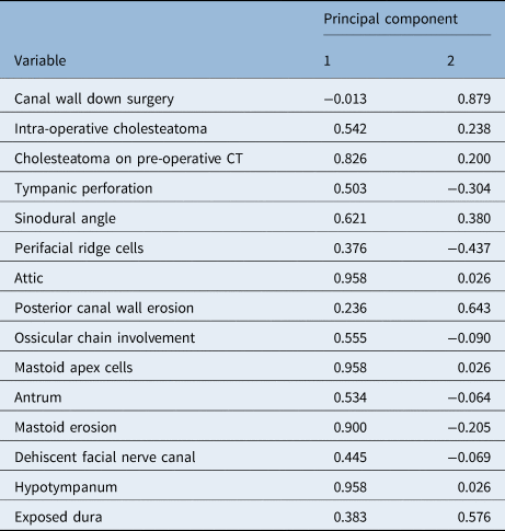
Principal component analysis was used to confirm the results of the binary logistic regression model and address possible confounds associated with the large number of variables. The results identified three primary components in our data set, with eigenvalue greater than 5 (Figure 1). Covariance co-efficient values in the first component encompassed most of the total variance, and showed significant correlation co-efficients for disease located in the attic (principal component 1 value = 0.958), mastoid apex (0.958) and hypotympanum (0.958), and mastoid erosion (0.900) on the pre-operative CT scan. The second principal component showed significant correlation co-efficients for performing canal wall down surgery, and posterior canal wall erosion (principal component 2 value = 0.643) and exposed dura (0.576) observed on the pre-operative CT scan (Table 6).
Principal component analysis identified three primary components in our data set, represented on the scree plot with eigenvalue greater than 5, with the first three components encompassing 67.6 per cent of total variance.

Principal component analysis*

* Describes a correlation matrix for the first two components, which encompassed 50.8 per cent of the total variance. Variables include disease localisation noted on pre-operative computed tomography scans, extent of surgery and intra-operative presence of cholesteatoma. CT = computed tomography
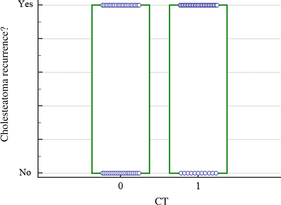
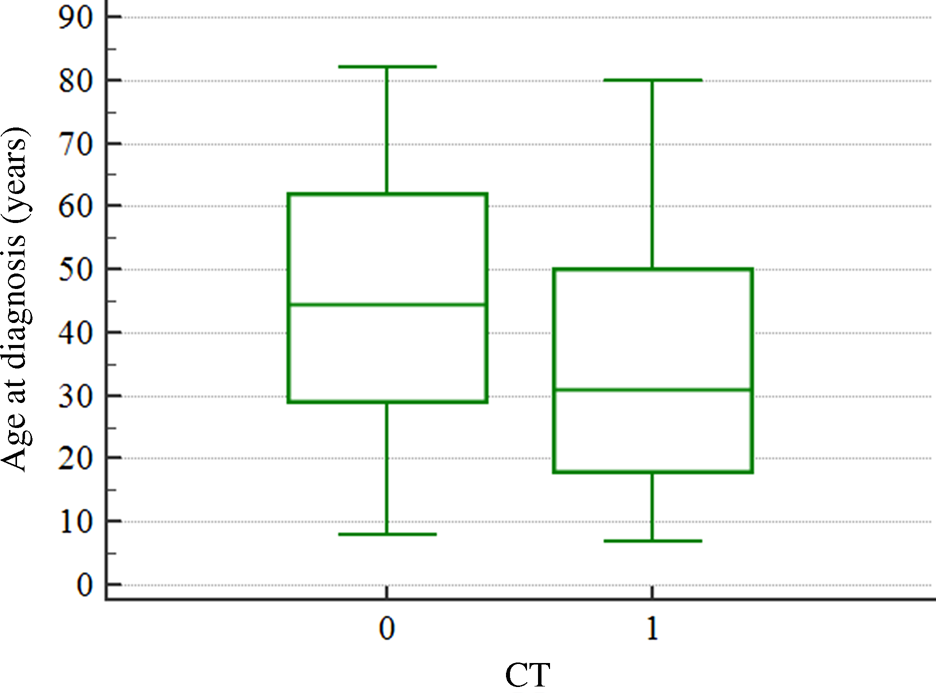
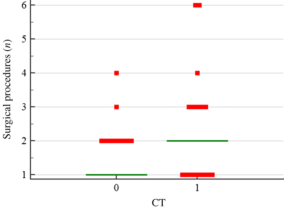
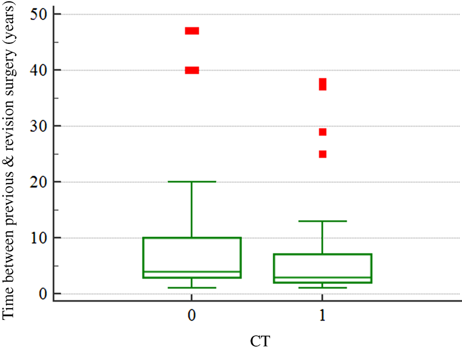
The matched pair analysis using the Kruskal–Wallis test showed that patients with pre-operative CT scans had a statistically: higher rate of cholesteatoma recurrence diagnosed pre-operatively (p = 0.043; Figure 2), younger age at diagnosis of recurrent disease (p = 0.044; Figure 3), higher number of revision surgical procedures (p = 0.0001; Figure 4), and shorter period of time between previous and revision surgical procedures (p = 0.043; Figure 5). There was no significant difference between type of surgery (canal wall up or canal wall down) and presence or absence of pre-operative CT scanning (p = 0.267).
Matched pair analysis showed that patients with pre-operative computed tomography (CT) scans had a statistically higher rate of cholesteatoma recurrence diagnosed pre-operatively (p = 0.043, Kruskal–Wallis test). 0 = no pre-operative CT scan; 1 = pre-operative CT scan available

Matched pair analysis showed that patients with pre-operative computed tomography (CT) scans had a statistically younger age at diagnosis of recurrent disease (p = 0.044, Kruskal–Wallis test). 0 = no pre-operative CT scan; 1 = pre-operative CT scan available

Matched pair analysis showed that patients with pre-operative computed tomography (CT) scans had a statistically higher number of revision surgical procedures (p = 0.0001, Kruskal–Wallis test). 0 = no pre-operative CT scan; 1 = pre-operative CT scan available

Matched pair analysis showed that patients with pre-operative computed tomography (CT) scans had a statistically shorter period of time between previous and revision surgical procedures (p = 0.043, Kruskal–Wallis test). 0 = no pre-operative CT scan; 1 = pre-operative CT scan available

Discussion
Revision surgery for chronic otitis media ideally results in a manageable and dry middle-ear cavity, but it is often plagued with high failure rates, with cholesteatoma recurrences found in up to 49 per cent of patients after previous cholesteatoma surgery.Reference Nadol6,Reference Veldman and Braunius7 Recurrent disease is commonly identified when initial surgery fails to address tegmental, perifacial, mastoid apex and sinodural mastoid cells.Reference Kasenõmm11,Reference Košec, Kelava, Ajduk, Ries, Trotic and Bedekovic12 However, clinical follow up using otoendoscopy and presenting symptoms may not identify a significant portion of patients with occult disease recurrence, especially after previous canal wall up surgery.Reference Yu, See, Ng, Low, Tan and Yuen8,Reference Campos, Mata, Reboll, Peris and Basterra9
Pre-operative CT imaging is a first-line option in middle-ear imaging because of its excellent spatial resolution and delineation of vital landmarks.Reference Campos, Mata, Reboll, Peris and Basterra9,Reference Coralles and Blevins10,Reference Gul, Akdag, Kinis, Yilmaz, Sengul and Teke14 It is especially useful in cases where the initial surgery was performed elsewhere, if complications are present, or when recurring disease cannot be properly assessed using otoendoscopy alone.Reference Coralles and Blevins10 It is used to predict the extension of the disease into the sinus tympani and the attic, with antral and peri-antral involvement.Reference Campos, Mata, Reboll, Peris and Basterra9 However, when interpreting CT scans, the inability to discriminate between cholesteatoma, granulation, fibrosis and thick mucus secretion in the middle ear must be taken into account; one should consider performing magnetic resonance imaging (MRI) as well, especially in the paediatric population.Reference Campos, Mata, Reboll, Peris and Basterra9,Reference Romano, Covelli, Confaloni, Rossi-Espagnet, Butera and Barbara15 Recurrent middle-ear cholesteatoma evaluated by MRI also represents a diagnostic challenge because of the small size of these tissues, the different kinds of surgical procedures, and the non-specific signal intensity characteristics on imaging. The presence of erosion caused by soft tissue on nearby bone structures is generally interpreted as indicating the presence of cholesteatoma.Reference Gul, Akdag, Kinis, Yilmaz, Sengul and Teke14,Reference Romano, Covelli, Confaloni, Rossi-Espagnet, Butera and Barbara15
Previously published studies have been concerned with comparing pre- and post-operative CT and MRI scans in relation to initial surgery for chronic otitis media. However, very few studies have addressed the much more complicated issue of whether pre-operative imaging is useful in revision surgery. While MRI is still widely regarded as the most accurate imaging method for assessing cholesteatoma, high-resolution CT may represent an alternative imaging tool in patients with contraindications for MRI or in practices where pre-operative MRI is not routinely performed. At the cost of a low radiation burden, CT represents a cheaper and faster examination, with the additional advantage of providing high-resolution anatomical details.Reference Lingam and Bassett16,Reference Khemani, Singh, Lingam and Kalan17 Advancing imaging protocols may further minimise radiation, and, in some instances, cone beam CT may be useful. However, cone beam CT may not capture areas of interest outside the field of view (beyond 4 × 4 cm classically).Reference Stutzki, Jahns, Mandapathil, Diogo, Werner and Güldner18 Cone beam CT may aid the surgeon's pre-operative plan and discussion of treatment options, which may be different from decisions made based on intra-operative findings or pre-operative imaging. In revision cases, the possibility of intra-operative complications and plan changes is higher, and the surgeon may be required to widen the extent of surgery beyond the original plan.
Several studies have assessed the value of diffusion-weighted MRI and concluded that a high signal on diffusion-weighted images does not always indicate the presence of cholesteatoma, with false-positive cases found in the post-operative period due to artefacts, bone dust, cartilage, tympanosclerosis, granulation tissue, proteinaceous fluid, purulent content, cholesterol granuloma or wax in the adjacent external auditory canal.Reference Romano, Covelli, Confaloni, Rossi-Espagnet, Butera and Barbara15 A recent meta-analysis of 1152 patients (both pre- and post-surgical cases) showed that the use of diffusion-weighted images alone led to 3.4 per cent of false-positive cases, with a lower specificity for post-operative controls.Reference Lingam and Bassett16
The principal question analysed in this study was whether pre-operative CT scans may present a useful tool in identifying localisations with high risk for recurrent disease, and in planning for revision surgery. The results confirm our hypothesis that CT can reliably identify recurrent disease. In the current study, CT identified 25 out of 31 cholesteatoma recurrences, with high sensitivity (80.65 per cent) and a high positive predictive value (92.59 per cent). This value is much higher than some of the results in the literature, which cite sensitivity and specificity as low as 42 per cent and 48 per cent, respectively.Reference Khemani, Singh, Lingam and Kalan17,Reference Songu, Altay, Onal, Arslanoglu, Balci and Ucar19,Reference Alzahrani, Alhazmi, Bélair and Saliba20 Pre-operative CT scans revealed correlations between the presence of cholesteatoma and attic disease, ossicular chain involvement, sinodural angle cells, opacified facial ridge cells and an obstructed antrum. In addition, the likelihood of performing canal wall down tympanomastoidectomy was higher in patients presenting with disease in the sinodural angle, erosion of the posterior canal wall and exposed dura on the pre-operative CT scan, which was further confirmed by principal component analysis results.
• Recurrence after initial surgery for chronic otitis media may be difficult to identify in the tympanic cavity and mastoid prior to revision surgery
• High-resolution computed tomography (CT) of the temporal bone may be vital in assessing disease and key anatomical characteristics pre-operatively
• This study analysed correlations between pre-operative CT imaging and intra-operative revision tympanomastoidectomy details
• Describing disease recurrence patterns and determining the reliability of such findings on pre-operative scans may aid decisions regarding type and extent of revision surgery
• Pre-operative CT identified cholesteatoma recurrences with 80.65 per cent sensitivity and 81.82 per cent specificity
• Pre-operative CT increased accuracy of diagnosing recurrent cholesteatoma, reduced time required to diagnose, and lowered age at diagnosis and revision surgery
Male sex was linked to a higher risk of cholesteatoma recurrence, which has been described earlier, and indicates that our sample is representative of the larger patient population.Reference Allam, Abdel Razek, Ashraf and Khalek21
The most common localisations of recurrent disease visible on pre-operative CT were tympanic perforation, ossicular chain involvement, posterior canal wall destruction, attic disease and antrum involvement. This correlated well with intra-operative findings (Table 3). These findings correspond to well-known disease recurrence patterns in published literature, confirming that failure to address these areas during initial or previous surgery may be a principal factor in disease recurrence.Reference Allam, Abdel Razek, Ashraf and Khalek21
The results of the matched pair analysis also indicate that performing pre-operative CT scans increases the rate of accurate diagnosis of recurrent cholesteatoma, reduces the time required to diagnose recurrent disease, and lowers the average age at diagnosis and corresponding revision surgery, without influencing the type of surgery. Analysis of correlations between pre-operative CT scans and intra-operative findings in revision surgery is clearly of value when planning the extent and type of surgery, and when determining which areas are high risk for subsequent close scrutiny, without introducing bias when choosing the type and extent of surgery required to eliminate recurrent disease.Reference Alzahrani, Alhazmi, Bélair and Saliba20
The habit of analysing pre-operative imaging must be a valuable tool in any surgeon's armamentarium, and should not be relegated exclusively to the radiologist. However, a capable surgeon should be able to navigate a temporal bone to ensure that the bone bed is clear and comprehensively drilled as part of any approach when removing cholesteatoma disease, irrespective of pre-operative scanning. The point of obtaining the CT scan prior to surgery is not to aid the surgeon in identifying areas in the ear where there is concern for recurrence. It might seem obvious that CT may help with surgical planning, but data supporting objective analysis of the CT scan in revision surgery are surprisingly scarce, and supporting its consistent use would help clinicians show value to third parties who may question the need for CT in revision cases. Knowledge of fine anatomical detail provides additional refinement in surgical technique, which is a decisive factor influencing post-operative outcomes.Reference Košec, Kelava, Ajduk, Ries, Trotic and Bedekovic12,Reference Faramarzi, Motasaddi-Zarandy and Khorsandi13 Recurrent cholesteatoma is associated with inaccurate pre- and intra-operative evaluation findings of the surgical field and a failure to address high-risk areas, putting patients at a higher risk of canal wall down procedures in revision surgery.Reference Košec, Kelava, Ajduk, Ries, Trotic and Bedekovic12 The correlations between CT scans and intra-operative findings identified in this study may be useful in follow up as well, as indicators of early recurrence, and for prompt timely surgical intervention in patients with otherwise occult disease.Reference Allam, Abdel Razek, Ashraf and Khalek21
When planning for surgery, it may be especially valuable to have in mind the significant correlations between disease involving the sinodural angle and posterior canal wall and the presence of exposed dura, as early indicators that a canal wall down procedure might be required (Table 5). Similar findings have been reported previously, with intra-operative findings identifying posterior external auditory canal wall erosion, mastoid erosion and residual cells along the facial ridge as reliable predictors of cholesteatoma recurrence, and associated with canal wall down procedures, but they have not been linked with pre-operative CT findings.Reference Kasenõmm11–Reference Faramarzi, Motasaddi-Zarandy and Khorsandi13,Reference Selwyn, Howard and Cuddihy22
Conclusion
Utilising readily available pre-operative CT imaging alongside a detailed assessment of possible cholesteatoma recurrence patterns by a radiologist may improve outcomes of revision surgery. Performing pre-operative CT scans increases the rate of accurate diagnosis of recurrent cholesteatoma, reduces the time required to diagnose recurrent disease, and lowers the average age at diagnosis and corresponding revision surgery. The CT will mostly accurately predict where to look for cholesteatoma recurrence, but will not accurately predict that there is no recurrence and therefore that the surgery is not indicated.
Competing interests
None declared