Introduction
Poultry are raised surrounded by pathogens that include viruses, fungi, bacteria, and parasites, and other stress factors that can affect the performance and health of birds. To cope with these pathogens and unhealthy conditions, birds have a well-defined immune system. Innate immunity senses these pathogens by their specific molecular structure and passes pathogen-specific signals to adoptive immune system for the initiation of signal dependent immune responses. The adaptive immune system is comprised of entities such as B cells which produce specific antibodies, CD4+ T cells for production of cytokines and immune regulation and CD8+ T cells to kill antigen infected cells. The major focus for this review is on vital cells of the innate immune system of poultry, the dendritic cells.
Dendritic cells were discovered by Canadian scientists Ralph Steinman and Zanvil Cohn in 1973 (Steinman and Cohn, Reference STEINMAN and COHN1973), who were subsequently awarded the Noble Prize in 2011. The first article that reported the avian dendritic cells from caecal tonsils was published by Olah and Glick (Reference OLAH and GLICK1979) and subsequently, three major subsets of dendritic cell were identified in chicken lymphoid organs and epidermis (Igyártó et al., Reference IGYÁRTÓ, LACKÓ, OLÁH and MAGYAR2006; Del Cacho et al., Reference DEL CACHO, GALLEGO, LILLEHOJ, LÓPEZ-BERNARD and SÁNCHEZ-ACEDO2009). Distinctive features of dendritic cells in immunity have now been reported after decades of scepticism. These are a diverse group of cells that are reknown for their antigen recognition roles. They are involved in other important immune functions like antigen presentation, initiation of adoptive immune responses, generation of immunological memory (Steinman, Reference STEINMAN1991).
Dendritic cells work as sentinels of the immune system and are present in every part of the body including skin, peripheral blood, mucosal surfaces, interstitial tissues, and in lymphoid and non-lymphoid tissue (Yasmin et al., Reference YASMIN, YEAP, TAN, HAIR-BEJO, FAKURAZI, KAISER and OMAR2015; Rehman et al., Reference REHMAN, MENG, UMAR, MUNIR and DING2016). Dendritic cells recognise the pathogens by pattern recognition receptors including toll like receptors (TLR), cytoplasmic nucleotide-binding oligomerisation domain (NOD) like receptors, retinoic acid-inducible gene I (RIG-I)-like receptors (RLRs) and C-type lectins, and initiate innate immune responses (Akira et al., Reference AKIRA, UEMATSU and TAKEUCHI2006). Pathogen activation causes many changes such as maturation, production of cytokines and the expression different co-stimulatory molecules (Banchereau and Steinman, Reference BANCHEREAU and STEINMAN1998; Liu and Nussenzweig, Reference LIU and NUSSENZWEIG2010).
In last decade, much work has been done on dendritic cell biology, function, and immune responses in avian species. Extensive reviews on dendritic cell phenotype and ontogeny (Nagy et al., Reference NAGY, BÓDI and OLÁH2016) and their regulation (De Geus and Vervelde, Reference DE GEUS and VERVELDE2013) have been published. However, review articles about immunological functions in poultry are scarce.
Biology of dendritic cells
Dendritic cells are derived from bone marrow derived haematopoietic stem cells precursors expressing leukocyte antigen CD45+. Diversified dendritic cells with specialised functions are formed through diverse ontogenic pathways, as reviewed by Merad et al. (Reference MERAD, SATHE, HELFT, MILLER and MORTHA2013). In a steady state these reside in blood and peripheral tissues. The phenotype of avian DCs is less understood because of a lack of specific markers but these can be cultured and activated by LPS. These activated DCs express CD11, C type lactin DEC205, activation marker of MHC II and CD40 (Wu et al., Reference WU, ROTHWELL, YOUNG, KAUFMAN, BUTTER and KAISER2010; De Geus et al., Reference DE GEUS, JANSEN and VERVELDE2012).
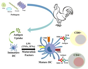
Immature dendritic cells work as immunological sensors and are activated directly by pathogens or other innate responses, signalling to the immune system to act against potentially harmful antigens ( Figure 1 ) (Ueno et al., Reference UENO, KLECHEVSKY, MORITA, ASPORD, CAO, MATSUI, DI PUCCHIO, CONNOLLY, FAY, PASCUAL, PALUCKA and BANCHEREAU2007). Acquired immunological response is highly dependent on the signal of these cells. Dendritic cells are activated by different type of signals, such as direct capture of antigens, pathogen invasion, innate immune cells, dying cells and microenvironment ( Figure 1 ). Immature DCs sense pathogen associated molecular patterns (PAMPs) through pattern recognition receptors (PRRs), including toll-like receptors (TLRs) cell surface C-type lectin receptors (CLRs), and intracytoplasmic nucleotide oligomerisation domain (NOD)-like receptors (NLRs) (Ueno et al., Reference UENO, KLECHEVSKY, MORITA, ASPORD, CAO, MATSUI, DI PUCCHIO, CONNOLLY, FAY, PASCUAL, PALUCKA and BANCHEREAU2007; Liang et al., Reference LIANG, YIN, QIN and YANG2015).
Dendritic cell maturation and activation of CD4+ and CD8+ T cells in response to pathogens. Schematic drawing depicting the differentiation of immature DC into mature DC by up-taking antigen. This process involved in morphological changes in mature DC as well as development of strong co-stimulatory molecules for the activation of T cells as well as pattern recognition sites on the surface of DC.

Adequate stimulation of the adaptive immune responses mainly depends on dendritic cell signals. Less scientific data is available about adaptive immune regulation in avian species compared to human and mice (Staines et al., Reference STAINES, YOUNG and BUTTER2013). Activated DCs express high level of the MHC I and MHC II which are recognised by the CD8+ and CD4+ T cells respectively. These cells are present at the antigen entry sites, from where DCs recognise them, then migrate to lymphoid organs and leads to the stimulation of naïve T cells (Liang et al., Reference LIANG, YIN, QIN and YANG2015). T cell maturation, in response of DCs signalling, is controlled by the microenvironment created by production of cytokine from DCs which direct activation toward Th1 or Th2 immune response immunity (Rissoan et al., Reference RISSOAN, SOUMELIS, KADOWAKI, GROUARD, BRIERE, DE WAAL MALEFYT and LIU1999).
DEC205 is a C-type lectin endocytic receptor of the mannose receptor family and is mostly known to involve antigen cross presentation (Cheong et al., Reference CHEONG, CHOI, VITALE, HE, TRUMPFHELLER, BOZZACCO, DO, NCHINDA, PARK and DANDAMUDI2010), recognition of apoptotic cells (Shrimpton et al., Reference SHRIMPTON, BUTLER, MOREL, EREN, HUE and RITTER2009) and enhances antigen presentation via MHC II+ve lysosomal compartments (Mahnke et al., Reference MAHNKE, GUO, LEE, SEPULVEDA, SWAIN, NUSSENZWEIG and STEINMAN2000). DEC205 was noted in spleen on the antigen trapping sites and thymus to educate T cells and also in bursa for diversification of B cells of chickens (Staines et al., Reference STAINES, YOUNG and BUTTER2013).
Antigen specific responses
response to viral diseases
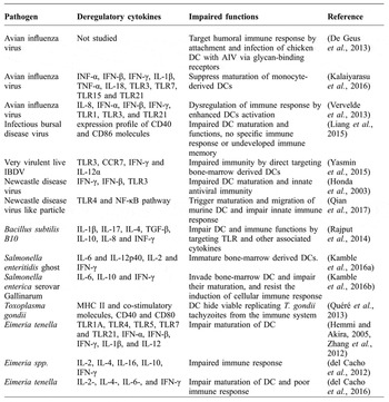
Dendritic cells have a undetermined role in viral infections, and viral interactions with dendritic cells have not been well understood in poultry (Yasmin et al., Reference YASMIN, YEAP, TAN, HAIR-BEJO, FAKURAZI, KAISER and OMAR2015). Many viral components can activate and promote the production of cytokines. The ability of DCs to escalate the recruitment of T cells, production of antibodies from B cells and signalling to other innate immune cells may help in production of strong cell mediated immune response and viral clearance from the infected tissue. However, virus and dendritic cell interactions do not always support the host and these may affect the maturation and functioning of dendritic cells. Highly pathogenic avian influenza (HPAI) compared to low pathogenic avian influenza (LPAI), increases the expression of IL-8, IFN-α, IFN-γ mRNA, viral RNA and in TLR1, 3, and 21 mRNA of dendritic cells which may lead to deregulation of immune response ( Table 1 ) (Vervelde et al., Reference VERVELDE, REEMERS, VAN HAARLEM, POST, CLAASSEN, REBEL and JANSEN2013).
Similarly, (Kalaiyarasu et al., Reference KALAIYARASU, KUMAR, SENTHIL KUMAR, BHATIA, DASH, BHAT, KHETAN and NAGARAJAN2016) found that monocyte derived DCs (chMoDCs) from chicken peripheral blood are the target of HPAI and cause the death of DCs, and this was greater for HPAI than LPAI. HPAI impair the activation of DCs and increase the expression of INFs and cytokines (IL-1β, IL-6, IL-18 and TNF-α factor) at the early phase of infection. However, (Rebel et al., Reference REBEL, PEETERS, FIJTEN, POST, CORNELISSEN and VERVELDE2011) found that chickens infected with LPAI or HPAI express the same level of viral RNA, cytokine mRNA and same influxes of KUL01+ macrophages and DCs, but the location of the virus varies in the lungs. Previously, Waithman and Mintern (Reference WAITHMAN and MINTERN2012) reported that the AI virus infection of human myeloid DCs inhibits maturation and impairs cross presentation of antigens via MHC1. AI virus infection in chicken DCs decreases the level of glycan-recognising antibodies, when measured by glycan array analysis ( Table 1 ) and there is an increasing trend in anti-glycan antibodies against specific glycan antigen which was observed by ELISA (De Geus et al., Reference DE GEUS, TEFSEN, VAN HAARLEM, VAN EDEN, VAN DIE and VERVELDE2013). Liang et al. (Reference LIANG, YIN, QIN and YANG2015) compared the effect of vaccine strain of inactive infectious bursal disease virus (IBDV) with live IBDV, and found that inactive IBDV have stronger capability to up-regulate expression of CD40 and CD86 molecules and stimulate naive T cells proliferation than live IBDV. This may be due to impairment of dendritic cells by IBDV (Liang et al., Reference LIANG, YIN, QIN and YANG2015). Infection of chBM-DCs with virulent live IBDV increases the expression of TLR3, Th1-like cytokines, IFN-γ and IL-12α, indicating they are the target of IBDV (Yasmin et al., Reference YASMIN, YEAP, TAN, HAIR-BEJO, FAKURAZI, KAISER and OMAR2015; Rehman et al., Reference REHMAN, MENG, UMAR, MUNIR and DING2016). Response of DCs to other poultry viruses is not clear and further studies regarding this interaction with e.g. Newcastle disease (ND), infectious bronchitis, infectious laryngotracheitis remains to be explored.
response to bacterial diseases
To protect the host, DCs are one of the most important cells to initiate innate and adoptive immune responses after encountering the bacterial pathogens and their products. DCs play a significant role in enhancing immune responses during bacterial infections. Stimulation of chi-BMDCs with Bacillus subtilis B10 (Bs), Saccharomyces boulardii (Sb), co-culture of Sb + Bs, lipopolysachride (LPS), and phosphate buffer saline (PBS) as a control group at three, six, and 12 hour intervals resulted in the change of phenotype and biological functions including engulfment, degradation and attachment of bacteria on the cells surface ( Table 1 ). Treatment of chi-BMDCs with LPS or beneficial bacteria increased the expression of TLR1, TLR2, TLR4, chicken specific TLR15, MHC-II, CD40, CD80 and CD86 compared to a control. Furthermore, higher concentration of IL-1b, IL-4, IL-10, IL-17, TGF-β, and lower levels of INF-γ and IL-8 were observed in beneficial bacteria treated groups (Rajput et al., Reference RAJPUT, HUSSAIN, LI, ZHANG, XU, LONG, YOU and LI2014). Treatment with vaccine candidate, S. enteritidis and its serovar Gallinarum lead to phenotypical and functional maturations of chi-BMDCs as well as increased expression of IL-6 and IL-12 cytokines. These bacteria upregulated the expression of CD40, CD80, and MHC-II from Chi-BMDCs ( Table 1 ) (Kamble et al., Reference KAMBLE, JAWALE and LEE2016a; Reference KAMBLE, JAWALE and LEE2016b). Chicken BMDCs from Fayoumi and leghorn chickens exhibit different expression of immune genes in response to LPS stimulation, including higher MHC-11 surface expression, phagocytic ability and production of NO (Van Goor et al., Reference VAN GOOR, SLAWINSKA, SCHMIDT and LAMONT2016).
The ideal result after bacterial engulfment from dendritic cells is the destruction of invading pathogen, and dendritic cells perform this efficiently (Rajput et al., Reference RAJPUT, HUSSAIN, LI, ZHANG, XU, LONG, YOU and LI2014; Kamble et al., Reference KAMBLE, JAWALE and LEE2016a; Reference KAMBLE, JAWALE and LEE2016b).
Role of different pathogens on dendritic cells by dysregulation in cytokines and their ultimate impaired function in maturation of dendritic cells and immune response.

response to parasitic diseases
Parasites have the ability to alter DC function allowing them to evade from lethal immune responses ( Table 1 ). Toxoplasma gondii tachyzoites are able to replicate in chicken splenic DCs without activating these cells, making them cellular carriers (Quéré et al., Reference QUÉRÉ, PIERRE, HOANG, ESNAULT, DOMENECH, SIBILLE and DIMIER-POISSON2013). Immunisation of chickens infected with Eimeria tenella with pulsed DCs or exosomes increased the numbers of cells expressing IL-2-,IL-16-, and IFN-γ, IgG and/or IgA. After E. tenella challenge, DCs pulsed or derived from exosome pulsed E. tenella immunised chickens, exhibited reduced oocyte shedding, lower mortality, better body weight gain and FCR compared to birds which were given antigen alone (Del Cacho et al., Reference DEL CACHO, GALLEGO, LEE, LILLEHOJ, QUILEZ, LILLEHOJ and SANCHEZ-ACEDO2011). These researchers described that E. tenella, maxima and acervulina loaded exosomes of DCs could boost the cellular secretions of IL-16 and the Th1cytokines, IL-2 and IFN-γ, increase antigen stimulated proliferative responses, and higher numbers of IgG and IgA-producing cells compared to non-immunised and infected birds (del Cacho et al., Reference DEL CACHO, GALLEGO, LEE, LILLEHOJ, QUILEZ, LILLEHOJ and SÁNCHEZ-ACEDO2012). Similar results were found when the chickens were vaccinated with serum exosomes containing parasite antigens (del Cacho et al., Reference DEL CACHO, GALLEGO, LILLEHOJ, QUILEZ, LILLEHOJ and SÁNCHEZ-ACEDO2016). It has been reported in mouse models that temporary depletion of DCs increased mortality during Toxoplasma gondii infection (Liu et al., Reference LIU, FAN, DIAS, ESPER, CORN, BAFICA, MACHADO and ALIBERTI2006; Yao et al., Reference YAO, WANG, ZHU, LUO, ZHU, FLIES, XU, RUFF, BROADWATER and CHOI2009). This field needs more attention to completely understand pathogenesis, designing effective vaccines and control of parasitic diseases of poultry.
response to fungal infections
Mycotoxins are one of the major cause of immunosuppression in poultry, leading to other bacterial and viral infections (Dhama et al., Reference DHAMA, CHAKRABORTY, VERMA, TIWARI, BARATHIDASAN, KUMAR and SINGH2013). Most common fungal diseases of poultry are aspergillosis and candidiasis. Mycotoxicosis is often caused by the aflatoxins, ochratoxins, trichothecene fusariotoxins and fusariotoxins and C-type lectins (Neumann and Jacobson, Reference NEUMANN and JACOBSON2010), TLR2, TLR3, TLR4, TLR7, TLR9, mannose receptors and NOD-like receptors are involved in sensing fungi and its metabolites (Roy and Klein, Reference ROY and KLEIN2012). Aflatoxin B1 increases the secretion of IL-1b and IL-6 from human CD11c+ DCs (Mohammadi et al., Reference MOHAMMADI, MEHRZAD, MAHMOUDI and SCHNEIDER2014). Aflatoxins upregulate the expression of CD25 and CD80/86, enhance antigen presenting capacity but have no effect on the phagocytic activity of porcine DCs (Mehrzad et al., Reference MEHRZAD, DEVRIENDT, BAERT and COX2015). However, aflatoxin B1 dysregulate the antigen presenting capacity of porcine DCs (Mehrzad et al., Reference MEHRZAD, DEVRIENDT, BAERT and COX2014). Similarly, deoxynivalenol (DON) and T-2 affect the function and maturation of DCs (Bimczok et al., Reference BIMCZOK, DOLL, RAU, GOYARTS, WUNDRACK, NAUMANN, DANICKE and ROTHKOTTER2007; Escrivá et al., Reference ESCRIVÁ, FONT and MANYES2015). Activated DCs present fungal peptides to T cells, and activated CD4 and CD8 cells produce IFN which stimulates macrophages, which kill fungus and fungus infected cells. Fungally infected macrophages are killed by the activated CD8 T cells. Experimental infection studies regarding the interaction between DCs and fungi and their toxins in poultry are limited and there is a need for studies to fully elucidate the pathogenesis and possible intervention strategies.
Exploitation of dendritic cells
The poultry industry mainly relies on the vaccination for the prevention and control of a wide range of pathogens (Marangon et al., Reference MARANGON, BUSANI and CAPUA2007). A more detailed understanding of antigen presentation mechanisms is needed to improve the vaccines (Mattsson et al., Reference MATTSSON, YRLID, STENSSON, SCHÖN, KARLSSON, RAVETCH and LYCKE2011; Wu and Kaiser, Reference WU and KAISER2011) and DCs are one of the most important cells involved in antigen presentation and immune regulation (Banchereau and Steinman, Reference BANCHEREAU and STEINMAN1998). Recent developments in the evaluation of DC biology and immune interactions revealed the possibilities for their use as immune modulators and adjuvants in vaccination. DCs can be exploited in two ways for the development of effective vaccines; one by their in vivo targeting and secondly by the generation of antigen loaded DCs (Ueno et al., Reference UENO, KLECHEVSKY, MORITA, ASPORD, CAO, MATSUI, DI PUCCHIO, CONNOLLY, FAY, PASCUAL, PALUCKA and BANCHEREAU2007).
Dendritic cells have been evaluated in the vaccination of ND, IBD, salmonellosis, and coccidiosis. Jiang et al. (Reference JIANG, HU, GUO, YANG, YE, SHI, LIU, YANG and WANG2015) compared the efficacy of ND vaccine by synthesising recombinant Lactobacillus plantarum expressing haemaglutinin–neuraminidase protein alone or fused along with DC targeting peptides. Results showed that the vaccine plus DC peptides significantly improved the Ig A and CD3+CD4+ T cells in the spleen and peripheral blood leukocytes. Inactive IBDV strongly upregulated DC surface expression of CD40 and CD86, while live IBDV did not have this ability (Liang et al., Reference LIANG, YIN, QIN and YANG2015; Rehman et al., Reference REHMAN, MENG, UMAR, MUNIR and DING2016) indicating the possibilities of DCs to increase the efficacy of IBDV vaccines. Salmonella vaccines interact with the chBM-DCs by activation, maturation and cytokine production which demonstrates the potential of antigen delivery via dendritic cells (Kamble et al., Reference KAMBLE, JAWALE and LEE2016a; Reference KAMBLE, JAWALE and LEE2016b).
Immunisation with E. tenella loaded DCs and E. tenella pulsed DCs derived exosomes efficiently protected the chicken challenged with E. tenella after 10 days of immunisation and reduced oocyte shedding, increased body weight gain and improved FCR and mortality in a chicken challenge model (del Cacho et al., Reference DEL CACHO, GALLEGO, LEE, LILLEHOJ, QUILEZ, LILLEHOJ and SÁNCHEZ-ACEDO2012). Similar results of protection against E. tenella, maxima, and acervulina challenge were observed by vaccinating the chicken with Emeria pulsed DCs derived exosomes (Del Cacho et al., Reference DEL CACHO, GALLEGO, LEE, LILLEHOJ, QUILEZ, LILLEHOJ and SANCHEZ-ACEDO2011). These authors observed an increase resistance in chickens against E. tenella infection after immunisation with antigen loaded caecal tonsil DC exosomes (del Cacho et al., Reference DEL CACHO, GALLEGO, LILLEHOJ, QUILEZ, LILLEHOJ and SANCHEZ-ACEDO2013). Fungal pulsed and RNA transfected DCs have been proven to enhance the activation of CD4+ cells, resistance to infection and decrease the toxin load in different tissues of mice (Bacci et al., Reference BACCI, MONTAGNOLI, PERRUCCIO, BOZZA, GAZIANO, PITZURRA, VELARDI, D'OSTIANI, CUTLER and ROMANI2002; Bozza et al., Reference BOZZA, PERRUCCIO, MONTAGNOLI, GAZIANO, BELLOCCHIO, BURCHIELLI, NKWANYUO, PITZURRA, VELARDI and ROMANI2003; Reference BOZZA, MONTAGNOLI, GAZIANO, ROSSI, NKWANYUO, BELLOCCHIO and ROMANI2004). However, dendritic cell targeted or loaded fungal vaccines remains to be explored for poultry.
DC surface receptor targeted vaccines can be generated by attaching DCs specific ligands with the antigens (Apostolopoulos et al., Reference APOSTOLOPOULOS, THALHAMMER, TZAKOS and STOJANOVSKA2013). It is well known that DCs can be targeted by activating TLR ligands which then show more potent immune response. Chemokine receptor targeting in DCs induces enhanced immune response both in vivo and in vitro. DC targeting studies show an increase in production of proinflammatory vaccines and promising results when these were targeted by poly lactide-co-glycolic acid nanoparticles base tumour vaccine, which silence the immunosuppressive gene of DCs (Heo et al., Reference HEO, CHO and LIM2014). Nanoparticle based targeting of DCs is an emerging trend for the development of more immune enhancer vaccines.
Chicken BM-DCs can be targeted by using CpG oligonucleotides which stimulate the maturation and expression of CD40 and CD 86 molecules (Fu et al., Reference FU, LIANG, KANG, LIN, YU and YANG2014). Oxidation of DCs can be prevented by bursopentine (Qin et al., Reference QIN, YIN, YU and YANG2015) which may be helpful in certain disease conditions. However, different methods and strategies for the production of clinically effective DCs based or targeted vaccines need to be developed in future.
Conclusions
DCs provide an essential link between the innate and adaptive immune responses. In recent years, with the emergence of new chicken DCs markers, it has become increasingly possible to culture chicken DCs in vitro. DCs are an attractive target for therapeutic manipulation of the immune system and to enhance insufficient immune responses against infectious diseases. Vaccination strategies involving DCs have become useful owing to the special properties of these cells in coordinating innate and adaptive immune responses. Insights into the roles of DCs in host antiviral defence may supply useful information for the development of novel preventive or therapeutic strategies against viral infections in poultry.