Introduction
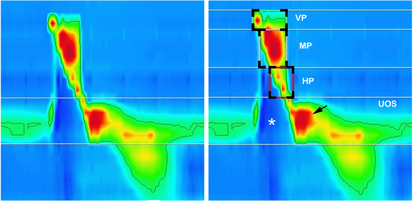
The introduction of pharyngeal high-resolution manometry (PHRM) has improved our understanding of pharyngeal swallow biomechanics. It offers high spatial and temporal resolution that allows for the objective measurement of pressures across various anatomical sites, including the pharynx, tongue base and upper oesophageal sphincter (UOS)Reference Ryu, Park and Kang1 (Fig 1), and improves our understanding of various swallowing pathologies affecting these regions.Reference Cock and Omari2
Pressure topography of a normal pharyngeal swallow. Various pressure measurements are represented by a colour gradient from blue (low pressure) to red (high pressure). The image on the right shows various anatomical sites, including the Velopharynx (VP), Mesopharynx (MP), hypopharynx (HP) and upper oesophageal sphincter (UOS). During a swallow, the baseline pressure of the UOS drops (*), leading to a change of colour. This is followed by transient hypercontractility (black arrow) to trigger an oesophageal peristalsis.

Rapid pressure changes during the pharyngeal swallowing phase are attributed to the striated muscle involved in the food bolus propulsion. Thus, conventional manometric techniques were limited in their ability to capture the complexity of pharyngeal function due to their low sensor density and difficulty in assessing transient pressure events.Reference Takasaki, Umeki, Enatsu, Tanaka, Sakihama and Kumagami3 PHRM has overcome these limitations by providing high fidelity quantitative measures of intraluminal pressure, enabling clinicians to identify UOS pressures and relaxation, subtle dysmotility patterns, differentiate between obstructive and propulsive disorders and estimate the risk of aspiration, offering guidance for therapeutic interventions.Reference Omari, Ciucci, Gozdzikowska, Hernández, Hutcheson and Jones4
In 2020, an independent PHRM International Working Group published recommendations for PHRM protocols and metrics.Reference Omari, Ciucci, Gozdzikowska, Hernández, Hutcheson and Jones4 At our centre, HRM has been used to enhance the diagnostic process and guide the management of patients with dysphagia. In this paper, we present an overview of our clinical experience with HRM, including our protocol, which is adapted from the PHRM International Working Group’s recommendations.Reference Omari, Ciucci, Gozdzikowska, Hernández, Hutcheson and Jones4
Materials and methods
Retrospective review of 43 patients who underwent PHRM study at our institute. This study has been approved and registered as a service evaluation project (reference 1110), and governance was provided via the Imperial College Healthcare NHS Trust (ICHT) Service Evaluation and Audit Governance Committee.
Protocol
All examinations are conducted using Laborie Solar™ GI High-Resolution Manometry (Enschede, Netherlands) and K83659-E-10107 catheter while the patients are seated with their head in a neutral position. Following the catheter insertion and accommodation, baseline measurements are recorded before introducing several consistencies. The comprehensive protocol is provided below:
1. Baseline measurements – measurements are taken while the patients are sitting in a neutral and steady position, without speaking or swallowing
2. 3 x trials of 10 millilitres (ml) saline to test International Dysphagia Diet Standardisation Initiative (IDDSI) = 0 consistency. For those unable to tolerate a 10-ml bolus due to severe dysphagia, the volume can be decreased to 5 ml.
3. 3 x trials of 5 mL of applesauce to test IDDSI = 4 consistency. This is useful in supporting the diagnosis of UOS dysfunction, as the PHRM abnormalities should become more pronounced with increased viscosity.
4. Some patients with swallowing issues related to solid food re given 3 x trials of cookie/biscuit to test IDDSI =7 regular consistency
5. If esophageal dysmotility is suspected, five screening swallows with 5 mL of saline are completed. The catheter may need to be advanced as the initial placement may not capture the lower oesophageal sphincter.
Analysis
For each pharyngeal swallow, eight parameters are measured and recorded: pharyngeal, velopharyngeal, mesopharyngeal and hypopharyngeal contractile integrals (PhCI, VCI, MCI and HCI), hypopharyngeal intrabolus pressure (H-IBP), integrated relaxation pressures (IRP) at 0.2s, upper oesophageal sphincter relaxation time (UOS-RT) and UOS maximum admittance (UOS-MaxAd).
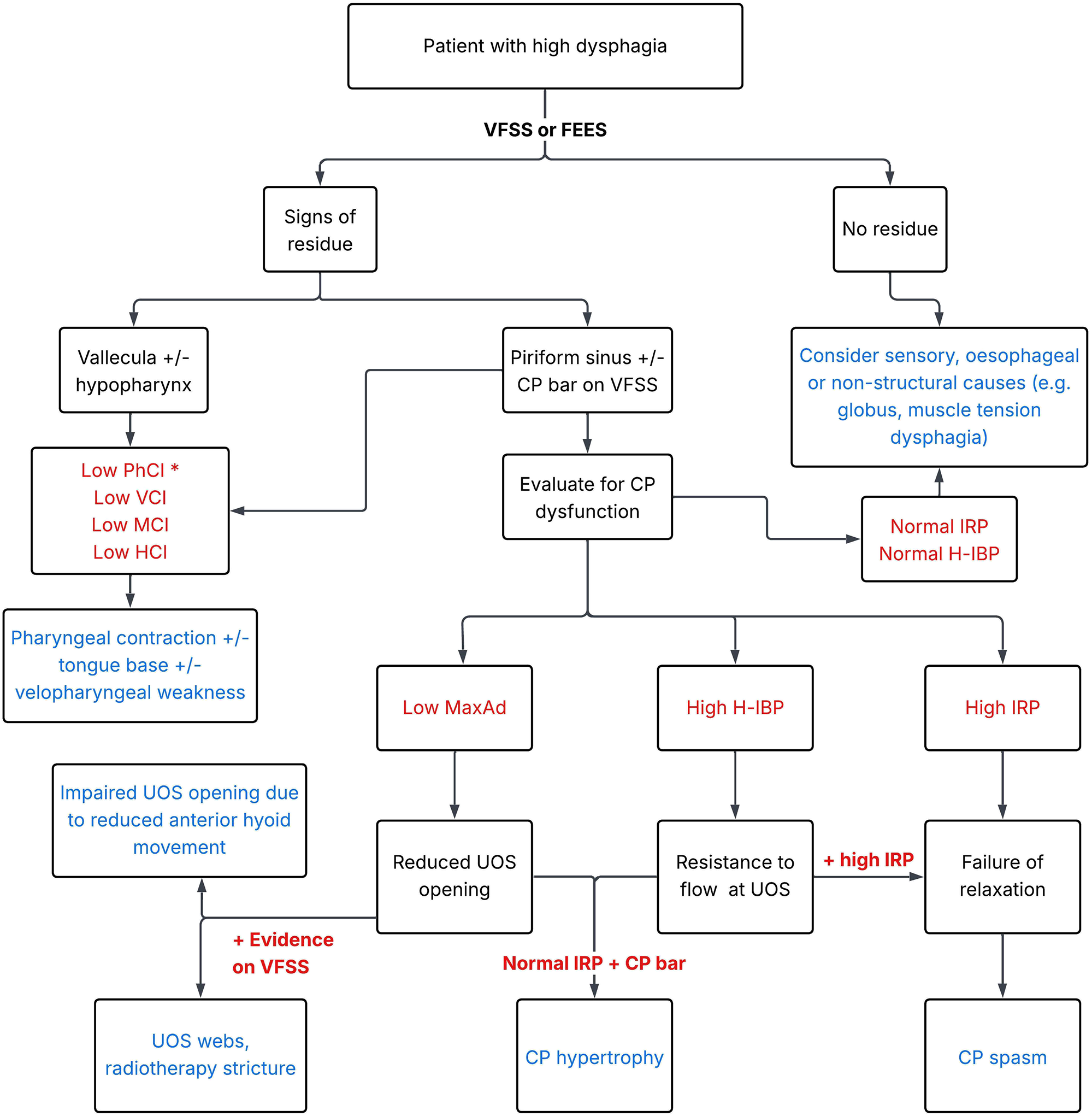
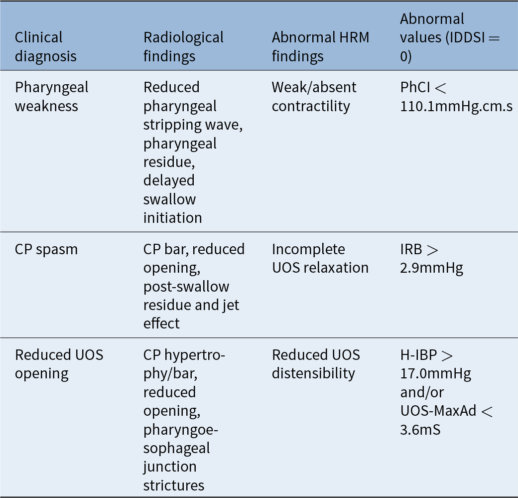
Four items are particularly utilised to interpret the study findingsReference Cock and Omari2 (Table 1 and Figure 2):
1. PhCI measures the overall pharyngeal contractile vigour. It is derived from considering the amplitude, duration and length of the pharyngeal contraction. A lower value indicates a hypo-contractile pharynx, while a higher value indicates hypercontractility.
2. IRP measures the decrease in UOS pressures during swallow, indicating UOS relaxation. An increase in IRP suggests incomplete UOS relaxation.
3. H-IBP measures the pressure 1 cm above the UOS at maximal distension. It indicates UOS resistance to bolus flow leading to increase pressure gradient across the pharyngo-oesophageal junction.
4. UOS-MaxAd measures the maximum admittance value during bolus flow across the UOS. Admittance is the inverse product of impedance and correlates with the luminal diameter area. A decrease in UOS-MaxAd may suggest reduced UOS distensibility. However, the results should be interpreted with caution in the presence of pharyngeal weakness as diminished bolus propulsion can reduce UOS-MaxAd.
Flowchart showing diagnosis algorithm in patients with high dysphagia combining both radiological and manometric findings. In the case of pharyngeal weakness (*low PhCI) all further HRM results should be interpreted with caution due to the risk of false negatives.

Interpretation of various PHRM findings

Note: Normative data are based on Omari et al.Reference Omari, Ross, Schar, Campbell, Thompson, Besanko, Lewis, Robinson, Farahani, Cock and Mossel11
Results
A total of 43 patients (M:25 and F:29) with an average age of 61 ± 21, who had PHRM as a part of their diagnostic workups, were included in this cohort. All patients whose previous radiologic swallowing assessments were not available at the time of data extraction and two patients with primarily oesophageal symptoms (one patient with oesophageal dysmotility and one patient with regurgitation sensation) were excluded from the study.
The procedure was well tolerated by all patients. The test was terminated in two patients (not included in this study cohort). One termination was due to a significant gag reflex on insertion of the catheter compounded by patient anxiety. The other patient was post-radiotherapy for hypopharyngeal squamous cell carcinoma with significant post-radiotherapy changes and UOS fibrosis. The catheter could not be passed into the oesophagus despite multiple attempts. There were no complications or adverse events recorded.
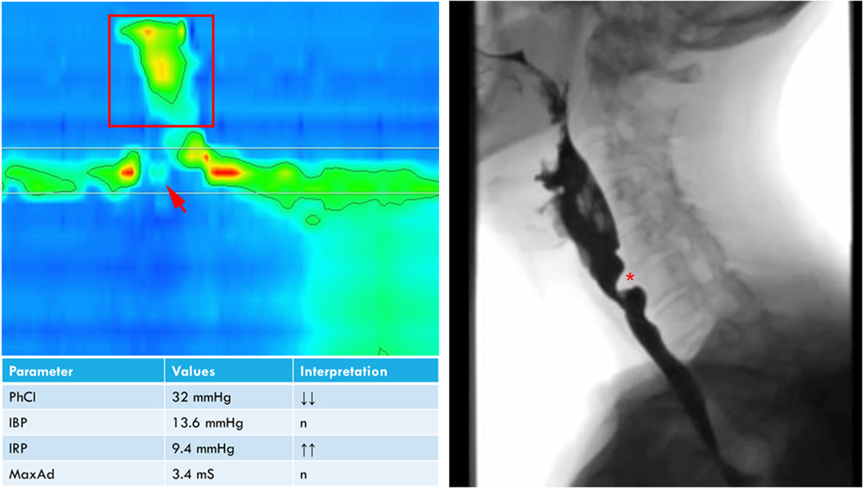
The most common indication for PHRM was oropharyngeal dysphagia, particularly in the presence of cricopharyngeal (CP) spasm and/or bar on either barium swallow test or video fluoroscopic studies (n = 18). Interpretation of the PHRM findings has been used to further delineate the patients into sub-categories depending on the presence of UOS hypertrophy and/or spasm (Figure 3), pharyngeal weakness and oesophageal dysmotility.
Representative clinical case of UOS spasm. Videofluoroscopy study shows CP bar (*) with limitation to bolus flow and pharyngeal residue. PHRM using 10ml normal saline (IDDSI=0) showed pharyngeal weakness (red square), and reduced UOS relaxation indicated by the high IRP values. This can be visually detected on the PHRM study as persistent high pressure area at the level of UOS during relaxation (red arrow). The patient went on to have dilation and botulinum toxin injection to the CP muscle and was referred to SLT for swallowing therapy to improve pharyngeal contraction.

The second most common indication is post-treatment evaluation of previous failed treatment attempts of CP bar (n = 6). In five of these patients PHRM results indicated additional pharyngeal weakness that was not previously diagnosed.
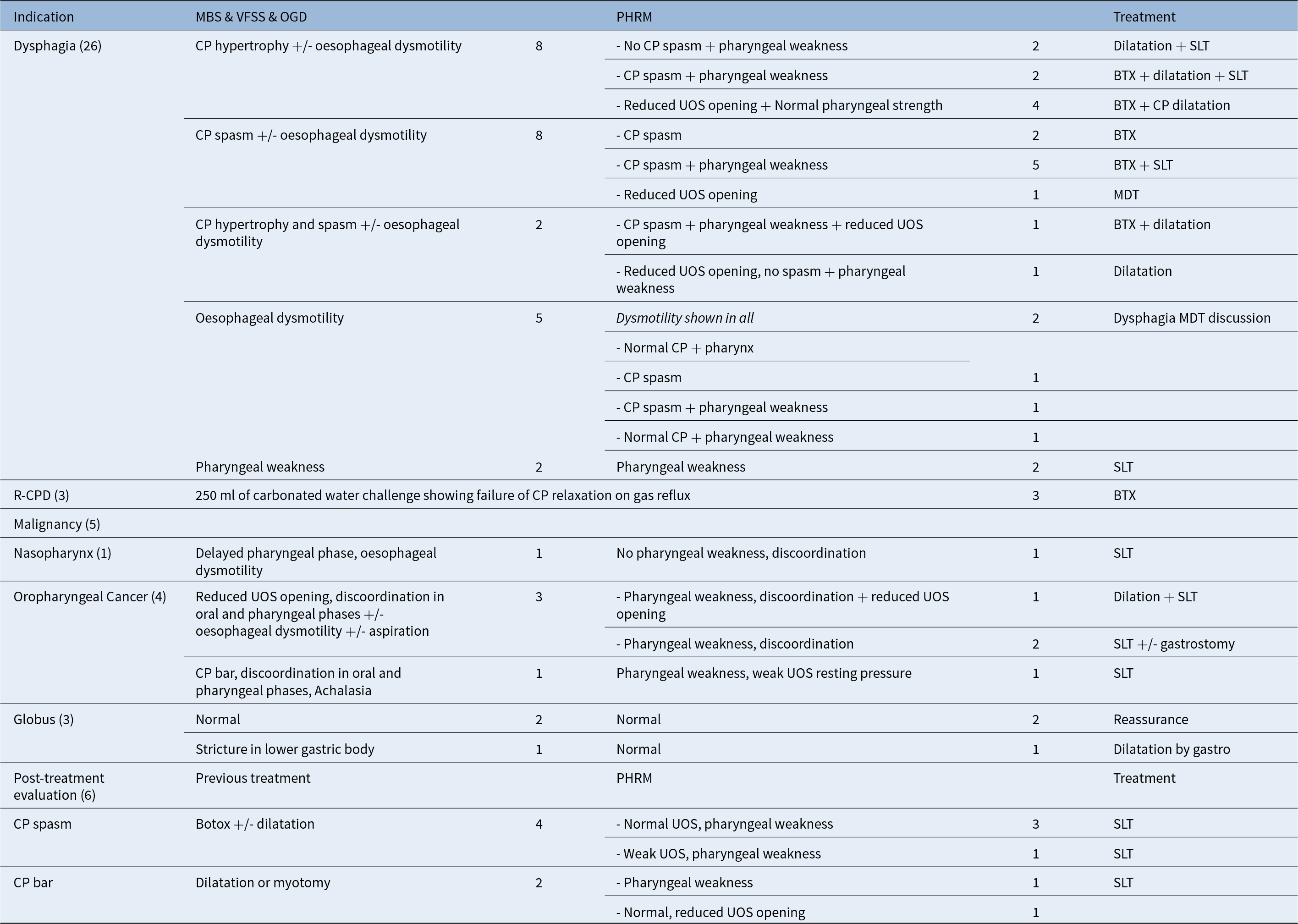
The indications, radiologic swallowing assessments, PHRM findings and recommended treatments are comprehensively detailed (Table 2).
The indications, radiologic swallowing assessments, PHRM findings and recommended treatments for our patient cohort

BSS = barium swallow study; BTX = botulinum toxin injection; CP = cricopharyngeus; MBS = modified barium swallow; MDT = McNeill Dysphagia Therapy Program; PHRM = pharyngeal high-resolution manometry; OGD = oesaphogo-gastro-duodenoscopy; R-CPD = retrograde cricopharyngeus dysfunction; SLT = speech and language therapist; UOS-IRP = upper oesophageal sphincter integrated relaxation pressure, VFSS = videofluoroscopic swallow study.
Discussion
The symptom of dysphagia is most commonly caused by pathologies of the upper digestive tract, including propulsive disorders, abnormal transport of the bolus due to inappropriate contractility, or disorders of the UOS, which arise from abnormal UOS opening.Reference Clavé and Shaker5
UOS opening is both a passive and active process. It relies on the actions of supra- and infra-hyoid muscles leading to hyolaryngeal elevation, the propulsive power of the pharyngeal contraction leading to distension pressure as the bolus advances and finally the active neuromuscular relaxation of the CP muscle.Reference Cook, Dodds, Dantas, Massey, Kern and Lang6 While UOS opening can be measured by UOS Max Admittance,Reference Omari, Ciucci, Gozdzikowska, Hernández, Hutcheson and Jones4 IRP is used to determine the relaxation of the UOS. PhCI is an overall measure of the vigour of the pharyngeal contraction and reduced values were found to indicate pharyngeal weakness.Reference Omari, Ciucci, Gozdzikowska, Hernández, Hutcheson and Jones4
In our practice, we use PHRM to assess the overall contractile function of the pharynx and UOS opening in patients with oropharyngeal dysphagia, the most common indication in our cohort (60 per cent; n = 26). Our findings suggest that PHRM was helpful in diagnosing CP spasm in 15 per cent and pharyngeal weakness in 50 per cent of the patients, who were previously undiagnosed based on radiological findings alone, and excluding CP spasm in 20 per cent of patients with a previous suspected diagnosis. CP spasm diagnosis is based on high IRP supported by other radiological (CP bar, piriform fossa residue or jet effect) and manometric findings (high IRP and/or low MaxAd). Cock et al.Reference Cock, Besanko, Kritas, Burgstad, Thompson and Heddle7 previously showed that radiological CP bars do not always obstruct the bolus transport and emphasised the significance of sensorial dysfunction and pharyngeal weakness as an explanation of dysphagia symptoms in the patients with CP bar with normal UOS MaxAd.
The second most common indication in our cohort is an evaluation of patients with previous unsuccessful surgical intervention of CP bar and/or spasm (balloon dilation +/- botulinum toxin injection). PHRM was helpful in diagnosing pharyngeal weakness in 83 per cent of these patients and they were referred for non-surgical swallowing interventions (exercises, compensatory strategies and diet modifications), avoiding further unnecessary surgeries.
Dysphagia is a common sequelae of head and neck cancer survivors. A recent review indicated that reduced PhCl, UOS dysfunction and dysmotilityReference Neijman, van Mierden, Karakullukcu, Hilgers, van den Brekel and van der Molen8 are the main factors causing dysphagia symptoms. Similarly, our findings also endorse pharyngeal weakness, reduced UOS opening and discoordination as the positive findings in our patient cohort of cancer survivors. Considering that complications of surgery on the UOS are common, diagnostic accuracy is extremely important in this patient group to avoid any unnecessary surgical intervention.
Retrograde CP dysfunction (R-CPD) is a condition characterised by a failure of CP relaxation with retrograde passage of air.Reference Bastian and Smithson9 It is a clinical diagnosis based on the presence of the following features: an inability to belch, abdominal bloating and discomfort, gurgling noises and excessive flatulence. The clinical diagnosis based on these features is described by Bastian and Smithson,Reference Bastian and Smithson9 with a resolution of symptoms after treatment with botulinum toxin injection to CP. In our cohort, PHRM confirmed failure of CP relaxation on gas reflux in all patients with suspected R-CPD. In our practice, we use clinical symptoms to diagnose R-CPD and offer botulinum toxin injections accordingly. We reserve PHRM for treatment non-responders, atypical history or patients with suspected multifactorial dysphagia.
Globus pharyngeus is a sensation of lump in the throat. Most common causes of globus sensation are gastroesophageal and laryngopharyngeal reflux, oesophageal dysmotility or pathologies affecting the UOS.Reference Järvenpää, Arkkila and Aaltonen10 In patients without any concurrent symptoms, such as pain and dysphagia, the mainstay of management is patient education and reassurance.Reference Järvenpää, Arkkila and Aaltonen10 The utility of PHRM in patients with globus can exclude UOS dysfunction and offer support and biofeedback.
• This article presents the first United Kingdom (UK) clinical experience with pharyngeal high-resolution manometry (PHRM) in a cohort of patients with dysphagia and related upper aerodigestive tract symptoms.
• Our protocol is adapted from the 2020 International Working Group published recommendations and focuses on four parameters: pharyngeal contractile integral, integrated relaxation pressure, hypopharyngeal intrabolus pressure and upper oesophageal sphincter (UOS) maximum admittance.
• This article aims to establish a practical diagnostic algorithm where PHRM can be used as an additional instrumental tool for patients with oropharyngeal dysphagia to increase diagnostic accuracy
• While there is an agreement in the literature on the HRM protocol, there is a lack of published evidence on how to translate these findings into clinical diagnosis and how to inform surgical decision-making, which this article aims to address.
• In our cohort, PHRM was helpful in diagnosing cricopharyngeal (CP) spasm in 15 per cent and pharyngeal weakness in 50 per cent of the patients, which were previously undiagnosed; and excluding CP spasm in 20 per cent of patients with a previous suspected diagnosis on imaging.
Conclusion
In our experience, PHRM can offer an additional detailed understanding of swallow physiology and UOS function that supplements instrumental swallowing evaluation tests. Furthermore, it has been helpful in reassuring globus patients and in aiding diagnosis and management in patients with persistent symptoms post-treatment for head and neck cancer, CP spasm and CP hypertrophy. Furthermore, it can be used as a definite diagnostic tool in patients with suspected R-CPD. In summary, the superior diagnostic capability of PHRM has aided in guiding the management of our patient cohort, allowing the delivery of more personalised care and avoiding unnecessary surgical procedures.
Aknowledgement
None.
Funding
The purchase of the High-Resolution Manometry equipment was funded via Imperial Innovate Grant II2122_17. M.C. (NIHR302221) is funded by the NIHR for research related to HRM. The views expressed in this publication are those of the authors and not necessarily those of the NIHR, NHS or the UK Department of Health and Social Care.
Conflict of interest
None.