Introduction
Infertility is increasingly acknowledged as a global public health issue by the World Health Organization, and a growing number of couples currently worry or have previously worried about infertility (De Neubourg et al., Reference De Neubourg, van Duijnhoven, Nelen and D’Hooghe2012; Barratt et al., Reference Barratt, Björndahl, De Jonge, Lamb, Osorio Martini, McLachlan, Oates, van der Poel, St John, Sigman, Sokol and Tournaye2017). Since the first in vitro fertilization (IVF) baby was born in 1978, more than 5 million IVF babies have been born worldwide, and the number is increasing exponentially (Faddy et al., Reference Faddy, Gosden and Gosden2018). Embryo culture is a crucial step for IVF and other assisted reproduction techniques, and the embryo culture medium has a wide influence on embryo development in terms of metabolism, morphology, epigenetic conditions, gene expression and developmental ability (Mani and Mainigi, Reference Mani and Mainigi2018). It has been reported that IVF babies have a higher risk of cancer, metabolic diseases, hypertension, and cardiovascular diseases, and this may be closely related to in vitro culture (Simonstein et al., Reference Simonstein, Mashiach-Eizenberg, Revel and Younis2014).
Many kinds of embryo culture medium are at this time available for humans and animals (Rossi et al., Reference Rossi, Di Nisio, Macchiarelli, Nottola, Halvaei, De Santis and Cecconi2019). Most research has focused on optimizing the composition of the culture medium, and only a few studies have tackled the culture medium changing procedure (de los Santos et al., Reference de Los Santos, Gámiz, de Los Santos, Romero, Prados, Alonso, Remohí and Dominguez2015). Studies have shown that in vitro cultured embryos can secrete some important factors into the medium that play an important role in regulating their development (Kim et al., Reference Kim, Lee, Kim and Jun2016; Lin et al., Reference Lin, Beckers, McCafferty, Gansemans, Szymanska, Pavani, Catani, Van Nieuwerburgh, Deforce, De Sutter, Van Soom and Peelman2019). At the same time, embryo metabolites or degradation products of the culture medium will produce some substances that are harmful to the embryo cultured in vitro (Shohael et al., Reference Shohael, Ali, Yu, Hahn, Islam and Paek2006). Renewal of the embryo culture medium may lead to the loss of some important secretions, while non-renewal of the embryo culture medium may lead to the accumulation of toxic substances.
Rabbits play an important role in reproductive biology (Fischer et al., Reference Fischer, Chavatte-Palmer, Viebahn, Navarrete Santos and Duranthon2012). Many pioneering achievements in reproductive biology come from research using rabbits (Alexandre Reference Alexandre2001; Fischer et al., Reference Fischer, Chavatte-Palmer, Viebahn, Navarrete Santos and Duranthon2012). In this study, the rabbit was used as a model to study the effect of changing the culture medium on embryo development in vitro, and our work will provide an important reference for the optimization of embryo culture.
Materials and methods
Animal
Here, 6–8-month-old rabbits were raised individually in cages at the Laboratory Animal Center of Xi’an Jiaotong University with unrestricted access to food and water. The temperature was maintained at c. 25°C, and the day:night ratio was 12 h:12 h. All procedures involved in the animal experiments were in line with animal ethics and welfare requirements to minimize animal suffering.
Collection of fertilized eggs
Here, 80 IU pregnant mare serum gonadotropin (PMSG; Shusheng, Ningbo, China) was injected subcutaneously, then hCG (SHUSHENG, Ningbo, China) 100 IU was injected intravenously 72 h later, and the female rabbits were immediately mated with male rabbits. At 20–22 h after mating, the female rabbits were anaesthetised, and the abdominal cavity was opened to separate the fallopian tube. A syringe loaded with 2 ml of Dulbecco’s phosphate-buffered saline (DPBS) containing 5% fetal bovine serum was inserted into the fallopian tube near the uterine segment, and the end of the fallopian tube near the ovary was placed in a centrifuge tube. Then the plunger of the syringe was pressed down to flush out the fertilized eggs. The fertilized eggs were collected under a stereomicroscope and transferred into DPBS containing hyaluronidase (5 mg/l).
In vitro culture
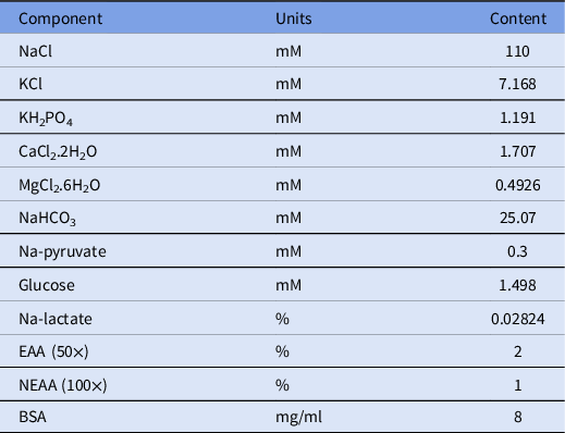
The fertilized embryos were treated with DPBS containing hyaluronidase for 5 min at 38.5°C, and then blown gently to remove the granulosa cells from the embryos. The embryos were then transferred into embryo culture medium covered with oil. The formula of the embryo culture medium is shown in Table 1. The amount of culture medium in each culture dish was 400 µl, the number of embryos in each dish was 40, and the culture environment was maintained at 38.5°C, at saturated humidity and with 5% CO2. Three groups were designated, a non-renewal group, a renewal group, and a half-renewal group. In the non-renewal group, embryo culture medium was not changed from day 1 to day 4. In the renewal group, embryo culture medium was completely changed on day 3. In the half-renewal group, 200 µl of old culture medium was drawn out on day 3, and replaced with 200 µl fresh culture medium.
Composition of embryo culture medium

ROS staining
Embryos were incubated for 20 min in serum-free medium containing 10 mM 2′,7′-dichlorodihydrofluorescein diacetate (DCHF-DA) at 37°C. Then the embryos were washed three times in serum-free medium, and examined and photographed using a Nikon Eclipse TI-s microscope (Nikon, Tokyo, Japan), and analyzed using Image Pro Plus software (Media Cybernetics, Bethesda, MD).
Apoptosis staining
A terminal deoxynucleotidyl transferase dUTP nick end labelling (TUNEL) kit (Promega, Madison, WI, USA) was used to analyze the apoptosis of blastocysts. Embryos were washed three times with 0.2% PBS-polyvinyl alcohol (PBS-PVA) for 5 min each time, and then fixed with 4% paraformaldehyde at room temperature (RT) for 30 min. Embryos were washed three times with 0.2% PBS-PVA for 5 min each time. The washed embryos were then equilibrated in E-buffer for 5–8 min, and then transferred to apoptotic dye solution containing 45 μl E-buffer, 5 μl nucleotide mix and 1 μl rTDT at 37°C for 1 h. The reaction was terminated by applying saline–sodium citrate (SSC) buffer for 15 min, and the embryos were washed three times with 0.2% PBS-PVA. Embryos were stained with 4′,6-diamidino-2-phenylindole (DAPI) for 3 min, then the embryos were washed three times with 0.2% PBS-PVA. Next the embryos were transferred to a glass slide, pressed and covered with glass, and sealed with nail polish. Samples were examined and photographed under a Nikon Eclipse TI-s microscope (Nikon), and analyzed using Image Pro Plus software (Media Cybernetics, Bethesda, MD, USA).
Counting total number of cells in blastocysts
Blastocysts were treated with DPBS containing hyaluronidase to remove the granulosa cells. Then the blastocysts were washed three times with 0.2% PBS-PVA for 5 min each time, and fixed with 4% paraformaldehyde at RT for 30 min. The blastocysts were treated with DAPI for 3 min, and then were washed three times with 0.2% PBS-PVA. After transfer onto a glass slide and being pressed with a coverslip, the blastocysts were examined and photographed under a Nikon Eclipse TI-s microscope (Nikon), and counted using Image Pro Plus software (Media Cybernetics).
Statistical analysis
SPSS software (SPSS Inc., Chicago, IL, USA) was used for statistical analysis. Blastocyst rate was analyzed using the chi-squared test. The relative level of ROS, apoptotic index of blastocysts, and blastocyst total cell number were analyzed using one-way analysis of variance (ANOVA). Each experiment was repeated more than three times.
Results
Effect of changing the culture medium on the ROS level of blastocysts
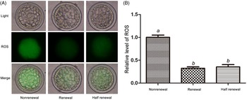
Embryos in the non-renewal, renewal and half-renewal groups were cultured for 4 days, and the relative level of ROS of the blastocysts was analyzed using DCHF-DA. The result showed that the ROS level of blastocysts in the non-renewal group was significantly higher than in the renewal group and the half-renewal group (P < 0.05), and that there was no significant difference between the renewal group and the half-renewal group (P > 0.05; Fig. 1).
Relative level of ROS in blastocysts in the non-renewal, renewal and half-renewal groups. (A) ROS staining in blastocysts. Upper row: bright field; middle row: green fluorescence indicating ROS; lower row: merged bright field and green fluorescence. (B) Quantification of ROS fluorescence intensity. Different letters above the bars indicate significant differences (P < 0.05).

Effect of changing the culture medium on the apoptotic index of blastocysts
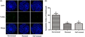
On day 4 blastocysts were collected and analyzed using a TUNEL kit in each group. The apoptotic index was defined as the number of apoptotic cells divided by the blastocyst total cell number. The result showed that the apoptotic index of blastocysts in the non-renewal group was significantly higher than in the renewal group and the half-renewal group (P < 0.05), and that the difference between the renewal group and the half-renewal group was not significant (P > 0.05; Fig. 2).
Apoptosis index of blastocysts in the non-renewal, renewal and half-renewal groups. (A) TUNEL assay of apoptotic blastomeres. Upper row: DAPI; middle row: green fluorescence indicating apoptotic cells; bottom row: merged DAPI and apoptotic cell. (B) Apoptosis index of blastocysts. Different letters above the bars indicate significant differences (P < 0.05).

Effect of changing the culture medium on the blastocyst ratio and blastocyst total cell number
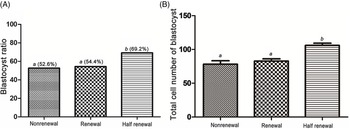
The blastocyst ratio and the blastocyst total cell number in each group were analyzed on day 4. The results showed that the blastocyst ratio (Fig. 3A) and blastocyst total cell number (Fig. 3B) were significantly higher in the half-renewal group than in the non-renewal group and the renewal group. The blastocyst ratio and the blastocyst total cell number in the non-renewal group and the renewal group showed no significant difference (P < 0.05).
Blastocyst ratio and blastocyst total cell number in non-renewal, renewal and half-renewal groups. (A) Blastocyst ratio on day 4 in non-renewal, renewal and half-renewal groups. (B) Total cell number of blastocysts in non-renewal, renewal and half-renewal groups. Different letters above the bars indicate significant differences (P < 0.05).

Discussion
Much progress has been made in the optimization of embryo culture medium, and two different approaches exist to embryo culture: one attitude is ‘let the embryo choose’, the other is ‘return to nature’ (Koscinski et al., Reference Koscinski, Merten, Kazdar and Guéant2018). Guided by the first tactic, culture medium’ formula was determined by experience, experiment and algorithm analysis, allowing the culture medium to support all nutrient requirements from the zygote to the blastocyst stage (Thouas et al., Reference Thouas, Jones and Trounson2003). For example, KSOM medium was developed based on this idea and has been successfully used in mice, rabbits and cattle (Liu et al., Reference Liu, Foote and Yang1995; Jin et al., Reference Jin, Kim, Im and Choi2000; Zhu et al., Reference Zhu, Walker and Maddocks2004; Men et al., Reference Men, Stone and Bryda2020). Under the guidance of ‘return to nature’, culture medium was synthesized by defining the different nutrients required at different stages of embryonic development, simulating the components of oviduct fluid or uterine fluid in vivo (Qu et al., Reference Qu, Wang, Zhang and Liu2020a). In the zygote and the first few cleavage stages, the metabolic activity of the embryo is relatively weak (Pantaleon et al., Reference Pantaleon, Ryan, Gil and Kaye2001). With the activation of the embryo’s genome, the embryo migrates from the fallopian tube to the uterus, and during this process, the metabolism of the embryo gradually increases (Yang et al., Reference Yang, Wang, Chen, Qi, Baker, Liu, Zhang and Han2020). G1/G2 culture media were designed based on this. G1 culture medium was used at the first few cleavages, and then embryos were transferred into G2 culture medium according to the changes of embryonic development and nutritional requirements (Hall-Woods et al., Reference Hall-Woods, Krisher, Lane, Gardner and Asa2000; Sanches et al., Reference Sanches, Pontes, Basso, Ferreira, Perecin and Seneda2013). For KSOM medium, there was no need to renew the culture medium, but for G1/G2 culture medium, renewal of the culture medium was necessary.
During its development, the embryo is in close contact with the microenvironment of the embryo culture medium or oviduct fluid (Sekirina and Neganova, Reference Sekirina and Neganova1995; Lee et al., Reference Lee, Lee, Lee, Park, Elahi, Lee, Park, Hyun and Lee2017; Chen and Schoen, Reference Chen and Schoen2019). Renewal of the culture medium may lead to the loss of these important factors, which is not conducive to embryonic development. conversely, with increasing duration of the culture, some nutrients will be consumed, and toxic substances will accumulate as a result of decomposition or metabolism (Kleijkers et al., Reference Kleijkers, van Montfoort, Bekers, Coonen, Derhaag, Evers and Dumoulin2016). In this study, three groups were designed: a non-renewal group, a renewal group and a half-renewal group. The ROS level, apoptotic index, embryonic development-related genes, blastocyst ratio and blastocyst total cell number were analyzed to study the effect of changing the culture medium on preimplantation embryos.
Previous studies have reported that the ROS level of embryos in vitro is higher than that of embryos developed in vivo (Amoushahi et al., Reference Amoushahi, Salehnia and Ghorbanmehr2018; An et al., Reference An, Peng, Cheng, Lu, Zhou, Zhang and Su2019; Qu et al., Reference Qu, Luo, Du, Zhang, Song, Yuan, Lin, Li and Liu2020b). Excessive ROS levels could damage lipid, protein and DNA, disturb gene expression, and block embryo development (Takahashi, Reference Takahashi2012; Alarifi et al., Reference Alarifi, Ali, Verma, Almajhdi and Al-Qahtani2014). At appropriate concentrations, ROS can be used as a signal molecule participating in the regulation of the cell cycle, cell differentiation and development (Yang et al., Reference Yang, Zhu, Zheng, Chai, Ji, Ren, Liu, Liu, Song, Li, Liu and Li2017). Treating the embryo in vitro with antioxidants, such as melatonin and glutathione, can effectively reduce ROS level and improve the ability of embryos to develop in vitro (Huang et al., Reference Huang, Pang, Hao, Du, Zhao and Zhu2018; Li et al., Reference Li, Gu, Lu, Zhao, Feng and Sun2018; Pang et al., Reference Pang, Jiang, Wang, Wang, Hao, Zhao, Du, Zhao, Wang and Zhu2019; Hicks et al., Reference Hicks, Mentler, Arena, Current and Whitaker2020). In this study, the ROS level of blastocysts was significantly higher in the non-renewal group than in the renewal group and the half-renewal group, and there was no significant difference between the renewal group and the half-renewal group. Previous studies have reported that the concentration of ammonia in the culture medium increased with culture time, which led to an increase of the embryonic ROS level (Kleijkers et al., Reference Kleijkers, van Montfoort, Bekers, Coonen, Derhaag, Evers and Dumoulin2016). The results indicated that oxides may accumulate in the culture medium during embryo culture, and the oxides might be removed or diluted by renewing culture medium, which could decrease the ROS level of the embryo.
In the process of embryonic development, many factors can lead to apoptosis (Qu et al., Reference Qu, Wang, Zhang and Liu2020a). The apoptotic ratio of blastocysts was significantly increased under endoplasmic reticulum stress or heat stress (Lin et al., Reference Lin, Lee, Oqani, Kim, Cho, Jeong, Baek and Jin2016). In a hyperoxic environment, embryos showed a higher apoptotic ratio than in a hypoxic environment (Lin et al., Reference Lin, Lee, Oqani, Kim, Cho, Jeong, Baek and Jin2016). Pesticides and other toxicants increased the apoptotic ratio and caused embryonic malformation (Jia et al., Reference Jia, Ding, Shen, Luo, Zhang, Zhou, Ding, Qu and Liu2019). There is a close relationship between the apoptotic ratio and embryonic development potential, and a higher apoptotic ratio is considered to have a lower developmental potential (Jia et al., Reference Jia, Ding, Shen, Luo, Zhang, Zhou, Ding, Qu and Liu2019). Adding antioxidants or anti-stress compounds to the culture medium can effectively reduce the apoptotic ratio and enhance the development of the embryo in vitro (Lin et al., Reference Lin, Lee, Oqani, Kim, Cho, Jeong, Baek and Jin2016). In this study, we found that the apoptotic ratio of blastocysts was significantly higher in the non-renewal group than in the renewal group and the half-renewal group, and that the difference between the renewal group and the half-renewal group was not significant, indicating that non-renewal of embryo medium may not be conducive to embryonic development, while changing the culture medium influenced the apoptosis of embryos in vitro.
The blastocyst ratio and blastocyst total cell number are important indexes to evaluate embryo development. Studies have shown that embryos with a higher blastocyst ratio and blastocyst total cell number have higher developmental potential (Jia et al., Reference Jia, Ding, Shen, Luo, Zhang, Zhou, Ding, Qu and Liu2019). In this study, the blastocyst ratio and the blastocyst total cell number in the half-renewal group were significantly higher than those in the renewal group and the non-renewal group. This further confirms our hypothesis that changing the culture medium may lead to the loss of important regulatory factors for embryos, while not changing the culture medium may lead to the accumulation of toxic substances, which are both not conducive to embryonic development. Half-renewal can alleviate the defects of non-renewal or renewal, and benefit embryo development.
In conclusion, we found that the culture medium changing procedure could affect the level of ROS, the apoptotic index of blastocysts, the development rate of blastocysts and the blastocyst total cell number. This study has a high value as reference for the optimization of embryo culture in vitro, and has important significance for assisted reproduction.
Financial support
This work was supported by a project funded by the China Postdoctoral Science Foundation under grant no. 2020TQ0240 and the Natural Science Foundation of Shaanxi Province under grant no. 2020PT-001 and grant no. 2021PT-039.
Conflicts of interest
The authors have no conflicts of interest to report regarding the present study.
Ethical standards
The Animal Care and Use Committee of Xi’an Jiaotong University approved all animal use procedures applied in this study.