





Non-technical Summary
The Devonian of the Basque Massifs in Western Pyrenees is a poorly known remote area in the boundary between Spain and France. Devonian invertebrate faunas have been known since the sixties, but they have received little attention. Herein, we describe two new crinoid species Eifelocrinus baztani n. sp. and Gastrocrinus canteri n. sp. from the late Emsian (Early Devonian, ca. 400 million years ago) of this area. The specimens are exquisitely preserved in association with a rich invertebrate fauna of trilobites, mollusks, bryozoans, and brachiopods. Eifelocrinus and Gastrocrinus are present in the classic Hunsrück Slate Lagerstätte from Germany reinforcing the connection of faunas between Spain and Germany during the Lower Devonian.
Introduction
Articulated Devonian crinoids from Spain are well known from the Cantabrian Zone, the Iberian Chains, and the central part of the Pyrenees (see summary in Ausich and Zamora, Reference Ausich and Zamora2019) but unreported from other areas. The Devonian from the west Pyrenees in the Basque area (mainly located in Navarra Country) is one of the most unexplored areas with Devonian rocks from Spain. The Basque Massifs (including Cinco Villas, Aldudes, and Oroz-Betelu = Quinto Real) (Fig. 1) are made of Paleozoic rocks from the Ordovician to the Permian including thick Devonian successions (DeFelipe et al., Reference DeFelipe, Pulgar and Pedreira2018). Juch and Schäfer (Reference Juch and Schäfer1974) studied the Devonian of the Cinco Villas Massif, proposed a stratigraphic framework with thick Lower and Upper Devonian siliciclastic rocks, and reported indeterminate crinoids in a faunal list from the “Arizakun Limestone” that was considered upper Emsian. Exploration of the “Arizakun Limestone” in this area has resulted in the discovery of new outcrops with a rich invertebrate fauna of echinoderms, mollusks, trilobites, and brachiopods that confirm the presence of important faunas (see below). The echinoderm fauna is rich in mitrate stylophorans and crinoids that appear fully articulated. Among the later Eifelocrinus baztani new species and Gastrocrinus canteri new species are here described based on beautifully articulated specimens.
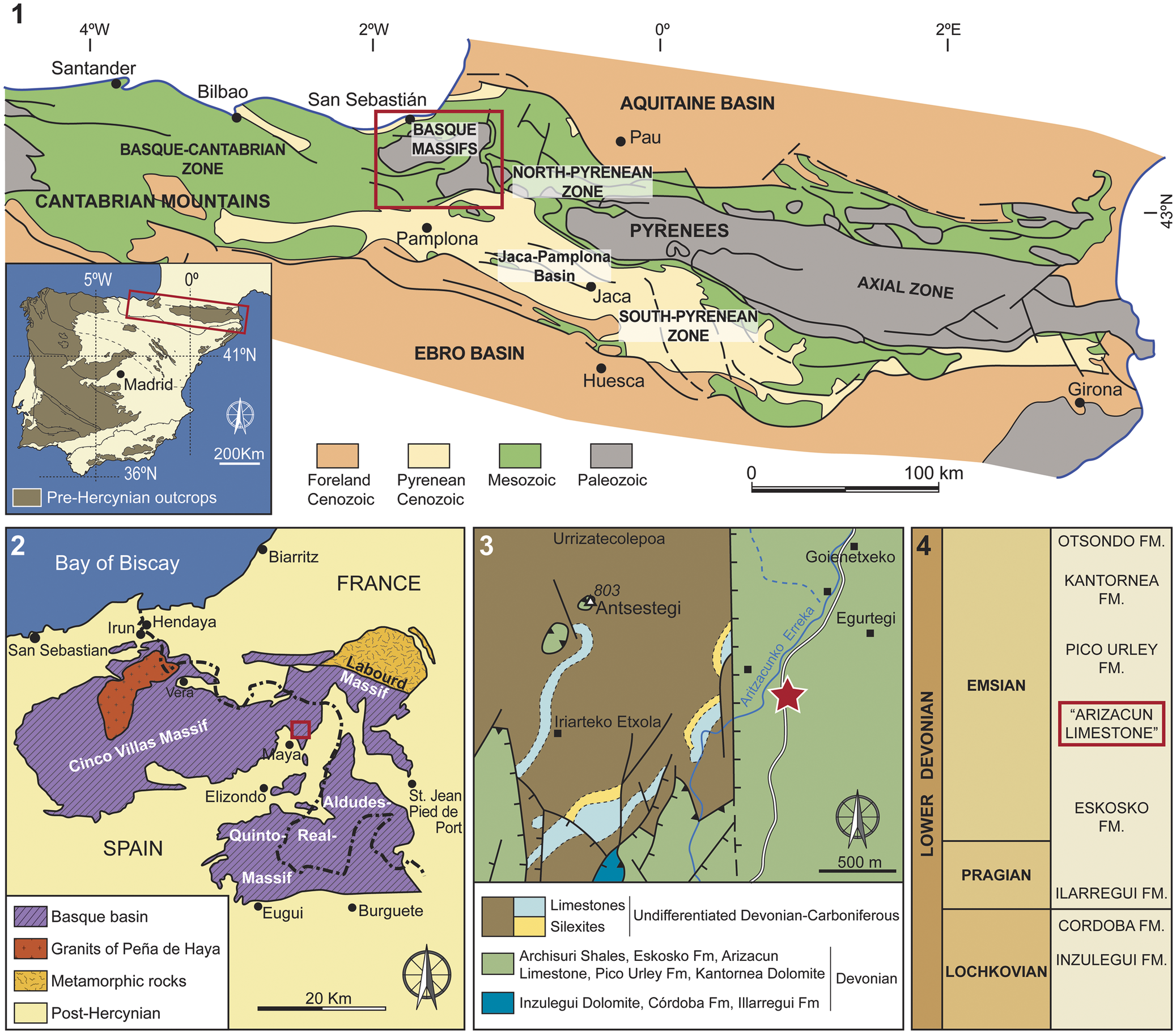
(1) Location of the Basque Massifs area in NE Spain (after Teixell, Reference Teixell1998, and DeFelipe et al., Reference DeFelipe, Pulgar and Pedreira2018). (2) Geographic and geologic setting of the Basque Massifs with location of the studied area in the Cinco Villas massif, in North Maya (modified from Juch and Schäfer, Reference Juch and Schäfer1974). (3) Geologic map of the Devonian and Carboniferous of the study area; red star indicates the sampled locality (modified from Juch et al., Reference Juch, Krausse, Müller, Requadt, Schafer, Sole and Villalobos1972). (4) Stratigraphy of the Devonian in the studied area; crinoids are from the “Arizacun Limestone” (after Sanz-López, Reference Sanz-López and Vera2004).

Figure 1. Long description
A four-panel scientific graphic.
Panel 1: A regional geologic map of Northeast Spain and Southwest France. An inset map shows the Iberian Peninsula with a red box over the Pyrenees. The main map shows the Basque-Cantabrian Zone to the West, the Pyrenees Axial Zone in the center, and the Ebro Basin to the South. A red box highlights the Basque Massifs. Legend categories include Foreland Cenozoic, Pyrenean Cenozoic, Mesozoic, and Paleozoic.
Panel 2: A detailed map of the Basque Massifs. The Cinco Villas Massif is on the West, and the Labourd and Quinto Real-Aldudes Massifs are to the East. A red square indicates the study area near Maya. Legend includes Basque basin, Granits of Peña de Haya, Metamorphic rocks, and Post-Hercynian.
Panel 3: A geologic map of the study area near Arizacunko Erreka. A red star marks the sampling site. Geologic units include Limestones, Silexites, Undifferentiated Devonian-Carboniferous, and various Devonian formations like Archisuri Shales and Inzulegui Dolomite. A 500 m scale bar is at the bottom right.
Panel 4: A stratigraphic column of the Lower Devonian. From bottom to top, the stages are Lochkovian (Inzulegui F M and Cordoba F M), Pragian (Ilarregui F M), and Emsian. The Emsian stage includes Eskosko F M, Arizacun Limestone (highlighted in a red box), Pico Urley F M, Kantornea F M, and Otsondo F M.
Eifelocrinus Wanner, Reference Wanner1916, and Gastrocrinus Jaekel, Reference Jaekel1895, are primarily known from the Emsian of Germany, with exceptions being Eifelocrinus cantabricus Hauser in Hauser and Landeta, Reference Hauser and Landeta2007 (Emsian or Emsian–Eifelian boundary, Spain) and Gastrocrinus sp. and Gastrocrinus aldudensis Le Maitre and Heddebaut, Reference Le Maitre and Heddebaut1962 (Early Devonian, France) (see Webster and Webster, Reference Webster and Webster2014). Eifelocrinus cantabricus is from the Santa Lucia Formation of northern Spain and is known from a single specimen composed of an aboral cup with several articulated primibrachials and several articulated columnals attached. Gastrocrinus sp. is based on a single anal sac from Mayenne (NW, France) (Le Menn, Reference Le Menn1985) and Gastrocrinus aldudensis is known from another anal sac from the Aldudes Massif (South France) (Le Maitre and Heddebaut, Reference Le Maitre and Heddebaut1962).
The new crinoids reported here, Eifelocrinus baztani n. sp. and Gastrocrinus canteri n. sp., extend the occurrence of these genera into the Western Pyrenees of Spain. Both Eifelocrinus and Gastrocrinus occur in the classic Hunsrück Slate Lagerstätte (Emsian of Germany), which is famous for exceptional fossil preservation (e.g., Bartels, et al., Reference Bartels, Briggs and Brassel1998). Perhaps counterintuitively, the moldic preservation of E. baztani n. sp. and G. canteri n. sp. significantly expands our understanding of the morphology of these genera (Fig. 2.3–2.5).
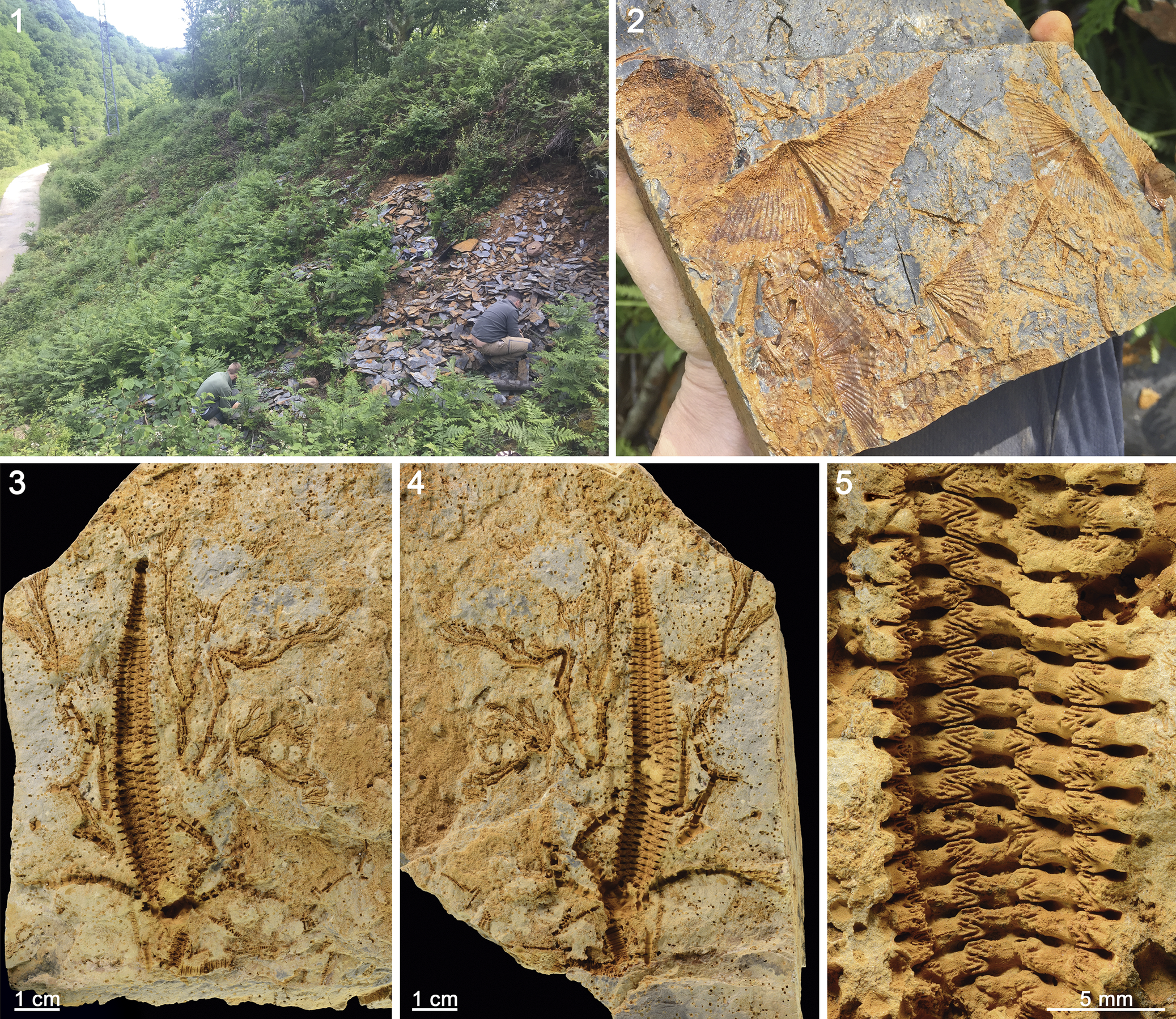
(1) Field photograph of outcrops that yielded the crinoids described here. (2) Moldic preservation of brachiopods (Euryspirifer pellicoi) and tentaculitids. (3–5) Gastrocrinus canteri n. sp. crown, MGM-3426D, holotype, part, counterpart, and detail of the anal sac.

Figure 2. Long description
Panel 1: A wide-angle photo of a lush, green hillside outcrop where two researchers are excavating loose, dark grey shale fragments.
Panel 2: A close-up of a hand holding a flat, grey rock slab containing several orange-brown moldic impressions of Euryspirifer pellicoi brachiopods, characterized by their wide, winged shell shapes and fine ribbing.
Panel 3: A macro photo of the holotype specimen M G M dash 3 4 2 6 D, showing the part of the crinoid crown preserved in a tan, porous matrix. The fossil displays a long, segmented central column with branching arms extending outward. A 1 cm scale bar is at the bottom left.
Panel 4: A macro photo of the counterpart of the same holotype specimen, showing the mirrored impression of the crinoid crown. A 1 cm scale bar is at the bottom left.
Panel 5: A high-magnification detail of the anal sac from the holotype. The structure consists of a vertical, ladder-like arrangement of small, interlocking ossicles with a zig-zag central pattern and lateral pores. A 5 mm scale bar is at the bottom right.
Location and stratigraphy
The Basque Massifs in the Western Pyrenees include several small massifs including Cinco Villas, Aldudes, and Oroz-Betelu = Quinto Real, with Paleozoic rocks. Heddebaut (Reference Heddebaut1970) and Juch and Schäfer (Reference Juch and Schäfer1974) studied the Devonian from the Cinco-Villas Massif located in the westernmost part of the Basque area (Fig. 1.2). They provided the first stratigraphic framework and faunal lists from the fossiliferous formations. The Emsian in this area is divided into five lithostratigraphic units with the age based on conodonts and brachiopods (Juch and Schäfer, Reference Juch and Schäfer1974; see Sanz-López, Reference Sanz-López and Vera2004, for a synthesis). In ascending stratigraphic order the units are: the Eskosko Formation, “Arizacun Limestone”, Pico Urley Formation, Kantornea Formation, and Otxondo Formation.
All the material described in the present paper comes from the “Arizacun Limestone”, which is a 50- to 200-meter-thick unit comprised mostly of dolostones with interbedded mudstones. The formation crops out in the sides of a small path that connects the small village of Maya with France to the northeast by crossing the mountains, in the Baztan valley (Navarra). Coordinates to the place are 43°14′31.4″N, 1°26′26.4″W. The invertebrate fauna is rich and was listed by Carls (in Juch et al., Reference Juch, Krausse, Müller, Requadt, Schafer, Sole and Villalobos1972) and García Alcalde (personal communication, September 2019). The outcrops are fully covered with vegetation, but access to some parts of the section allowed sampling of the mudstones interbedded with the dolostones characteristic of the “Arizacun Limestone”. The invertebrate fauna recognized includes the coral Pleurodyctium problematicum Goldfuss, Reference Goldfuss1829, trilobites (Homalonotidae indet., Asteropyge (Kaiserops?) cf. A. (K.?) diadema Richter, Reference Richter1909, Bryozoa indet., Ostracoda indet., indeterminate bivalves, tentaculitids, echinoderms (crinoids and mitrate stylophorans), and the brachiopods Euryspirifer pellicoi (Verneuil and d’Archiac, Reference Verneuil and D’Archiac1845), Leptostrophiella explanata (Sowerby, Reference Sowerby1842), Plicathyris ezquerrai (Verneuil and d’Archiac, Reference Verneuil and D’Archiac1845), Schizophoria beaumonti (Verneuil, Reference Verneuil1850), Plebejochonetes sp., and Fascistropheodonta indet. (Fig. 2.2). These faunas are indicative of the upper Emsian (Juch et al., Reference Juch, Krausse, Müller, Requadt, Schafer, Sole and Villalobos1972).
Material and methods
Fossil crinoids in the studied area are preserved articulated with complete calyxes, arms, and part of the stem. In some instances, disarticulated columnals are rare. Fossils are concentrated in certain layers, with other layers apparently unfossiliferous. When present, shelly faunas are abundant and well preserved. Tentaculitids are common and have a high degree of orientation in some cases indicating unidirectional currents. All the invertebrates are preserved as natural molds. Horizontal trace fossils are abundant in certain levels.
Specimens were casted with latex prior to photography and whitened with ammonium chloride sublimate (Figs. 3–5). All measurements are in mm; * indicates a measurement was incomplete.
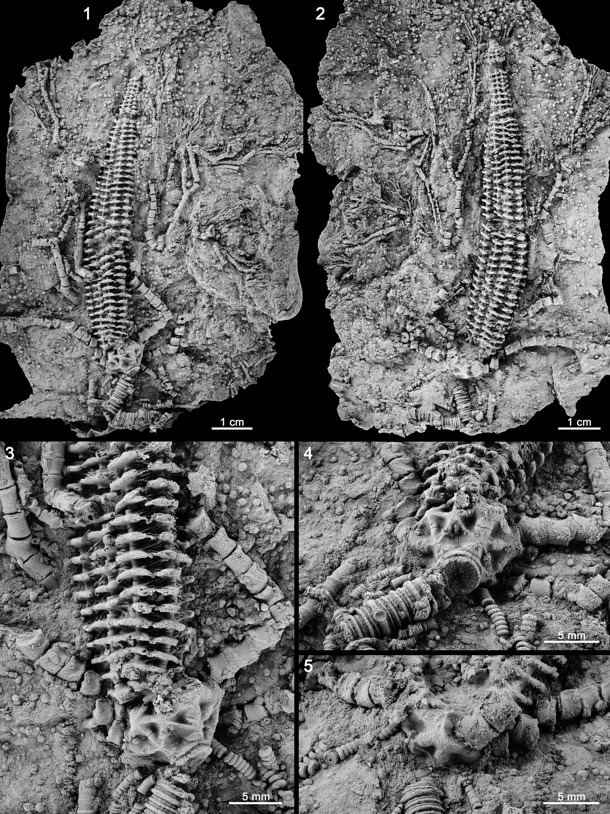
Gastrocrinus canteri n. sp. crown, holotype, MGM-3426D. (1) Part, note distal arm branching. (2) Counterpart. (3) Enlargement of aboral cup and proximal anal sac, note subcircular radial facet with axial canal and strong articular ridge, part. (4) Oblique view of proximal column and aboral cup, CD interray, note radice scars on nodal plates, part. (5) Radial plate with proximal brachials attached, note the radial facet, counterpart. All specimens are latex casts whitened with ammonium chloride sublimate.

Figure 3. Long description
A multi-panel series of grayscale photos showing latex casts of a crinoid fossil.
* Panel 1 (top-left): Full view of the part specimen showing a long, vertically oriented crown with distal arm branching. A 1 cm scale bar is at the bottom right.
* Panel 2 (top-right): The counterpart specimen mirroring the crown structure. A 1 cm scale bar is at the bottom right.
* Panel 3 (bottom-left): Close-up of the aboral cup and proximal anal sac. The cup shows a subcircular radial facet with a distinct axial canal and a strong articular ridge. A 5 mm scale bar is at the bottom right.
* Panel 4 (middle-right): Oblique view focusing on the C D interray. It shows the proximal column and aboral cup with visible radice scars on the nodal plates. A 5 mm scale bar is at the bottom right.
* Panel 5 (bottom-right): Detailed view of a radial plate with proximal brachials still attached, highlighting the texture of the radial facet. A 5 mm scale bar is at the bottom right.
Repository and institutional abbreviation
Specimens are deposited in the Museo Geominero, Instituto Geológico y Minero de España-CSIC, Madrid, Spain under acronym MGM.
Systematic paleontology
The superfamilial classification used here follows Wright et al. (Reference Wright, Ausich, Cole, Peter and Rhenberg2017) and Wright (Reference Wright2017). Family-level classifications follow Moore and Teichert (Reference Moore and Teichert1978). Morphologic terminology follows Webster (Reference Webster1974), Ubaghs (Reference Ubaghs, Moore and Teichert1978), Ausich et al. (Reference Ausich, Wright, Cole and Sevastopulo2020), Ausich (Reference Ausich2021), Donovan (Reference Donovan2021), and Ausich and Donovan (Reference Ausich and Donovan2023).
Class Crinoidea Miller, Reference Miller1821
Subclass Pentacrinoidea Jaekel, Reference Jaekel1918
Infraclass Inadunata Wachsmuth and Springer, Reference Wachsmuth and Springer1885
Parvclass Cladida Moore and Laudon, Reference Moore and Laudon1943
Magorder Eucladida Wright, Reference Wright2017
Family Botryocrinidae Wachsmuth and Springer, Reference Wachsmuth and Springer1886
Genus Gastrocrinus Jaekel, Reference Jaekel1895
Type species
Poteriocrinus patulus Müller, Reference Müller1859; monotypy.
Included species
Gastrocrinus brancai Schmidt, Reference Schmidt1914; G. canteri Zamora and Ausich n. sp.; G. dahmeri Schmidt, Reference Schmidt1942; G. drevermanni Schmidt, Reference Schmidt1914; G. eupelmatus (Schmidt, Reference Schmidt1915); G. follmanni Schmidt, Reference Schmidt1914; G. giganteus Schmidt, Reference Schmidt1934; G. leunisseni Franke, Reference Franke2012; G. patulus (Müller, Reference Müller1859); and G. rugosus Schmidt, Reference Schmidt1914.
Gastrocrinus canteri new species
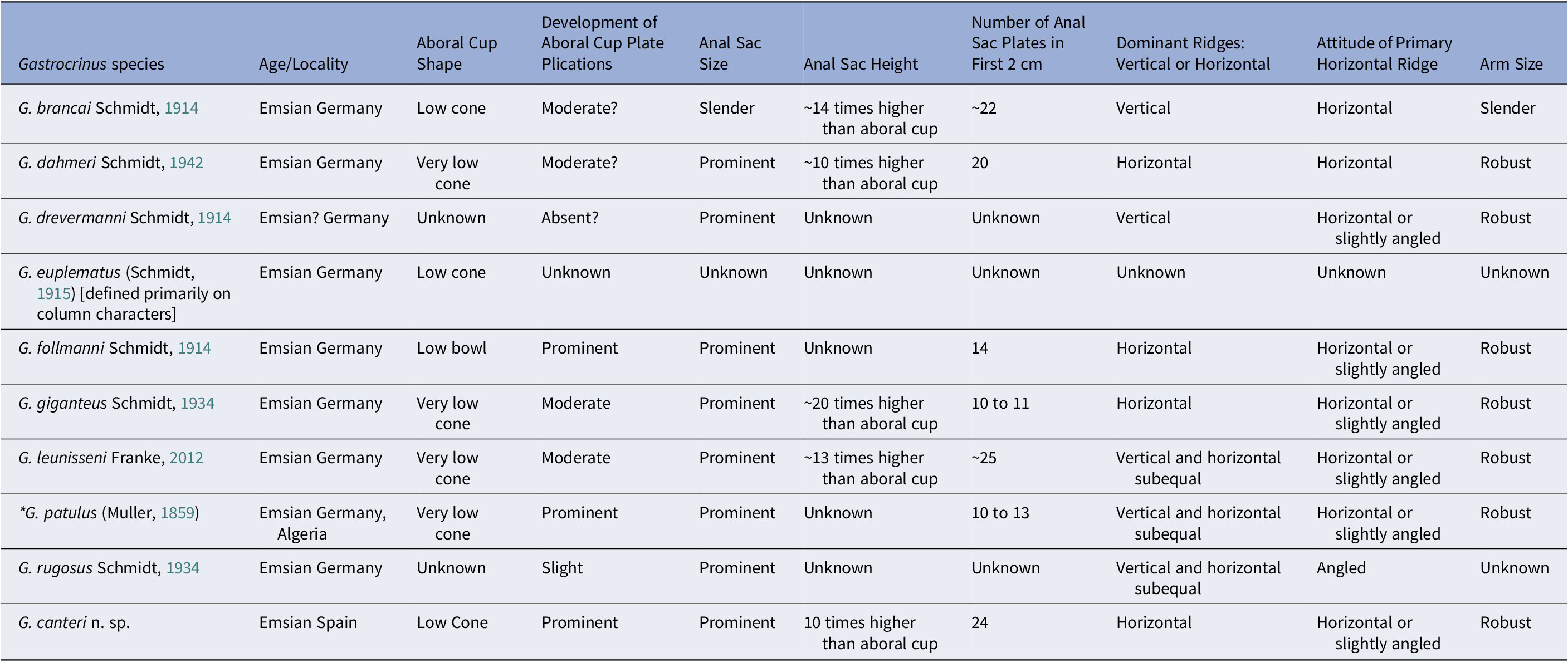
Figures 2.3–2.5, 3–5; Table 1
Characters that differentiate species of Gastrocrinus

Table 1. Long description
The table consists of 10 columns and 11 rows including the header. The columns are: Gastrocrinus species, Location/Age, Aboral Cup Shape, Development of Aboral Cup Plate Plications, Anal Sac Size, Anal Sac Height, Number of Anal Sac Plates in First 2 cm, Dominant Ridges (Vertical or Horizontal), Attitude of Primary Horizontal Ridge, and Arm Size.
* G. brancai: Emsian Germany; Low cone; Moderate?; Slender; ~14 times higher than aboral cup; ~22 plates; Vertical dominant ridges; Horizontal primary ridge; Slender arms.
* G. dahmeri: Emsian Germany; Very low cone; Moderate?; Prominent; ~10 times higher than aboral cup; 20 plates; Horizontal dominant ridges; Horizontal primary ridge; Robust arms.
* G. drevermanni: Emsian? Germany; Unknown shape; Absent?; Prominent; Unknown height; Unknown plates; Vertical dominant ridges; Horizontal or slightly angled primary ridge; Robust arms.
* G. euplematus: Emsian Germany; Low cone; Unknown plications; Unknown sac size, height, and plates; Unknown ridges; Unknown arms.
* G. follmanni: Emsian Germany; Low bowl; Prominent; Prominent; Unknown height; 14 plates; Horizontal dominant ridges; Horizontal or slightly angled primary ridge; Robust arms.
* G. giganteus: Emsian Germany; Very low cone; Moderate; Prominent; ~20 times higher than aboral cup; 10 to 11 plates; Horizontal dominant ridges; Horizontal or slightly angled primary ridge; Robust arms.
* G. leunisseni: Emsian Germany; Very low cone; Moderate; Prominent; ~13 times higher than aboral cup; ~25 plates; Vertical and horizontal subequal; Horizontal or slightly angled primary ridge; Robust arms.
* G. patulus: Emsian Germany and Algeria; Very low cone; Prominent; Prominent; Unknown height; 10 to 13 plates; Vertical and horizontal subequal; Horizontal or slightly angled primary ridge; Robust arms.
* G. rugosus: Emsian Germany; Unknown shape; Slight; Prominent; Unknown height; Unknown plates; Vertical and horizontal subequal; Angled primary ridge; Unknown arms.
* G. canteri n. sp.: Emsian Spain; Low Cone; Prominent; Prominent; 10 times higher than aboral cup; 24 plates; Horizontal dominant ridges; Horizontal or slightly angled primary ridge; Robust arms.
Types
The holotype, MGM-3426D, is a complete specimen lacking the distal stem and preserving both part and counterpart. The paratype, MGM-3430D, is also a nearly complete specimen but the calyx is crushed.
Other material
MGM-2427D, MGM-2428D (part and counterpart), MGM-3429D (part and counterpart), MGM-3431D, MGM-3432D (part and counterpart), and MGM-3433D (part and counterpart).
Diagnosis
Gastrocrinus with a low cone-shaped aboral cup; prominent development of plicated aboral cup plates, anal sac prominent, ~10 times higher than aboral cup height; ~24 anal sac plates in first 2 cm of height; horizontal or slightly angled ridges are the dominant anal sac ridges, arms robust.
Occurrence
“Arizacun Limestone” (Baztan Valley, Navarra, NE Spain).
Description
Crown small in size (Figs. 2, 3.1, 5). Aboral cup low cone-shaped (height/width ~0.57); plates with prominent ridges as part of plate plications connecting to like plications on adjacent plates; and deep, triangular depressions at plate triple junctions (Fig. 3.4). Infrabasal circlet ~20% of aboral cup height. Infrabasal plates presumably five, visible in lateral view, ~2.0 times wider than high. Basal circlet ~30% of aboral cup height. Basal plates presumably five, hexagonal or heptagonal, as high as wide (Fig. 3.3); BC basal plate mostly beneath C radial plate; CD basal plate sutured above to D radial plate, anal X, and radianal (not in sutural contact with C radial plate) (Fig. 3.4). Radial circlet height ~50% of aboral cup height. Radial plates five, pentagonal, height/width ratio ~0.89. Radial facets peneplenary, subcircular, strongly declivate (Fig. 3.5), occupies ~77% of distal radial plate width; axial canal penetrates through center of radial facets and a prominent articular ridge across width of facet and on either side of axial canal (Fig. 3.3).
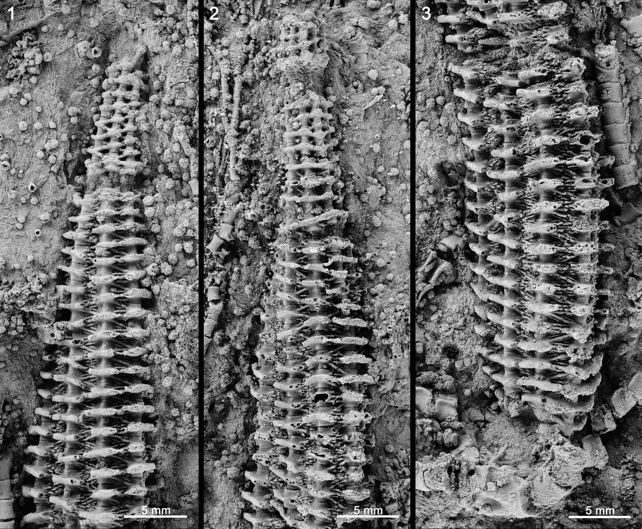
Three posterior plates in aboral cup. Radianal smaller than radial plates pentagonal, sutured beneath to upper shoulders of BC and CD basal plates; sutured laterally to anal X and C radial plate, and sutured above to right sac plate (Fig. 3.4). Anal X plate sutured beneath with D radial plate, CD basal plate, and radianal; above sutured to anal sac plates and right sac plate. Right sac plate partially in aboral cup, sutured below to the radianal and above to anal sac plates. Anal sac very high (~10 times higher than aboral cup height), gradually tapering cone (Fig. 3.1, 3.2), composed of ~8 columns of highly plicate plates (Fig. 4.1–4.3). Anal sac plates strongly convex laterally. Plates of adjacent columns alternate (Fig. 4.2). Perpendicular to the anal sac height a thick longitudinal ridge connects to similar ridges on plates of the adjoining columns of anal sac plates. Small ridges radiate from the side of anal sac column plates and also connect to similar ridges on adjoining column anal sac plates ( Fig. 4 ) forming “rhomb-like” endothecal structures.
Gastrocrinus canteri n. sp., holotype MGM-3426D, note the plicate nature of the anal sac ridges and lack of pores into the interior of the anal sac (except where broken), compare to Figure 2.5. (1) Enlargement of distal and medial anal sac, part. (2) Enlargement of distal and medial anal sac, counterpart. (3) Enlargement of proximal and medial anal sac, counterpart. All specimens are latex casts whitened with ammonium chloride sublimate.

Figure 4. Long description
A three-panel series of high-resolution grayscale photographs showing whitened latex casts of the holotype M G M-3426D. Each panel includes a 5 mm scale bar in the bottom right corner.
* Panel 1: An enlargement of the distal and medial anal sac part. The structure is elongated and tapering, composed of horizontal ridges with a plicate, folded texture. The ridges are stacked vertically, narrowing significantly toward the distal tip. The surface is solid without visible pores.
* Panel 2: An enlargement of the distal and medial anal sac counterpart. This view shows the same tapering structure as panel 1 but from the opposing side of the fossil mold. The horizontal ridges appear more deeply recessed, highlighting the skeletal lattice. A small, bulbous structure is visible at the very distal tip.
* Panel 3: An enlargement of the proximal and medial anal sac counterpart. This panel focuses on the wider, lower section of the sac. The horizontal ridges are broader and more robust than in the distal sections. The plicate nature of the ridges is highly visible, appearing as sharp, wavy horizontal lines. To the right of the main sac, a fragmented cylindrical stem or columnal section is visible in the matrix.
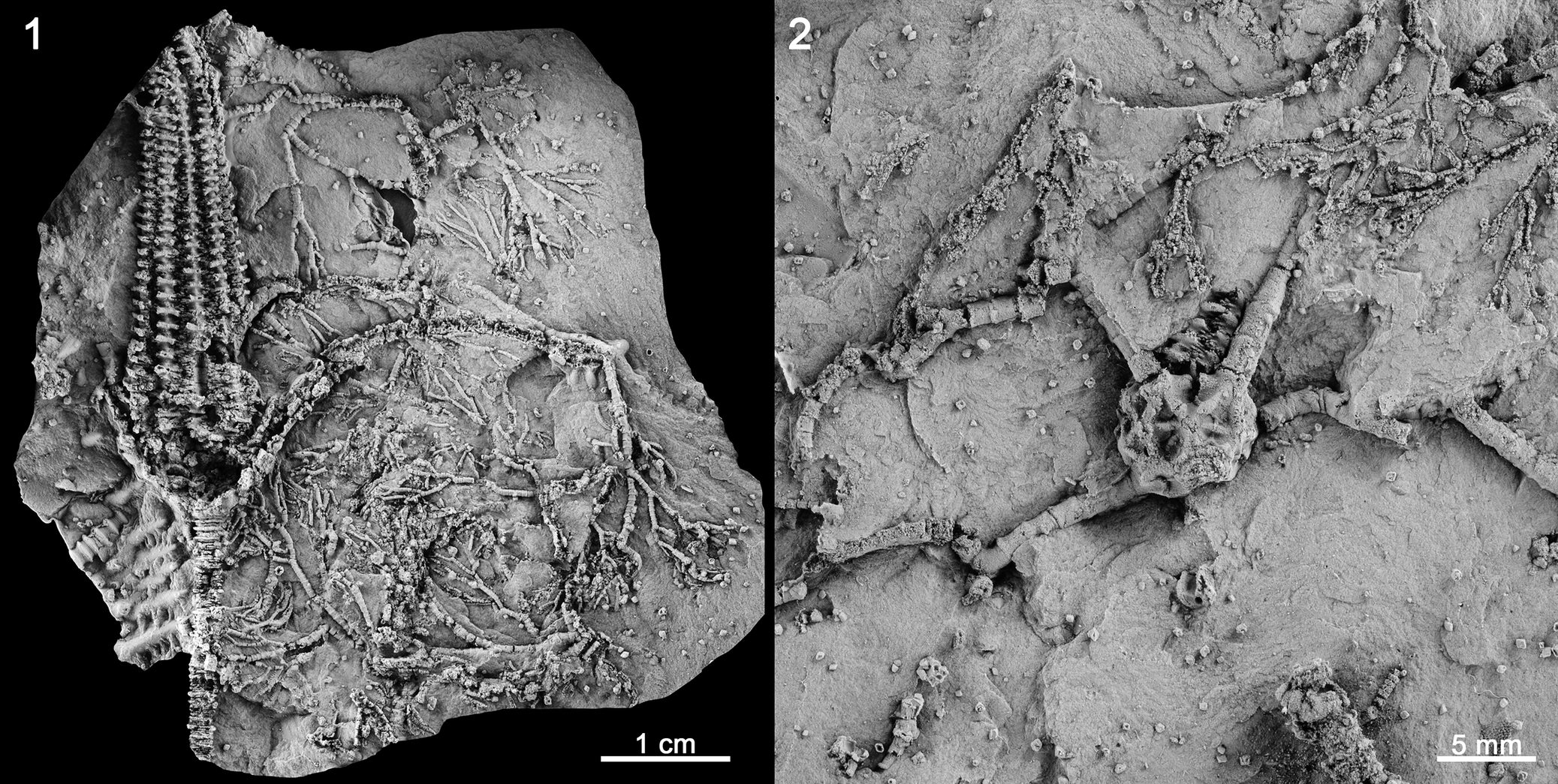
Arms poorly preserved, nonpinnulate, irregularly dichotomous branching in proximal arm with at least 4 major branches per ray (probably more) (Fig. 3.1, 3.2). Distal arms very narrow, branch many times with poor isotomy (Fig. 5). Brachials rectilinear uniserial proximally. Number of primibrachials more than 10. Primibrachials as high as wide, secundibrachials and above higher than wide, widest part of brachial plates at suture with adjacent plates, sides of brachials convex.
Gastrocrinus canteri n. sp. (1) Crown with crushed calyx, note dense distal arm branching, paratype, MGM-3430D. (2) Calyx with partial proximal anal sac and arms, specimen, MGM-3427D. All specimens are latex casts whitened with ammonium chloride sublimate.

Figure 5. Long description
A two-panel grayscale photo of latex casts of crinoid fossils.
Panel 1 (Left): Labeled with the number 1 in the top-left corner. It shows a large specimen, M G M dash 3430 D. At the left side is a thick, segmented stem or proximal column. Moving right, the central calyx is crushed and flattened. Extending from the calyx is a dense network of branching distal arms that spread across the rock matrix. A scale bar at the bottom right indicates 1 c m.
Panel 2 (Right): Labeled with the number 2 in the top-left corner. This is a closer view of specimen M G M dash 3427 D. At the center is a bulbous, textured calyx with visible plate structures. Extending upward from the calyx is a partial proximal anal sac. Several long, segmented arms radiate outward from the calyx into the surrounding matrix. A scale bar at the bottom right indicates 5 m m.
Proximal column holomeric, heteromorphic, and circular. Proximal column with nodal-internodal pattern N3231323; more internodals distally. Nodals with radices, much wider and higher than internodals; expanded, protruding radice facet (Fig. 3.4).
Etymology
The species name refers to Antonio García Cantero who collected this crinoid. He also helped during field work.
Measurements
MGM-3426D holotype: aboral cup height, 6.5 mm; aboral cup width, 9.5 mm; anal sac height, 57.0* mm. MGM-3430D, paratype: crown height, 105.0* mm; aboral cup height, 7.0* mm; aboral cup width, 18.0* mm; anal sac height, 57.0* mm.
Remarks
The large anal sac, cone-shaped aboral cup, subcircular peneplenary radial facets, arrangement of CD interray plates, sparsely branched proximal and medial arms, and densely branched distal arms align this new species with Gastrocrinus. Previously, Gastrocrinus was primarily known from the Emsian Hunsrückshiefer in Germany, although it has also been reported from other Emsian units in Germany and from Algeria and France (Webster and Webster, Reference Webster and Webster2014). Gastrocrinus aldudensis from the close Aldudes Massif in France (Fig. 1.2) is only known from an isolated anal sac, so comparison to other species is not possible. Herein, we consider G. aldudensis a nomen dubium, and this specimen should be referred to as Gastrocrinus sp. Several Devonian crinoid faunas have been described from Spain (e.g., Breimer, Reference Breimer1962; Hauser and Landeta, Reference Hauser and Landeta2007; Hauser, Reference Hauser2012; see a summary in Ausich and Zamora, Reference Ausich and Zamora2019), but G. canteri n. sp. is the first report of this genus from Spain.
Characters that differentiate species of Gastrocrinus are aboral cup shape, degree of development of plications of the aboral cup plates, relative size of the anal sac, anal sac height, number of plates in the first 2 cm of the anal sac, ridges on anal sac plates are dominant, the attitude of the primary horizontal ridges on anal sac plates, and relative arm size. Gastrocrinus canteri n. sp. is most similar to G. brancai and G. follimanni. Gastrocrinus brancai has a low cone-shaped aboral cup; moderate development of plicated aboral cup plates; slender anal sac, ~14 times higher than aboral cup height; ~22 anal sac plates in the first 2 cm of anal sac height; vertical ridges are the dominant anal sac ridges; additional horizontal ridges on anal sac plates, and slender arms. Gastrocrinus follmanni has a low bowl-shaped aboral cup; moderate development of plicated aboral cup plates; anal sac prominent, height relative to aboral cup unknown, ~14 anal sac plates in the first 2 cm of anal sac height; horizontal ridges are the dominant anal sac ridges additional ridges on anal sac oriented horizontal or slightly angled; and robust arms. In contrast, G. canteri n. sp. has a low cone-shaped aboral cup; prominent development of plicated aboral cup plates; anal sac prominent, ~10 times higher than aboral cup; ~24 anal sac plates in the first 2 cm of anal sac height; horizontal ridges are the dominant anal sac ridges, oriented horizontally or slightly oblique; and robust arms. All species assigned to Gastrocrinus are compared in Table 1.
The anal sac of Gastrocrinus has endothecal “rhomb-like” structures formed by the small plications at the lateral margins of two adjacent anal sac plates. Where broken, it is evident that the smaller and perhaps larger plications of these endothecal structures were open to the interior of the aboral cup, but there is no evidence that these endothecal structures directly connected the interior of the specimen to open sea water. Hence, these “rhomb-like” structures are similar to those of the anal sac in several other eucladids (e.g., Aesiocrinus Miller and Gurley, Reference Miller and Gurley1890; see Lane, Reference Lane1975).
The distinctive arms of Gastrocrinus are quite unusual. The proximal two-thirds of the arms have widely spaced, poor isotomy branching, whereas the distal third of the arms are densely branched with poor isotomy. The result is a very open filtration fan in the lower portion of the arms and very dense branching in the upper third.
Family Mastigocrinidae Jaekel, Reference Jaekel1918
Genus Eifelocrinus Wanner, Reference Wanner1916
Type species
Ptilocrinus dohmi Wanner, Reference Wanner1916; original designation.
Included species
Eifelocrinus baztani Zamora and Ausich n. sp.; E. bifurcatus Haarmann, Reference Haarmann1920; E. cantabricus Hauser in Hauser and Landeta, Reference Hauser and Landeta2007); E. dohmi (Wanner, Reference Wanner1916); E. hefteri Schmidt, Reference Schmidt1942; E. pennula Schmidt, Reference Schmidt1942; E. pulvinatus Schmidt, Reference Schmidt1942; and E. rhenanus Jaekel, Reference Jaekel1918.
Eifelocrinus baztani new species
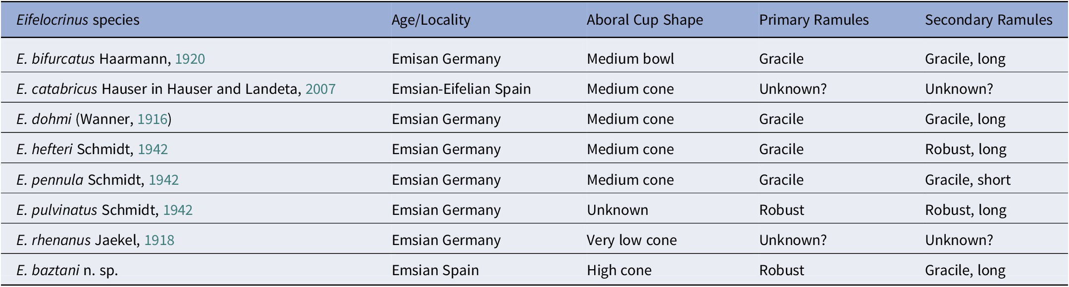
Characters that differentiate species of Eifelocrinus
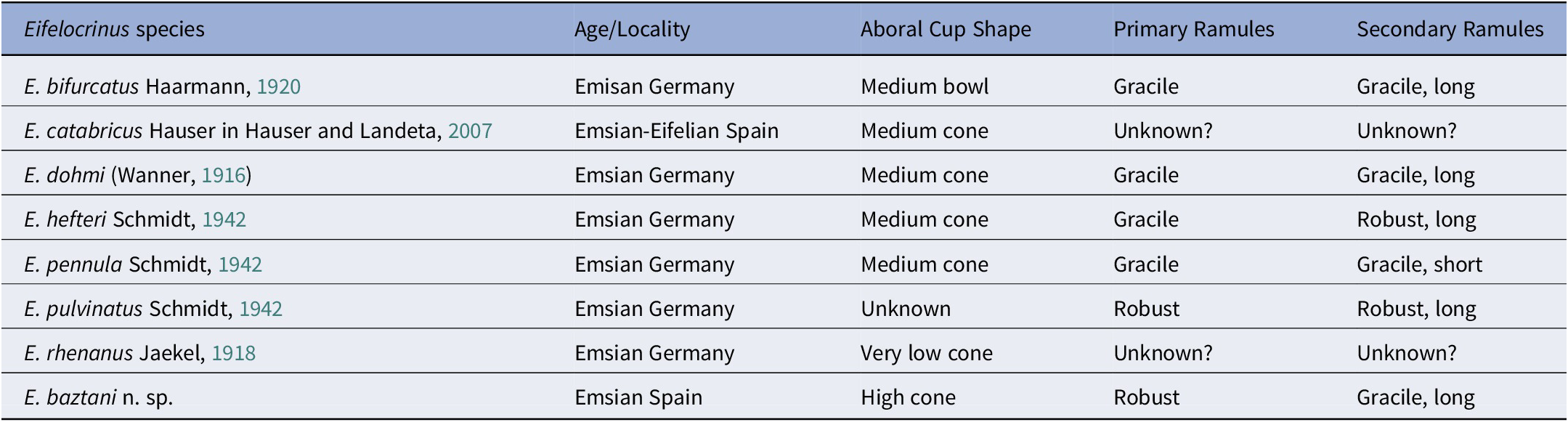
Table 2. Long description
The table contains five columns and eight rows of data for Eifelocrinus species.
* E. bifurcatus (Emisan Germany): Medium bowl cup, gracile primary ramules, gracile and long secondary ramules.
* E. catabricus (Emsian-Eifelian Spain): Medium cone cup, unknown primary and secondary ramules.
* E. dohmi (Emsian Germany): Medium cone cup, gracile primary ramules, gracile and long secondary ramules.
* E. hefteri (Emsian Germany): Medium cone cup, gracile primary ramules, robust and long secondary ramules.
* E. pennula (Emsian Germany): Medium cone cup, gracile primary ramules, gracile and short secondary ramules.
* E. pulvinatus (Emsian Germany): Unknown cup shape, robust primary ramules, robust and long secondary ramules.
* E. rhenanus (Emsian Germany): Very low cone cup, unknown primary and secondary ramules.
* E. baztani n. sp. (Emsian Spain): High cone cup, robust primary ramules, gracile and long secondary ramules.
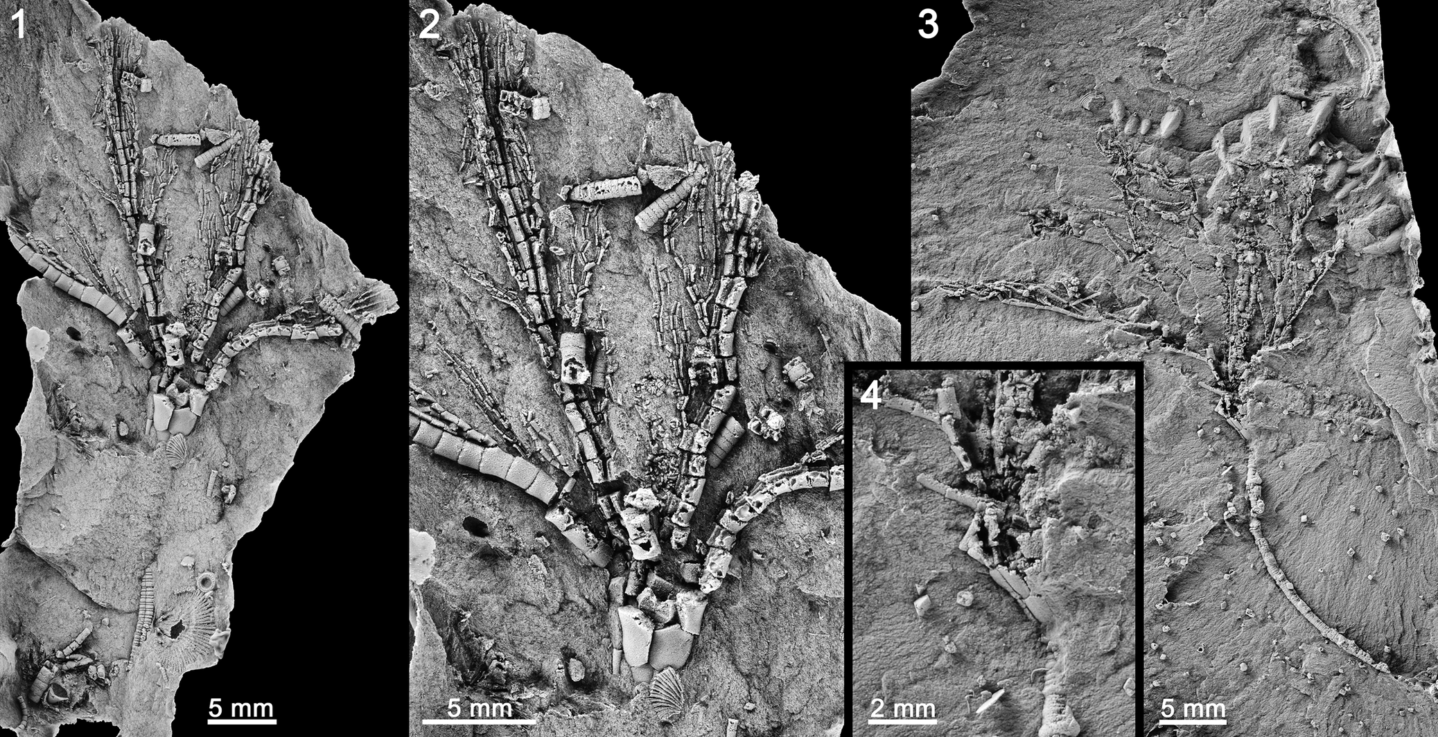
Incomplete crowns of Eifelocrinus baztani n. sp. (1) Crown and part of column, holotype MGM-3435D, (2) enlargement of aboral cup and proximal anal sac. ?Eifelocrinus baztani n. sp. (3) Crown, arms, and column, MGM-3434D, (4) enlargement of aboral cup of specimen illustrated in (3). All specimens are latex casts whitened with ammonium chloride sublimate.

Figure 6. Long description
The figure consists of four panels labeled 1 through 4.
* Panel 1: A wide view of holotype M G M-3435D. At the bottom, a segmented column extends upward into a cup-like base. From this base, multiple branching arms fan out toward the top and sides. A 5 mm scale bar is at the bottom right.
* Panel 2: A close-up enlargement of the aboral cup and proximal anal sac of the holotype from panel 1. The individual plates of the cup are visible at the center base, with the complex, textured anal sac structure extending upward between the branching arms. A 5 mm scale bar is at the bottom left.
* Panel 3: A view of specimen M G M-3434D. It shows a long, curved column at the bottom right leading to a crown with spreading arms at the top. The fossil is embedded in a textured rock matrix. A 5 mm scale bar is at the bottom right.
* Panel 4: An inset enlargement of the aboral cup from the specimen in panel 3. It focuses on the junction where the column meets the cup plates and the initial branching of the arms. A 2 mm scale bar is at the bottom left.
Type
The holotype, MGM-3435D, is a complete specimen (one part only) preserving the cup, arms, and stem.
Other material
MGM-3434D is a partial specimen.
Diagnosis
Eifelocrinus with a high cone-shaped aboral cup, robust primary ramules, and gracile and long secondary ramules.
Occurrence
“Arizacun Limestone” (Baztan Valley, Navarra, NE Spain).
Description
Crown small in size. Aboral cup plates broadly convex with smooth plate sculpturing (Fig. 6.2). Aboral cup high bowl-shaped. Infrabasal plate circlet ~28% of aboral cup height; infrabasal plates presumably five, higher than wide, visible in lateral view. Basal plate circlet ~39% of aboral cup height (Fig. 6.4); basal plates presumably five, hexagonal, higher than wide (height/width ratio ~1.3). Radial plate circlet ~33% of aboral cup height; radial plates presumably five, approximately as high as wide, pentagonal. Radial facets subcircular to ellipsoidal, peneplenary (~90% of radial plate width), gently declivate, deep adoral groove, a linear articular ridge along the width of the facet below adoral groove.
Posterior plates of aboral cup not preserved.
Five arms; with ramulate branching (Fig. 6.1–6.3). First ramule branches from ~4th primibrachial, subsequent branches at irregular intervals. Individual primary ramules bear ramules approximately every other ramular with secondary ramules always branching on the adaxial side of primary ramules. Brachials and ramulars rectilinear uniserial, higher than wide, sides straight.
Middle column may be pentameric, pentalobate, heteromorphic (Fig. 6.1, 6.4). Nodal-internodal pattern N-1.
Etymology
The name baztani refers to the Baztan valley in the Basque massifs where the material has been collected.
Measurements
MGM-3435D, holotype: Crown height, 30.4* mm; aboral cup height, 5.4* mm; aboral cup width, 4.4* mm; basal plate height, 2.4* mm; basal plate width, 1.9* mm; radial plate height, 2.2 mm; radial plate width 2.2* mm.
Remarks
Eifelocrinus is a distinctive eucladid with variously shaped aboral cups, five arms with primary ramules that have secondary ramules branching only on the adaxial side of the first-order ramules, and a pentalobate column. Based on these defining characters MGM-3435D can be confidently assigned to Eifelocrinus, despite the fact that the aboral cup is poorly preserved and the posterior interray is not known.
A second specimen (MGM-3434D) (Fig. 6.3, 6.4) is tentatively assigned to E. baztani n. sp. The aboral cup of this second specimen is nearly one-half the size of the E. baztani n. sp. holotype. The aboral cup is also partially disarticulated, the arms are much thinner and poorly preserved, it has a long length of column, and an anal sac preserved. The aboral cup is very similar to that of E. baztani n. sp., but the thin arms are too poorly preserved to discern whether the arm branching pattern is that of Eifelocrinus. The anal sac of MGM-3434D is cylindrical, narrow, and high but shorter than the arms (Fig. 6.3). It probably consists of 4–5 columns of small, convex plates. A long portion of the column is preserved with pentalobate proximal columnals and circular columnals in the middle portion of the column.
Seven species of Eifelocrinus have been described previously, all but one were described from the Emsian of Germany (Webster and Webster, Reference Webster and Webster2014). The single exception is E. cantabricus, which was described from the Emsian Santa Lucia Formation (Ausich and Zamora, Reference Ausich and Zamora2019) in northern Spain (see Hauser in Hauser and Landeta, Reference Hauser and Landeta2007). The holotype and only known specimen of E. cantabricus is an aboral cup with a few proximal primibrachials and proximal column. The height of the arms on this specimen is insufficient to preserve the characteristic ramule branching of Eifelocrinus.
Characters that differentiate species of Eifelocrinus are the aboral cup shape, character of the primary ramules, and character of the secondary ramules. Eifelocrinus baztani n. sp. is most similar to E. dohmi and E. pulviatus. Eifelocrinus dohmi has a medium cone-shaped aboral cup, gracile primary ramules and gracile and long secondary ramules. Eifelocrinus pulviatus has gracile primary ramules and robust and gracile and long secondary ramules (the shape of the aboral cup is not known). In contrast, E. baztani n. sp. has a high cone-shaped aboral cup, robust primary ramules, and gracile and long secondary ramules.
Discussion and concluding remarks
Devonian crinoids from Spain are well documented from the Cantabrian Zone (Breimer, Reference Breimer1962), Iberian Chains (Ausich and Zamora, Reference Ausich and Zamora2019), and central Pyrenees (Haude, Reference Haude1992). With the description of E. baztani n. sp. and G. canteri n. sp., we expand the distribution of complete articulated crinoids to the western Pyrenees. Both genera also have been described from the Lower Devonian of Germany. Gastrocrinus is also present in the Devonian of France. These data reinforce the observations from other authors on the similarities of echinoderm faunas between the Iberian Peninsula and France (Le Menn, Reference Le Menn1985) and Germany (Smith et al., Reference Smith, Reich and Zamora2013) during the Lower Devonian.
A striking character of G. canteri n. sp. is the deeply plicated plates on the anal sac. Although not common, similar structures occur in other eucladids, including Botryocrinus Angelin, Reference Angelin1878 (middle Silurian–Middle Devonian, Europe and North America), Proctothylacocrinus Kier, Reference Kier1952 (Middle Devonian, North America), Gilmocrinus Laudon, Reference Laudon1933 (Lower Mississippian, North America), and Aesiocrinus Miller and Gurley, Reference Miller and Gurley1890 (Lower Pennsylvanian, North America). The structure on Aesiocrinus is quite different than that on Gastrocrinus, but one of the functions proposed by Lane (Reference Lane1975) for the plicated plates and canals was respiration. Although untestable, this is a reasonable interpretation for Gastrocrinus.
Acknowledgments
We appreciate comments from two referees L. Cole and an anonymous reviewer that greatly improved the resulting manuscript. Corrections from associate editor A. Pisera are also appreciated. This work was supported by the Spanish Ministry of Science, Innovation and Universities (PID2025-169859NB-I00), co-financed by the European Regional Development Fund, project “Aragosaurus: Recursos Geológicos y Paleoambientales” (E18_17R) funded by the Government of Aragon.
This paper would be not possible without the help of A. García Cantero (Spain) who collected most of the specimens and made them available for study. He also assisted in the field work. One specimen of Eifelocrinus was donated by G. Gaspar Zuasti (Spain). First excursion to the area was possible thanks to M. Angel Apesteguia who showed us some of the outcrops in May of 2008. J. García Alcalde (Oviedo University, Spain) provided identification of brachiopods associated with crinoids.
Competing interests
The authors declare none.

 Open access

