Implications
The results show that there is a strong correlation between the high-grain (HG) diet feeding pattern and the occurrence of laminitis, which may damage the health of ruminants and cause great economic losses. The trigger of laminitis may be associated with the increased levels of lipopolysaccharide (LPS) in the rumen and peripheral blood, confirmed that the effect of HG feeding on ruminants is systemic, including but not limited to the rumen. The present study also provides a theoretical basis for the nutritional prevention of laminitis.
Introduction
Due to the shortage of high-quality pastures and to efforts to achieve ruminants’ full potential for production, ruminants are often fed diets containing a high proportion of grains, particularly in the current intensive production system. However, this feeding pattern often leads to the occurrence of rumen acidosis and causes the damage to animal health (Khafipour et al., Reference Khafipour, Krause and Plaizier2009a). As an important organ for ruminants to adapt to HG diet feeding, rumen has received a great deal of attention from researchers, and results have showed that HG diets alter the state of rumen fermentation and cause rumen metabolic disorders in dairy cows and goats (Saleem et al., Reference Saleem, Ametaj, Bouatra, Mandal, Zebeli, Dunn and Wishart2012; Hua et al., Reference Hua, Tian, Tian, Cong, Luo, Geng, Tao, Ni and Zhao2017). In addition to rumen acidosis, HG diet feeding also triggers laminitis (Nocek Reference Nocek1997), which is characterized by diffuse, non-infectious inflammation of the dermal layers inside the foot. Laminitis is a common disease in ruminants subjected to intensive industry (Bicalho et al., Reference Bicalho, Machado and Caixeta2009); however, to date, the exact mechanism of the occurrence of laminitis induced by HG diet feeding is not clear.
Keeping rumen fermentation and the bacterial community in a normal and stable condition is important for maintaining the health and production of ruminants. Previous studies have shown that HG diet feeding causes the accumulation of volatile fatty acids (VFAs) and alters the composition and function of the rumen bacterial community (Fernando et al., Reference Fernando, Purvis, Najar, Sukharnikov, Krehbiel, Nagaraja, Roe and Desilva2010). During the HG diet feeding, rumen LPS contents increased and this may be associated with the dramatic fluctuations of gram-negative bacteria, such as Bacteroidetes (Khafipour et al., Reference Khafipour, Li, Plaizier and Krause2009b). Once the rumen epithelium has been impaired, the rumen LPS can translocate across the epithelial tissue into the peripheral circulation, and hence causes a systemic inflammatory response (Khafipour et al., Reference Khafipour, Krause and Plaizier2009a; Steele et al., Reference Steele, Croom, Kahler, AlZahal, Hook, Plaizier and McBride2011). The pro-inflammatory substance, LPS, has the ability to damage the capillaries of the lamellae in the foot and cause haemorrhage, inflammation and lameness in bovines (Nocek Reference Nocek1997). Hence, the LPS in peripheral circulation may be a triggering factor of laminitis in ruminants.
Matrix metalloproteinases (MMPs) are a subfamily of zinc-dependent proteinases known to be responsible for degradation of the extracellular matrix in lamellar tissues (Hendry et al., Reference Hendry, Knight, Galbraith and Wilde2003), and their activities are regulated by tissue inhibitors of metalloproteinases (TIMPs). Among the MMPs, MMP-2 (gelatinase A) and MMP-9 (gelatinase B) have been reported to be able to degrade the basement membrane type IV collagen in the lamellar tissue of hooves (Hendry et al., Reference Hendry, Knight, Galbraith and Wilde2003). Matrix metalloproteinases, MMP-2 and MMP-9, are secreted as inactive pro-forms that are activated by extracellular proteinases, such as membrane type-1 MMP (MT1-MMP) (Sato et al., Reference Sato, Takino, Kinoshita, Imai, Okada, Stetler Stevenson and Seiki1996). Previous studies have revealed that the carbohydrates (starch) overload models of laminitis in horses is related to the up-regulation of pro-inflammatory cytokine expression (Leise et al., Reference Leise, Faleiros, Watts, Johnson, Black and Belknap2011) and the increased activity of MMP-2 and MMP-9 in lamellar tissues (Kyaw-Tanner and Pollitt, Reference Kyaw-Tanner and Pollitt2004), and it is believed that the mechanism is involved in increased concentrations of LPS in plasma (Li et al., Reference Li, Jiang, Wang, Li, Fan, Liu, Wang, Pan and Gao2015). If these results can be applied to ruminants, laminitis could be linked to variations in MMPs and pro-inflammatory cytokines in the lamellar tissues and changes in LPS levels in the blood of ruminants fed HG diets.
Here, we hypothesized that HG diet feeding alters the composition and functions of rumen microbial community, and causes the translocation of rumen LPS into blood, which further alters the histological structure and induces inflammation in lamellar tissues in hooves. Thus, in the current study, we used goats to establish the laminitis model and then investigate the effects of HG diet feeding on rumen fermentation parameters, the composition and functions of rumen microbiota, LPS levels in rumen and blood, as well as the histological structure and the mRNA expression of genes coding for pro-inflammatory cytokines and MMPs in the lamellar tissue of goats.
Material and methods
Animal experimental design
The detail animal design has been described previously (Zhang et al., Reference Zhang, Ye, Liu and Mao2017). Briefly, 12 8-month-old institutionally owned male goats (Boer×Yangtze River Delta White) were selected and used for the experiment. Before the experiment, all goats were fed hay diet and free access to water for 2 weeks and there were no significant differences in BWs of goats (27±3 kg) of two groups. The animal experiment was carried out in November, December and January in 2015. During this period, the humidity is about 60% to 70%, the hours of light were about 10 h, and the temperature is about 5°C to 15°C. After adaptation, 12 goats were randomly divided into two groups: one group was fed a hay diet (0% grain; n=6); the other group was fed HG diet (56.5% grain; n=6). Water was available ad libitum. Each goat per day was fed 900 g of dry matter diet in equal amounts at 0800 and 1700 h for 7 weeks. The diets used in the present experiment were formulated to meet the nutrient requirements of goats (NY/Y816-2004; Ministry of Agriculture of China, 2004). The Supplementary Table S1 presents the nutrient composition of feeding diets in this study.
Sample collection
On days 47, 48 and 49, all goats were measured heart rate and rectal temperature. Furthermore, they were clinically examined daily for signs of laminitis, such as inflammation of the claw and pain in the foot (Greenough et al., Reference Greenough, Vermunt, McKinnon, Fathy, Berg and Cohen1990). As an assessment of whether lame or not, gait is used on locomotion and weight-shifting as an indication of the animal favouring one or more limbs is used.
On day 50, 4 to 5 h after the feeding, blood was drawn from the jugular vein of each goat into heparinized vacutainers (Becton Dickinson, Rutherford, NJ, USA). The goats were then humanely euthanized and the abdominal cavity opened through a ventral midline incision for careful removal of the gastrointestinal tract. Immediately following slaughter, rumen content samples from each goat was collected; its pH value was determined, and it was then strained through four layers of cheesecloth. The first portion of the rumen fluid samples was deposited in 25% (wt/vol) metaphosphoric acid and then stored at −20°C until its VFA and lactic acid concentration was assessed. The second portion of the rumen fluid samples used for ruminal LPS analysis was centrifuged at 13 000×g for 40 min at 4°C. The supernatant was transferred into a sterile glass tube using a disposable 0.22-μm pyrogen-free filter and boiled for 30 min, and then finally stored at −20°C until analysed. The third portion of the rumen fluid samples was stored at −20°C until DNA extraction.
The hooves were sectioned with a band saw, according to previously described protocol (Pollitt Reference Pollitt1996). After slaughter, a dermal-epidermal tissue of hooves was cut to ~1.0×0.5×0.1 cm. These specimens were deposited in 4% paraformaldehyde (Sigma, St. Louis, MO, USA), and were used for microscopic study. The methods for obtaining the lamellar tissues have been described previously (Johnson et al., Reference Johnson, Kreeger, Keeler, Ganjam and Messer2000). Within 5 min after slaughter, the lamellar tissue of each hoof of goats were collected and frozen in liquid nitrogen for RNA extraction. The collection site of lamellar tissue was consistent and the right position was the midpoint between the horizontal plane of the coronary band and the ground surface of the dorsal side of the foot.
Rumen fermentation parameters and lipopolysaccharide concentration
Rumen pH was measured using a portable pH meter (HI 9125; Hanna Instruments, Woonsocket, RI, USA). Concentrations of rumen VFAs were determined using capillary column GC. The limulus amebocyte lysate assay (Xiamen Houshiji, Ltd, Xiamen, China) was used to determine the free LPS concentration in the rumen fluid and blood samples. A microplate spectrophotometer (Spectramax 190; Molecular Devices Corporation, Sunnyvale, CA, USA) at a wavelength of 405 nm was used to measure the optical density.
DNA extraction, Illumina MiSeq sequencing, bioinformatics analyses and metagenome prediction
A 1-ml sample of thawed rumen fluid was used for DNA extraction. Samples were first subjected to mechanical lysis of the microbial cells using a bead-beating method. In brief, bead beating of samples was conducted in a mini-bead beater (BioSpec Products, Bartlesville, OK, USA) for 2 min twice, and each interval should be cooled on ice. After bead beating, samples were incubated at 70°C for 20 min and centrifuged at 12 000×g for 10 min, and then to phenol–chloroform extraction. The extracted DNA solution was assessed by a NanoDrop spectrophotometer (Nyxor Biotech, Paris, France) and was then stored at −80°C until the follow-up experiment.
A total of 12 DNA samples were subjected to Illumina MiSeq sequencing, and the PCR reactions were conducted using the bacterial universal primers 338 F (5'-barcode-ACTCCTRCGGGAGGCAGCAG-3') and 806 R (5'-GGACTACCVGGGTATCTAAT-3'), which covered the V3 to V4 regions of the bacterial 16S rRNA genes. Sequencing reads were processed using Quantitative Insights into Microbial Ecology (QIIME) pipeline, followed by the process of quality filtering. With the remaining sequences, operational taxonomic units (OTUs) classification with a threshold of 97% sequence similarity was performed using UPARSE (version 7.1 http://drive5.com/uparse/), and de novo chimera detection and removal was conducted using UCHIME with default parameters. To obtain the representative sequences’ phylogenetic relationship, multiple sequence alignment was conducted using PyNAST and Green Gene ‘Core Set’ in the database. Community diversity and richness were determined using the following estimators: ACE, Chao 1, the Shannon index and the Simpson index. The unweighted principal coordinate analysis (PCoA) was carried out using the MOTHUR programme (version 1.29). The analysis of similarities (ANOSIM) in the PRIMER 5 software was used to determine whether there was statistical significance in rumen bacterial composition of goats of two groups.
Metagenome functional predictions of bacterial composition
The bioinformatics software PICRUSt was adapted in the present study to predict the function of the rumen bacterial community. PICRUSt uses information of the reference genome databases and ancestral state reconstruction to complete the functional prediction with 16 S rRNA gene as a marker, and it performed functional analysis using Kyoto Encyclopedia of Genes and Genomes (KEGG) database in the present study. After that, the differences in predicted functions of rumen bacterial community were measured by a principal component analysis (PCA) using the SIMCA-P (14.0) software package (Umetrics, Umea, Sweden) and the ANOSIM analysis.
Microscopic study, RNA extraction and quantitative real-time PCR
Paraformaldehyde-fixed samples were dehydrated with alcohol, and embedded in paraffin. The sections (about 6 µm thickness) were stained using haematoxylin–eosin. The morphology of the dermal-epidermal tissue of hooves was observed and photographed using light microscopy (Nikon Eclipse 801; Nikon, Tokyo, Japan).
The RNA extraction of the lamellar tissue samples was conducted using TRIzol (Takara Bio, Otsu, Japan). A NanoDrop spectrophotometer (ND-1000UV-Vis; Thermo Fisher Scientific, Madison, WI, USA) was used to quantify the RNA concentration. A 1.4% agarose–formaldehyde gel electrophoresis was used to determine the integrity of RNA samples. The reverse-transcription of total RNA (1 μg) was conducted using a PrimeScript® RT Reagent Kit with gDNA Eraser (Takara Bio) following the standard operating instructions.
The primers for pro-inflammatory cytokines interleukin-1β (IL-1β), interleukin-6 (IL-6), tumour necrosis factor-α (TNF-α) and glyceraldehyde-3-phosphate dehydrogenase (GAPDH) used were described by Liu et al. (Reference Liu, Xu, Liu, Zhu and Mao2013) and primer sets of MMP-2, MMP-9, MT1-MMP and TIMP-1 were designed using the Primer 3 computer program (Whitehead Institute, Cambridge, MA, USA). Information about the used primers (primer sequences and amplicon sizes) is presented in the Supplementary Table S2. The quantitative real-time PCR of all genes in the present study was conducted using ABI 7300 real-time PCR system (Applied Biosystems, Foster City, CA, USA). Gene expressions of pro-inflammatory cytokines and MMPs were normalized using housekeeping gene GAPDH, and the relative expression was calculated using the
 $$2^{{{\minus}\Delta \Delta C_{T} }} $$
method.
Statistical analysis
Data of rumen fermentation parameters, LPS, bacterial community and gene expressions were analysed using the independent sample t test in SPSS software (SPSS version 18; SPSS, Inc.). Significance was considered when P<0.05. For data about bacterial composition and gene families in the rumen bacterial community predicted by PICRUSt, the P-values obtained by the t test were further adjusted with a false discovery rate (FDR) analysis, and FDR P<0.1 was considered as significance. A double hierarchical analysis was performed using the unweighted pair-group method with Manhattan distance in the Number Cruncher Statistical System software (2007; NCSS, Kaysville, UT, USA), based on the relative abundance of genera. Pearson’s correlation test by GraphPad Prism 5 was used to analyse the correlations between cytokines mRNA expression and MMPs mRNA expression in the lamellar tissue. Significance was considered when P<0.05.
Results
Clinical signs
The heart rates and rectal temperatures of all goats were found to be within the normal ranges (data not shown). One of the six goats fed the HG diets exhibited early signs of laminitis, with a slight reaction of weight-shifting and swelling over the coronary band.
Effect of high-grain feeding on rumen fermentation parameters and lipopolysaccharide concentration in blood
As reported previously (Zhang et al., Reference Zhang, Ye, Liu and Mao2017), HG diet feeding decreased (P<0.05) ruminal pH and increased (P<0.05) the concentrations of acetate, propionate, butyrate, valerate, total VFAs, lactic acid and free LPS concentration in the rumen fluid. Moreover, the free LPS level in the blood of the HG group was significantly higher (P<0.05) than that in the hay group (0.62 vs. 0.21).
The composition of the rumen bacterial communities
After quality control, 487 325 reads were obtained from 12 rumen samples. The estimators of community richness and diversity included Chao 1, Ace, Shannon index and Simpson index were 542±189, 531±189, 3.82±0.62 and 0.07±0.04, respectively. The results showed that the total sequences produced from all the goats comprised 16 phyla, 15 of which existed in both groups. As Supplementary Figure S1 shows, Bacteroidetes (51.14%) and Firmicutes (45.84) were the most abundant phyla in the rumen bacterial community, accounting for about 97% of the total effective bacterial sequences, followed by Actinobacteria (0.91%), Tenericutes (0.85%), Spirochaetes (0.27%), TM7 (0.21%), Verrucomicrobia (0.18%), Proteobacteria (0.17%) and Synergistetes (0.14%). The average abundance of other phyla (such as Chloroflexi, wp2, Fibrobacteres and SR1) was<0.1% of the total bacterial community.
At the genus level, these bacterial sequences comprised 75 taxa, and 73 and 65 genera were found in the hay-fed group and HG-fed group, respectively. In Supplementary Figure S2, the top 50 bacterial genera are presented in a heat map. The dominant genera were Prevotella (25.96%), unclassified Bacteroidales (17.49%), unclassified Clostridiales (13.02%) and Ruminococcus (10.63%), followed by Butyrivibrio (5.26%), unclassified Ruminococcaceae (5.26%), unclassified S24-7 (4.12%), unclassified Lachnospiraceae (2.78%), unclassified BS11 (2.62%), Succiniclasticum (2.29%) and Bulleidia (1.99%). The average abundance of other genera was <1% of the total bacterial community.
Effect of high-grain diet feeding on the richness and diversity of rumen bacteria
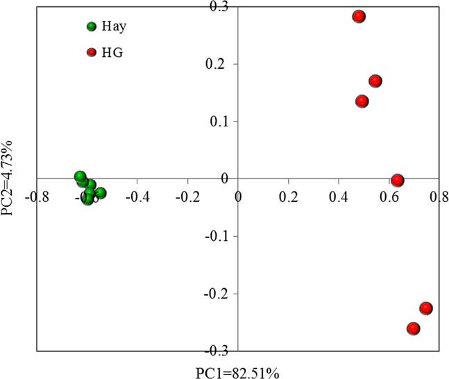
The rarefaction curves are presented in Supplementary Figure S3. The number of OTUs in each sample gradually increased with the growth of sequence numbers, and eventually reached a plateau. This result indicated that the number of samples in the present study was reasonable, and the bacterial sequences produced by the 16S rRNA sequencing reflected the abundance and diversity of the rumen bacterial community in the present study. The estimators of bacterial community richness (Ace and Chao 1) and diversity (the Shannon and Simpson indices) are summarized in the Supplementary Table S3. At the 0.03 dissimilarity, the Ace, Chao 1 and Shannon indexes were significantly lower (P<0.05) and the Simpson index was significantly higher (P<0.05) in the HG-fed group than in the hay-fed group. For further analysis of the difference in bacterial community diversity among samples, the PCoA based on unweighted UniFrac distance metrics (Figure 1) and ANOSIM were conducted. The PCoA plots showed a separation between the samples from hay-fed and HG-fed groups, and the result of ANOSIM demonstrated that this difference reached a statistically significant level (R=1, P=0.002).
(Colour online) Principal coordinate analysis (PCoA) of the rumen bacterial community in the hay-fed and high-grain (HG)-fed group. The PCoA plots were constructed using the unweighted UniFrac method.

Rumen bacterial taxonomic compositions in high-grain-fed goats as compared with hay-fed goats
At the phylum level, we observed a higher relative abundance of Actinobacteria (FDR P=0.083) and a lower relative abundance of Tenericutes (FDR P=0.083), Verrucomicrobia (FDR P=0.002), Proteobacteria (FDR P=0.008), WPS-2 (FDR P=0.009) and Fibrobacteres (FDR P=0.083) in goats fed HG diets compared with those fed hay diets (Supplementary Figure S1). However, there were no significant differences in the relative abundance of other phyla including the most abundant phyla Bacteroidetes (FDR P=0.419) and Firmicutes (FDR P=0.374) between the hay-fed and HG-fed goats.
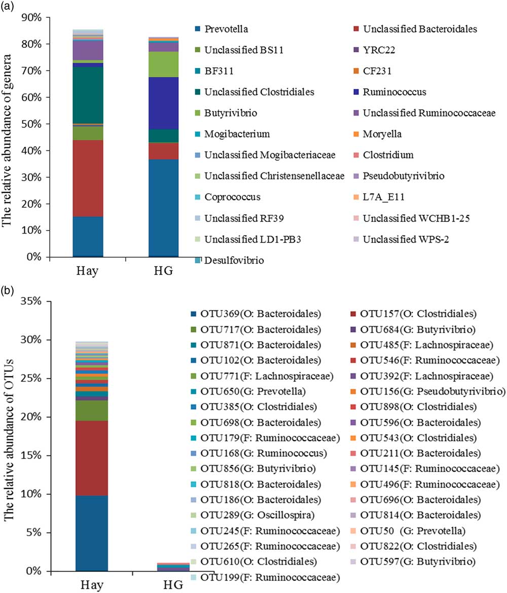
Figure 2a represented the genera that were predominant (the relative abundance of at least one group>0.1%) and significantly affected (FDR P<0.1) by dietary treatments. We observed that the majority of the changed genera belonged to the phyla Bacteroidetes and Firmicutes. In general, compared with hay diet feeding, HG diet feeding increased (FDR P<0.1) the relative abundance of Prevotella, Ruminococcus, Butyrivibrio, Mogibacterium and Moryella, and decreased (FDR P<0.1) the relative abundance of Clostridium, YRC22, BF311, CF231, Pseudobutyrivibrio, Coprococcus, L7A_E11 and Desulfovibrio. In addition, 10 unclassified taxa (such as unclassified Bacteroidales, unclassified BS11, unclassified Clostridiales and unclassified Ruminococcaceae) were affected by dietary treatments, and their relative abundance was lower (FDR P<0.1) in the goats fed HG diets than in the goats fed hay diets.
Effects of high-grain diet on the composition of rumen bacterial community. (a) The composition of rumen bacterial community at the genus level. Only the genera that were predominant (the relative abundance>0.1% in at least one group) and were significantly affected (false discovery rate (FDR) P<0.1) by dietary treatments were presented. (b) The composition of rumen bacterial community at the operational taxonomic unit (OTU) level. Only the OTUs that were predominant (the relative abundance>0.1% in at least one group) and were significantly affected (FDR P<0.1) by dietary treatments were presented.

At the OTU level, we listed the species that had a dominant position (representing>0.1% of all sequences in at least one group) in rumen bacterial community and were also significantly affected (FDR P<0.1) by treatments (Figure 2b). Among them, we found that 35 OTUs were significantly affected (FDR P<0.1) by dietary treatments, and these OTUs accounting for 29.82% and 1.09% in the hay-fed and HG-fed goats, respectively. For example, compared with the hay diet feeding, HG diet feeding increased (FDR P<0.1) the relative abundance of OTU392 (F: Lachnospiraceae), OTU650 (G: Prevotella), and OTU211 (O: Bacteroidales) and decreased the relative abundance of OTU369 (O: Bacteroidales), OTU157 (O: Clostridiales), OTU717 (O: Bacteroidales), OTU684 (G: Butyrivibrio), OTU871 (O: Bacteroidales) and OTU485 (F: Lachnospiraceae).
Predicted functional profiles of the rumen bacterial community
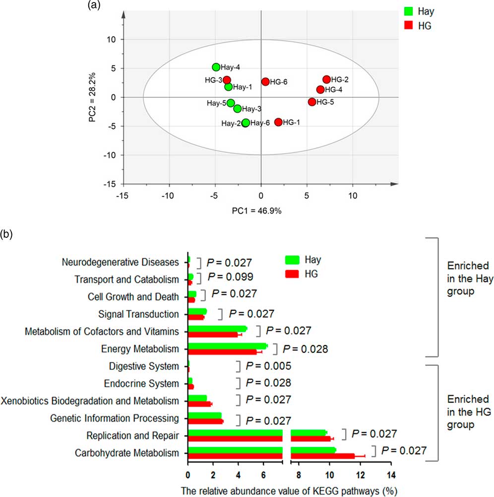
The functional profiles of the rumen bacterial community were predicted using PICRUSt, and the results identified a total of 39 gene families in all samples at the second tier (Supplementary Figure S4). Among the predicted KEGG pathways, most of the sequences were assigned to gene families carbohydrate metabolism (average 10.95%), membrane transport (average 10.36%), amino acid metabolism (average 10.19%), replication and repair (average 9.87%) and translation (average 6.31%). To further explore the implications of the effects of HG diet feeding on rumen bacterial functions, PCA was conducted and results showed that hay-group samples gathered together, and separated from HG-group samples (except the sample HG-3; Figure 3a). This separation was supported by ANOSIM analysis (R=0.474, P=0.011), which suggested a significant difference across different dietary treatments. As Figure 3b shows, compared with hay diet feeding, HG diet feeding increased the abundance of gene families carbohydrate metabolism (FDR P=0.027), replication and repair (FDR P=0.027), genetic information processing (FDR P=0.027), xenobiotics biodegradation and metabolism (FDR P=0.027), endocrine system (FDR P=0.028), and digestive system (FDR P=0.005), whereas decreasing the abundance of gene families energy metabolism (FDR P=0.028), metabolism of cofactors and vitamins (FDR P=0.027), signal transduction (FDR P=0.027), cell growth and death (FDR P=0.027), transport and catabolism (FDR P=0.099), and neurodegenerative diseases (FDR P=0.027). In addition, we also mapped the affected KEGG orthology groups (Supplementary Figure S5). Furthermore, we analysed the relative abundance of the predicted carbohydrate-active enzymes, and results showed that the relative abundance of some high-abundant enzymes (the average relative abundance ≥0.1% in at least one group) was affected by dietary treatment (Supplementary Table S4). For example, compared with hay diet feeding, HG diet feeding increased the relative abundance of α-N-arabinofuranosidase, ribose 5-phosphate isomerase B and DNA polymerase IV, whereas decreased the relative abundance of beta-galactosidase, hexosaminidase and α-glucosidase.
(Colour online) Functional diversity of the rumen bacterial community. (a) Principal component analysis (PCA) of bacterial functional diversity in hay and high-grain diet (HG) group. (b) Effects of HG diet feeding on the abundance of Kyoto Encyclopedia of Genes and Genomes (KEGG) pathways of rumen bacterial community of goats. Only the significantly affected KEGG pathways by dietary treatments are shown.

Effect of high-grain feeding on histology of lamellar tissue in hooves
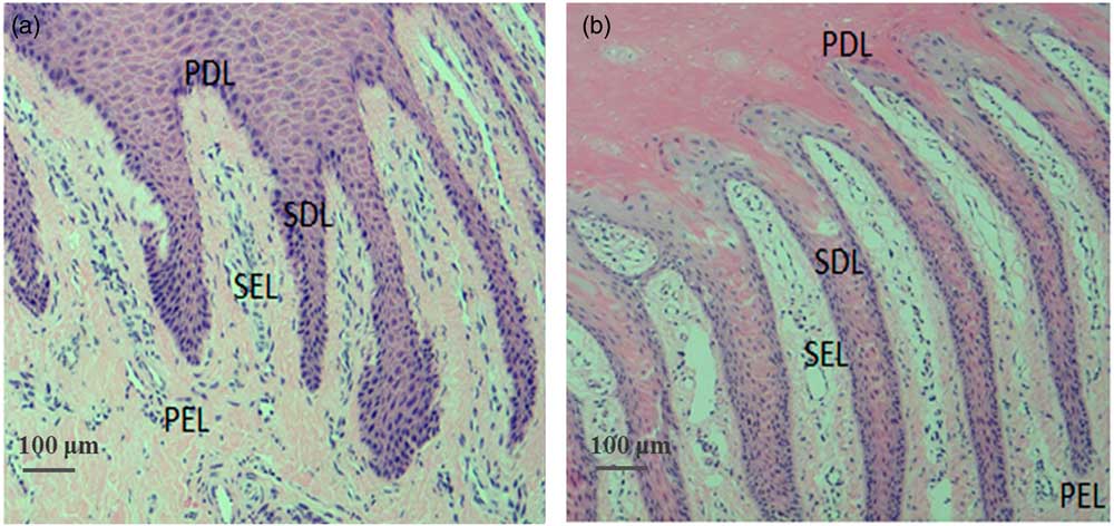
The morphology of lamellar tissue in hooves is shown in Figure 4. Compared with the hay group (Figure 4a), the secondary epidermal lamellae were straightened and elongated and the secondary dermal lamellae exhibited retraction in the goats fed an HG diet (Figure 4b). Moreover, for the HG-fed goats, between the primary dermal lamellae and the primary epidermal lamellae, more cells presented an amorphous mass alternating.
(Colour online) Histologic appearance of the dermal and epidermal lamellae in hooves of goats fed a hay diet (a) or a high-grain diet (b). SEL=secondary epidermal lamellae; SDL=secondary dermal lamellae; PDL=primary dermal lamellae; PEL=primary epidermal lamellae.

Effect of high-grain feeding on cytokines and matrix metalloproteinases mRNA expression in the lamellar tissue
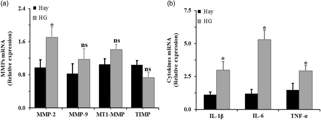
As Figure 5 shows, HG diet feeding significantly increased the relative mRNA expression of IL-1β (P=0.03), IL-6 (P<0.001) and TNF-α (P=0.03) compared with hay diet feeding (Figure 5b). Moreover, compared with hay-fed goats, HG-fed goats had higher mRNA expression of MMP-2 (P=0.001) and exhibited no impact on the mRNA expression of MT1-MMP (P=0.053), TIMP-1 (P=0.09) or MMP-9 (P=0.35) in lamellar tissues (Figure 5a).
Effects of high-grain diets on relative mRNA expression of matrix metalloproteinases (MMPs) and pro-inflammatory cytokines in lamellar tissues of goats (n=6). (a) Quantitative real-time PCR (qRT-PCR) analysis of interleukin-1β (IL-1β), interleukin-6 (IL-6) and tumour necrosis factor-α (TNF-α) mRNA expression in lamellar tissues in response to dietary change. (b). Quantitative real-time PCR analysis of MMPs mRNA expression in lamellar tissues in response to dietary change. MT1-MMP=membrane type-1 MMP; TIMP=tissue inhibitors of metalloproteinases; ns, not significant. *P<0.05.

Correlation analysis
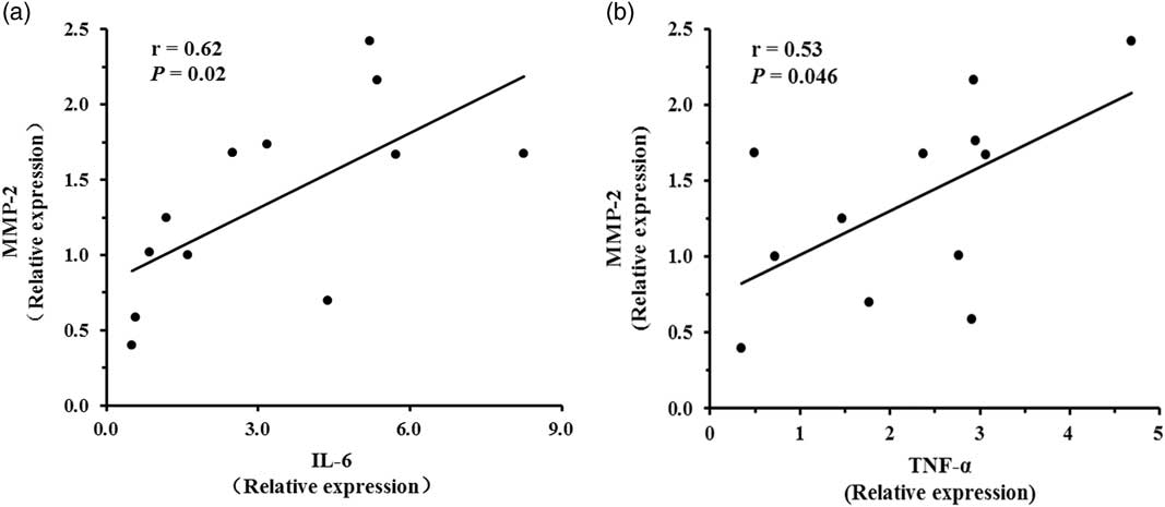
As Figure 6 shows, IL-6 (P=0.02; r=0.62; Figure 6a) and TNF-α (P=0.046; r=0.53; Figure 6b) expression were positively associated with MMP-2 expression in the lamellar tissues. There was no significant correlation between the levels of IL-1β mRNA expression and MMP-2 expression (P=0.087; r=0.51) in the lamellar tissues (data not shown).
Correlations between cytokine interleukin-6 (IL-6) (a) and tumour necrosis factor-α (TNF-α) (b) mRNA expression and matrix metalloproteinase-2 (MMP-2) mRNA expression.

Discussion
High-concentrate diet feeding, which results in higher rumen VFA production and more energy supply to the host, is a common practice in the current intensive ruminant industry. However, HG diet feeding is associated with the incidence of acidosis, and some complications, such as laminitis, usually occur with the onset of this metabolic disease (Plaizier et al., Reference Plaizier, Khafipour, Li, Gozho and Krause2012). Laminitis is a kind of aseptic inflammation disease in the dermal layers of the hooves (Nocek Reference Nocek1997), and it is rapidly becoming the most prevalent disease in the dairy industry and feedlots for beef cattle and goats (Nocek Reference Nocek1997). Hence, the relationship between HG diet feeding and laminitis has received increasing attention in recent years. In this study, we found that HG diet feeding significantly increased the concentrations of VFAs and lactic acid, which corresponded to a significant reduction in rumen pH. The pH decline induced by HG diets has been shown to result from an imbalance between the accumulations of acids (such as VFAs and lactic acid) and the absorption and reduced buffering capacity of rumen fluid (Owens et al., Reference Owens, Secrist, Hill and Gill1998).
In the present study, the alterations in rumen fermentation parameters reflected, from another point of view, that the rumen bacterial community in the HG group had undergone dramatic changes. A certain degree of bacterial community richness and diversity is important for maintaining the functional diversity of bacteria and the health of the gastrointestinal tract, and the reduced estimators of human gut have been shown to be correlated with metabolic diseases (Manichanh et al., Reference Manichanh, Rigottier-Gois, Bonnaud, Gloux, Pelletier, Frangeul, Nalin, Jarrin, Chardon, Marteau, Roca and Dore2006). In the present study, HG diet feeding reduced both the richness and diversity of bacterial community, with lower Ace, Chao 1 and Shannon index, and a higher Simpson index in the HG-fed group. This observation was consistent with the results obtained by Fernando et al. (Reference Fernando, Purvis, Najar, Sukharnikov, Krehbiel, Nagaraja, Roe and Desilva2010) and Plaizier et al. (Reference Plaizier, Li, Tun and Khafipour2016), and these changes were considered to be associated with the increased starch content in the rumen fermented substrates during HG diet feeding. Therefore, the reduced richness and diversity may compromise the functionality and stability of rumen bacteria, and hence reduce protection or contribution for the host.
In addition to these changes in richness and diversity, the composition of the rumen bacterial community in the HG-fed and hay-fed goats was also significantly different, as indicated by the clear separation of sample points from two groups on the PCoA plots. This observation was in agreement with the findings of Hua et al. (Reference Hua, Tian, Tian, Cong, Luo, Geng, Tao, Ni and Zhao2017). In general, the changes were mainly reflected in an increase in the relative abundance of genera Prevotella, Ruminococcus and Butyrivibrio, and a decrease in the relative abundance of unclassified Bacteroidales, unclassified Clostridiales, unclassified Ruminococcaceae and the phylum Fibrobacteres. The members of Prevotella show functionally and genetically diverse and are involved in the breakdown of starch and fibre (Stevenson and Weimer Reference Stevenson and Weimer2007), and the alteration in Prevotella has been reported by many studies and seems to be a typical occurrence during HG diet feeding (Fernando et al., Reference Fernando, Purvis, Najar, Sukharnikov, Krehbiel, Nagaraja, Roe and Desilva2010). However, the effect of HG diet feeding on the relative abundance of Ruminococcus and Butyrivibrio was inconsistent with the results of the above studies, in which the fluctuation of the species in these two genera appeared to depend on the adaptation time of HG diets or the severity of acidosis (Khafipour et al., Reference Khafipour, Li, Plaizier and Krause2009b; Fernando et al., Reference Fernando, Purvis, Najar, Sukharnikov, Krehbiel, Nagaraja, Roe and Desilva2010). In addition, some species in these two genera were also shown to exhibit amylolytic activities (Cotta Reference Cotta1988; Klieve et al., Reference Klieve, O’Leary, McMillen and Ouwerkerk2007), which suggested that these two genera may play an important role in adaptation to HG diets. Moreover, our results of PICRUSt predictions also showed that the genes responsible for replication and repair and genetic information processing were up-regulated in the HG group. This may be explained by the mass proliferation of gram-negative bacteria or amylolytic bacteria, which was confirmed in the present study, suggesting that the bacterial cell replication functions were enhanced in the HG group. Corresponding to the increase of starch-degradation bacteria, the relative abundance of fibre-degradation bacteria such as the phylum Fibrobacteres was reduced during HG diet feeding, and this result was consistent with the earlier study (Fernando et al., Reference Fernando, Purvis, Najar, Sukharnikov, Krehbiel, Nagaraja, Roe and Desilva2010). Fibrobacteres is the major cellulolytic bacteria within the rumen, so a decrease in its relative abundance in the rumen of the HG group was expected. It is noteworthy that several unclassified bacteria such as unclassified Bacteroidales, unclassified Clostridiales, unclassified Ruminococcaceae, were also decreased in the HG group. The exact functions of these unclassified bacteria are not clear, and hence more research is needed to determine whether they play an important role in the process of fibre degradation.
Lipopolysaccharide is the cell wall component of gram-negative bacteria and integrates closely with the bacteria surface. Increased rumen LPS concentration due to increased proportions of grains in rations is well documented (Emmanuel et al., Reference Emmanuel, Dunn and Ametaj2008). It has been suggested that the LPS generated in the rumen results from the process of rapid growth or death of gram-negative bacteria. At present, there is a paucity of data on the exact source of rumen LPS, but it is certain that its production is associated with severe fluctuations in the amount or composition of gram-negative bacteria. Indeed, in this study, the relative abundance of Bacteroidetes (the main gram-negative bacteria in rumen) was decreased by 8% in the HG-fed group when compared with the hay-fed group (55.04% v. 47.25%, not significant). Moreover, the gram-negative bacteria appeared to undergo the dramatic changes at the genus level. In general, HG diet feeding significantly increased the relative abundance of Prevotella and significantly decreased the relative abundance of unclassified Bacteroidales, unclassified BS11, YRC22, BF311 and CF231. In addition, the relative abundance of the other gram-negative bacteria Proteobacteria and Fibrobacteres, which are also considered major contributors to the free LPS pool in the rumen (Khafipour et al., Reference Khafipour, Li, Tun, Derakshani, Moossavi and Plaizier2016), also exhibited a decline in the HG group. Hence, increased rumen LPS concentration resulting from HG diet feeding was expected in the present study.
Results from the morphology and ultrastructure of the rumen epithelium revealed that the rumen epithelium of HG-fed goats showed severe injury and cellular junction erosion when compared with the hay-fed goats, and this result has been reported previously (Zhang et al., Reference Zhang, Ye, Liu and Mao2017). The study of Liu et al. (Reference Liu, Xu, Liu, Zhu and Mao2013) demonstrated that HG diet feeding caused disruption of ruminal epithelial tight junctions, and this may be responsible for the translocation of rumen LPS into peripheral blood. Consistent with these studies, our data showed that HG diet feeding caused higher LPS level in the rumen and blood of goats. The translocation of rumen LPS into peripheral blood and then triggered the systemic inflammation (Emmanuel et al., Reference Emmanuel, Dunn and Ametaj2008). Hence, LPS was considered to be an important contributor to the damage and local inflammation in the capillaries of the lamellae in hooves (Nocek Reference Nocek1997). Indeed, our data showed that HG diet feeding increased the relative mRNA expression of IL-1β, IL-6 and TNF-α in the lamellar tissue. Our findings, combined with the above reports, indicated that the development of laminitis was associated with rumen LPS translocation into peripheral blood during HG feeding.
Despite the lack of evidence on the underlying mechanism of laminitis development, laminitis in horses is believed to be related to the increased expression and activity of metalloproteinase enzymes in the lamellar tissue (Webster et al., Reference Webster, Knott and Tarlton2005). A previous study showed that laminitis in horses could be induced by MMPs (Kyaw-Tanner and Pollitt Reference Kyaw-Tanner and Pollitt2004). However, to our knowledge, information about changes in MMPs in ruminants during HG diet feeding has not been reported. In this study, the data showed that HG diet feeding up-regulated MMP-2 mRNA expression in goats, which agreed with a previous study (van Eps and Pollitt, Reference van Eps and Pollitt2004) showing that laminitis up-regulated MMP-2 gene expression in the hooves of horses. A possible explanation for this result could be increased pro-inflammatory cytokine expression. Indeed, some previous studies showed that inflammation and metalloprotease expression was closely intertwined, and MMP expression could be up-regulated by inflammatory mediators, such as IL-1β and IL-6 (Kossakowska et al., Reference Kossakowska, Edwards, Prusinkiewicz, Zhang, Guo, Urbanski, Grogan, Marquez and Janowska-Wieczorek1999). In this study, correlation analysis demonstrated that IL-6 and TNF-α were positively correlated with MMP-2 mRNA expression, indicating that activation of the inflammatory response may contribute to the process of laminar damage. In this study, there was no significant difference in MMP-9 mRNA expression between the two groups. This result was inconsistent with a previous report (Clutterbuck et al., Reference Clutterbuck, Harris, Allaway and Mobasheri2010) that showed that experimentally induced laminitis increased MMP-9 mRNA expression in the hooves of horses. A possible explanation for this could be the differences among species.
Previous studies have revealed that the lamellar pathology of horse laminitis is related to up-regulation of MT1-MMP and TIMP-1 (Loftus et al., Reference Loftus, Johnson, Belknap, Pettigrew and Black2009; Nourian et al., Reference Nourian, Asplin, McGowan, Sillence and Pollitt2009). Of these, MT1-MMP is the major activator of pro-MMP-2, and it can also degrade the basement membrane collagen type IV (Bassi et al., Reference Bassi, Lopez De Cicco, Cenna, Litwin, Cukierman and Klein-Szanto2005). Therefore, the up-regulation of MT1-MMP in the basement membrane may induce the occurrence of laminitis. In this study, HG feeding did not affect MT1-MMP mRNA expression, implying that HG diets may make only a small contribution to the development of HG-induced laminitis in goats. As mentioned above, the TIMP-1 in lamellar tissues maintains equilibrium between MMP activity and inhibition. In the present study, the expression of TIMP-1 mRNA was not affected by HG feeding in lamellar tissues of goats, and this finding was in keeping with a previous report (Webster et al., Reference Webster, Knott and Tarlton2005) that showed that diets did not affect the TIMP mRNA expression in dairy cattle. Moreover, these findings also suggested that the change in the expression of MMP-2 in lamellar tissues could play a stronger role in the development of laminitis in goats fed an HG diet.
In conclusion, we found that long-term HG diet feeding altered the pattern of rumen fermentation, with decreased rumen pH and increased levels of VFAs, lactic acid and LPS. HG diet feeding altered the relative abundance of the gram-negative bacteria in the rumen bacterial community and increased the LPS concentration in rumen fluid. HG diet feeding also resulted in a higher level of LPS in the blood, and further affected the mRNA expression of pro-inflammatory cytokines and MMP-2 in the lamellar tissues. Overall, these results suggested that HG diet feeding altered the ruminal microbial community and resulted in higher levels of LPS in the blood, which further activated the host inflammatory response and caused damage to the laminar tissues of goats.
Acknowledgements
The present study was supported by the Natural Science Foundation of China (31372339).
Declaration of interest
The authors declare they have no actual or potential conflict of interest that could inappropriately influence in this work.
Ethics statement
The experimental design and sampling procedures were approved by the Animal Care Committee of Nanjing Agricultural University in accordance with the Regulations for the Administration of Affairs Concerning Experimental Animals (approved by the State Council on 31 October, and promulgated by Decree no. 2 of the Chinese Science and Technology Committee on 14 November 1988).
Software and data repository resources
The 16S rRNA sequence data in the present study has been submitted to the NCBI Sequence Read Archive database (SRA; http://www.ncbi.nlm.nih.gov/Traces/sra/) under accession no. SRP113101.
Supplementary material
To view supplementary material for this article, please visit https://doi.org/10.1017/S175173111800040X