Introduction
The currently accepted term for a papillary tumour of the petrous bone is endolymphatic sac tumour; however, the true origin of this tumour remains controversial. We describe a case of endolymphatic sac tumour confined to the middle ear; this tumour was histologically identical to endolymphatic sac tumour but did not arise from the endolymphatic sac.
Case report
A 58-year-old woman was referred by her general practitioner to the ENT out-patients clinic, with a one-year history of right-sided tinnitus. There was no associated hearing loss or dizziness, although audiometric testing did reveal mild, asymmetrical, sensorineural impairment.
On examination, the right tympanic membrane was seen to bulge and had a slight reddish tinge, raising the suspicion of a glomus tumour.
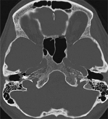
High resolution, axial computed tomography (CT) revealed a small, polypoidal mass within the right hypotympanum extending into the epitympanum (attic) (Figure 1). No associated bone destruction or ossicular disruption was demonstrated, and the inner ear structures, especially the vestibular aqueduct, were entirely normal. The jugular foramen was also normal. Appearances were highly suspicious of a glomus tympanicum with no evidence of a jugular component.
One-millimetre, axial computed tomography scan showing a soft tissue density tumour in the right middle ear (arrow).

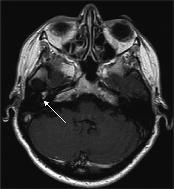
Subsequent gadolinium-enhanced, high resolution magnetic resonance imaging (MRI) confirmed the CT findings, showing a 9 mm, hypo- and epitympanic mass (Figure 2). This was avidly enhancing, and its site and homogeneous enhancement were felt to be in keeping with a glomus tympanicum. Once again, there was no evidence of jugular vein involvement and no abnormality in the region of the endolymphatic sac.
Post-gadolinium, axial magnetic resonance imaging scan showing an enhancing right middle-ear tumour (arrow).

As the mass was radiologically confined to the middle ear, surgery was deemed to be the treatment of choice. At tympanotomy, tumour was identified anterior to the handle of the malleus, filling the anterior hypotympanum with small extensions into the anterior epitympanum and down the eustachian tube. The tumour bled slightly on contact but was relatively easily cleared by potassium titanyl phosphate laser dissection, leaving the ossicular chain intact.
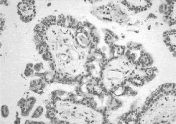
However, the histopathology specimens did not show features of a glomus tumour. Instead, the tissue consisted of fragments of a papillary tumour composed of branching tissue fronds. Each frond had a delicate, well vascularised connective tissue core and a covering of single- or double-layered, cuboidal epithelium (Figure 3). In places, the surface epithelial cells were more columnar, with eosinophilic cytoplasm, but there was minimal cytological atypia. Occasional calcospherites (psammoma bodies) and scattered intra-epithelial microcysts were evident. Despite the tumour having arisen within the middle ear rather than the endolymphatic sac, the histological picture was identical to that of a low grade papillary carcinoma of the endolymphatic sac, i.e. an endolymphatic sac tumour (previously termed ‘aggressive papillary middle-ear tumour’).
Photomicrograph showing the tumour to be composed of multiple, delicate tissue fronds with cytologically bland, cuboidal and columnar epithelium (H&E; ×20).

At follow up, the patient still complained of persistent, right-sided tinnitus. However, a combination of CT and MRI imaging nine months post-operatively, and a further MRI at 21 months, showed no evidence of tumour recurrence. Again, no abnormalities were visualised in the endolymphatic sac region.
Discussion
Endolymphatic sac tumours are rare, locally aggressive, papillary tumours of the petrous bone, which may be sporadic or associated with von Hippel–Lindau disease.Reference Kempermann, Neumann and Volk1 Their origin is controversial but, as the name suggests, they are thought to arise from the endolymphatic sac.Reference Luff, Simmons, Malik, Ramsden and Reid2, Reference Devaney, Ferlito and Rinaldo3 Other possible sites of origin include the epithelia of the middle ear and the mastoid air cells.Reference Pollak, Bohmer, Spycher and Fisch4, Reference Richards and Clifton5 Endolymphatic sac tumours were previously grouped with adenomatous tumours of the middle ear, until 1984 when Hassard and colleagues described an extradural papillary lesion that was adherent to the endolymphatic sac.Reference Hassard, Boudreau and Cronn6 In 1989, Heffner reviewed the light and electron microscopic and immunohistochemical features of 20 papillary-cystic tumours of the petrous bone, and concluded that they were low grade adenocarcinomas likely to be of endolymphatic sac origin.Reference Heffner7
The term ‘endolymphatic sac tumour’ was first proposed by Li et al. in 1993, and approximately 60 cases have since been reported using this nomenclature.Reference Li, Brackmann, Lo, Carberry and House8–Reference Jeijneveld, Hanlo, Groenewoud, Jansen, van Overbeeke and Tulleken11 Endolymphatic sac tumours are now thought to be distinct from middle-ear adenomas, which cause middle-ear masses but are rarely associated with bone destruction.Reference Van Thong, Rao and Mikaelian12 The histology of adenomas is usually that of mixed pleomorphic tumour, rather than the papillary cellular pattern seen in endolymphatic sac tumours.Reference Murphy, Geisinger and Bergman13 Carcinoid tumours are considered by some to be a subtype of mixed pleomorphic adenomas. These tumours may be locally invasive and metastasise, but they only secrete small amounts of neuropeptides and consequently tend not be associated with systemic symptoms.Reference Torske, Lester and Thompson14, Reference Ramsey, Nadol, Pilch and McKenna15
The endolymphatic sac is a complex structure of interconnecting spaces in the distal vestibular aqueduct, which ends in the epidural space of the posterior cranial fossa, close to the sigmoid sinus.Reference Lo, Daniels, Chakeres, Linthicum, Ulmer and Mark16 The sac cannot be identified on CT imaging, but the osseous vestibular aqueduct is easily identified on high resolution CT. The endolymphatic sac can be visualised with high resolution MRI. In our case, the vestibular aqueduct was entirely normal on CT, with no evidence of expansion or erosion; furthermore, no mass was identified in the endolymphatic sac region on MRI, as one would expect if this tumour had arisen from that site.
The epithelium of the endolymphatic sac is neuroectodermal in origin, whereas the middle ear, the site of glomus tympanicum tumours and mixed middle-ear pleomorphic adenomas, is lined by epithelium of endodermal origin.
Cross-sectional imaging of endolymphatic sac tumours shows avidly enhancing, vascular tumours centred around the petrous temporal bone.Reference Richards and Clifton5, Reference Hassard, Boudreau and Cronn6, Reference Torske, Lester and Thompson14 Initially, endolymphatic sac tumours involve the adjacent dura and endolymphatic duct, from where the lesion extends to the vestibule, semicircular canals, mastoid and middle-ear cavity. When tumours extend into the middle ear, they may appear as a bluish mass through an intact tympanic membrane, mimicking a glomus tumour. Further growth results in complete replacement of the mastoid and petrous pyramid by tumour. Although the tumours are locally aggressive, metastases are rare.
Early diagnosis is essential as complete excision is curative, but cure in cases of advanced tumour is virtually impossible. Patients may require further surgery to resect recurrent disease, and therefore follow-up imaging has an important role in tumour surveillance. Radiotherapy has been used in the treatment of inoperable tumours but its use is controversial and surgery is considered to be the preferred treatment option, possibly preceded by pre-operative embolisation for larger tumours.Reference Luff, Simmons, Malik, Ramsden and Reid2, Reference Richards and Clifton5 In our case, the tumour was completely excised, and therefore adjunctive radiotherapy was considered unnecessary.
To the best of our knowledge, endolymphatic sac tumour has not previously been described located solely within the hypo- and epitympanum. The tumour in our case presumably arose from neuroectodermal tissue in the middle ear – either neural crest cells or ectopic deposits.
Abnormal soft tissue demonstrated radiologically in the middle ear has a wide differential diagnosis. Glomus tympanicum tumours, vascular tumours of neural crest origin, classically present with hearing loss or pulsatile tinnitus. Cholesteatomas occur due to benign overgrowth of squamous epithelium and can cause destruction of the petrous temporal bone. Metastases to the middle ear are unusual and most commonly arise from colonic, thyroid, renal or breast primaries. Vascular anomalies (e.g. an aberrant internal carotid artery) can mimic a tumour, but the vessel can usually be identified on CT entering the middle ear.
• Endolymphatic sac tumours are rare, locally aggressive, papillary tumours of the petrous bone
• Endolymphatic sac tumour can arise solely within the hypo- and epitympanum of the middle ear and not from the endolymphatic sac; this case supports the view that the classification has been oversimplified
In our patient, there were no clinical, radiological or histological features to support these alternative diagnoses. The presentation of this patient's lesion in the middle ear provides a further challenge to the unproven and controversial concept that the endolymphatic sac is the site of origin of such tumours. This suggests that the classification of all these papillary tumours as endolymphatic sac tumours is an oversimplification, given the fact that, as in this case, they may not arise from the endolymphatic sac. Low grade, malignant, papillary tumour of the petrous temporal bone may therefore be more appropriate terminology.
Conclusion
This case was unusual in that a tumour histologically identical to an endolymphatic sac tumour arose not from the endolymphatic sac but from the middle ear. Moreover, this tumour mimicked a glomus tympanicum in its site of origin and enhancement pattern.