Introduction
Metallic implants (i.e., bone plates, screws, and Kirschner wire) have been widely used in orthopedic fields due to their significant mechanical properties and biocompatibility [Reference Hanawa1, Reference Bir, Khireddine, Touati, Sidane, Yala and Oudadesse2]. However, bone bonding or osteoconductivity is still challenging for most clinical cases since the material-dependent factors (i.e., chemical bonding associated with structure of the material) play an important role in the tissue response [Reference Ganser, Thompson, Tami, Neuhoff, Steiner and Ito3, Reference Nordström and Muñoz4]. Thus, to overcome the failure at bone and implant interface, coating of bioactive ceramics on metallic implants is suggested by a variety of techniques, including plasma spraying, sol–gel, electrophoretic deposition, laser ablation, hydrothermal, and biomimetic methods [Reference Xia, Lindahl, Lausma, Borchardt, Ballo, Thomsen and Engqvist5, Reference Pham, Dinh, Nguyen, Nguyen, Kergourlay, Grossin, Bertrand, Pebere, Marcelin, Charvillat and Drouet6, Reference Lina, Hsub, Linb and Sun7, Reference Babu, Manwatkar, Rao and Kumar8, Reference Valanezahad, Ishikawa, Tsuru, Maruta and Matsuya9, Reference Mehboob, Awais, Khalid, Ch, Siddiqi and Rehman10]. Among all these methods, biomimetic coating method is quite simple and cheap and has a low processing temperature with the ease of coating complex shapes [Reference Xia, Lindahl, Lausma, Borchardt, Ballo, Thomsen and Engqvist5]. In recent years, several studies reported that simulated body fluids (SBFs), a solution with ion concentrations and pH value similar to those of human plasma, provide a calcium-deficient (not stoichiometric) apatite layer grown on the surface of the implant [Reference Xia, Lindahl, Lausma, Borchardt, Ballo, Thomsen and Engqvist5, Reference Lina, Hsub, Linb and Sun7, Reference Kokubo and Takadama11, Reference Oyane, Kim, Furuya, Kokubo, Miyazaki and Nakamura12]. It is well known that biomimetic method produces hydroxyapatite (HA) coatings from SBF by precipitation of CaP at room temperature, which has been proven to enhance the cell proliferation and differentiation [Reference Maviş, Demirtaş, Gümüşderelioğlu, Gündüz and Çolak13, Reference Wu, Chang, Zhai and Ni14]. Thus, the biomimetic layer coated implants are expected to develop bone bonding properties [Reference Demirtaş, Kaynak and Gümüşderelioğlu15]. However, with this method, precipitation takes days to weeks due to the supersaturation levels in the SBF recipes, which commonly do not allow for rapid deposition rates. Thus, previous studies reported that synthesis of HA from supersaturated aqueous solutions like 10× SBF (10 times concentrated SBF) promoted the rapid deposition (2–24 h) of HA, which is advantageous due to low cost and simplicity [Reference Maviş, Demirtaş, Gümüşderelioğlu, Gündüz and Çolak13, Reference Demirtaş, Kaynak and Gümüşderelioğlu15, Reference Tunçay, Demirtaş and Gümüşderelioğlu16].
Strontium has received a lot of interest due to its biological role in bone metabolism, by effectively promoting bone formation and reducing bone resorption [Reference Dahl, Allain, Marie, Mauras, Boivin, Ammann, Tsouderos, Delmas and Christiansen17, Reference Marie18]. Although the main mechanism of strontium in bone-related cells has not been fully explored yet, previous studies have shown the effect of strontium on preosteoblastic cell replication, suggesting that Sr2+ ions activate the Wnt/β-catenin signaling pathway, which stimulates osteogenesis and differentiation into osteoblasts [Reference Pereiro, Rodriguez-Valencia, Serra, Solla, Serra and Gonzalez19, Reference Boyd, Rutledge, Randolph and Meenana20, Reference Wong, Lu, Chan, Cheung, Luk, Lu, Rabie, Deng and Leong21, Reference Ni, Lu, Chiu, Li, Fong and Luk22]. Thus, Sr2+ is announced as a promising enhancer of bone-related implants, and recently, studies were performed for the development of strontium-substituted HA ceramics, which are used for coatings [Reference Xia, Lindahl, Lausmaa, Borchardt, Ballo, Thomsen and Engqvist23, Reference Xue, Hosick, Bandyopadhyay, Bose, Ding, Luk, Cheung and Lu24]. Due to the similar chemical properties of Ca2+ and Sr2+, Sr2+ can be easily introduced as a natural substitute for calcium in HA, and strontium-incorporated HA (HASr) coatings have been successfully obtained using several methods, such as biomimetic precipitation, plasma spray, co-deposition sputter, and pulsed laser deposition [Reference Pereiro, Rodriguez-Valencia, Serra, Solla, Serra and Gonzalez19, Reference Boyd, Rutledge, Randolph and Meenana20, Reference Xia, Lindahl, Lausmaa, Borchardt, Ballo, Thomsen and Engqvist23, Reference Xue, Hosick, Bandyopadhyay, Bose, Ding, Luk, Cheung and Lu24]. Coating of implants is not only motivated by the development of bone bonding or osteoconductivity but also being extensively investigated to avoid bacterial proliferation and biofilm formation, which lead to endoprosthesis infections [Reference Albers, Hofstetter, Siebenrock, Landmann and Klenke25, Reference Melaiye and Youngs26]. Hence, silver-coated implants are suggested due to the well-known antimicrobial activity of silver [Reference Ciobanu, Massuyeau, Constantin and Predoi27–Reference Mirzaee, Vaezi and Palizdar30].
Recently, a few studies have reported dual coating of strontium and silver in HA on titanium implants [Reference Fielding, Roy, Bandyopadhyay and Bose31]. Fielding et al. have incorporated Ag2O and SrO into plasma-sprayed HA coatings [Reference Fielding, Roy, Bandyopadhyay and Bose31]. Huang et al. investigated silver- and strontium-doped HA coatings on titanium nanotubes fabricated by electrodeposition and reported suitability of the biomaterial for orthopedic applications [Reference Huang, Zhang, Zhang, Qiao, Zhang, Jia, Han, Gao, Xiao and Yang32]. Geng et al. successfully prepared silver- and strontium-incorporated HA coatings on Ti, which were fabricated via one-step hydrothermal method [Reference Geng, Wang, Zhuo, Li, Huang and Ma33]. However, according to our knowledge, there is no available information on providing HASr coatings on 316L stainless steel (316L SS) bone plates using biomimetic method induced by concentrated SBF-like solutions (10× SBF), and there is no available information of the ion release mechanism.
Nanomaterials not only would offer several advantages over conventional drug therapies but also expected to overcome side effects of dosing and toxicity. In this study, first, it is aimed to achieve multilayered form of strontium-incorporated and silver-coated bone-like nano-assembled apatite coatings on 316L SS bone plates, which can provide successful bone–implant interaction without causing implant infection in clinical applications. The significance and originality of this study arise from the fact that multilayered HA coatings sometimes fail efficient ion release for the osteogenesis or antimicrobial activity of the implanted area, and therefore, the release mechanisms should be determined. This study is motivated by the need to investigate release kinetics of Sr2+ and Ag+ from multilayered biomimetic bones plates due to safety concerns (i.e., possible side effects) associated with the long-term application of strontium and silver in local application. Therefore, investigation of the release profiles of Sr2+ and Ag+ is the intention of this study by researching mathematical models to evaluate release mechanisms.
In the present study, first, HASr coatings on 316L SS bone plates were maintained via a simple, cheap, time-reducing, and efficient biomimetic method with 10× SBF, then silver is coated as a second layer. The morphological, surface, and physicochemical properties of the coatings were subsequently investigated and analyzed. The coated layers are reservoirs of Sr2+ and Ag+, and the release mechanisms are determined according to several mathematical methods.
Results and discussion
In this study, it is aimed to enhance the biological and chemical properties of HA by incorporating strontium to produce HASr coatings. Then, Ag was added to biomimetic coating, as a second layer, to produce HASr-Ag coatings on 316L SS bone plates. In the present study, we used modified recipe of 10× SBF, developed previously [Reference Maviş, Demirtaş, Gümüşderelioğlu, Gündüz and Çolak13], to synthesize HASr biomimetic coatings, which could support bone growth more than pure HA does [Reference Tavares, Resende, Quitan, Castro, Granjeiro and Soares39]. Thus, not only Sr was incorporated into the HA structure by biomimetic method, but also 316L SS bone plates were coated with Sr-doped HA and Ag by means of multilayer coatings. However, previously it was reported that an alkali pretreatment is essential to establish and induce a bioactive HA layer on the surface of 316L SS [Reference Lina, Hsub, Linb and Sun7]. Thus, 316L SS bone plates were first alkali treated to form an amorphous sodium chrome pickle layer as an inter-compound for ceramic and metal linkage [Reference Lina, Hsub, Linb and Sun7].
Morphological observation
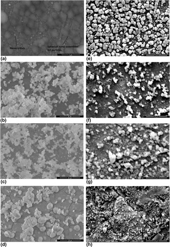
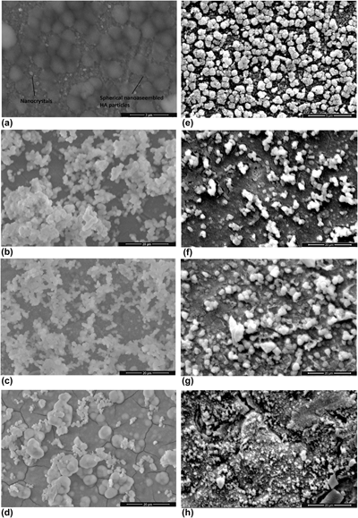
The surface morphology of HA coatings on bone plates was observed by SEM given in Fig. 1. During precipitation by 10× SBF biomimetic approach, ions in the SBF tend to form nanosized amorphous calcium phosphate precursors. After nano-HA crystals are formed, the growth of apatites continues through these nucleation zones and particles came together to form micrometer-sized spherical nanoaggregates [Reference Demirtaş, Kaynak and Gümüşderelioğlu15]. As shown in Figs. 1(a) and 1(b), nano-assembled HA structures (cauliflower structure) are formed as uniform aggregates (0.5–2 μm). Although the same morphologies were observed on HASr coatings [Figs. 1(b)–1(d)], HA clusters were nonuniform aggregates. Moreover, as shown in Fig. 1(d), it was observed that HA clusters were much more irregular in HASr3 coatings than in other coatings, forming large clusters away from nano-patterns. This result may be due to the Ca/P molar ratio (1.16, which was determined by XRF analysis) of apatites formed compared to Ca/P molar ratio of stoichiometric HA (1.67) [Reference Demirtaş, Kaynak and Gümüşderelioğlu15]. HA morphologies in HA-Ag coatings were observed to be intact [Figs. 1(e)–1(h)]. Especially, the bright areas indicate the presence of silver coating.
SEM images of (a) HA, (b) HASr1, (c) HASr2, (d) HASr3, (e) HA-Ag, (f) HASr1-Ag, (g) HASr2-Ag, and (h) HASr3-Ag coatings on 316L SS bone plates.

EDAX analysis and elemental mapping
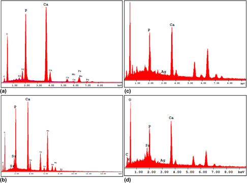
Figure 2 shows the spectra obtained from HA, HASr, HA-Ag, and HASr-Ag coatings on bone plates. The Sr and Ag peaks [Figs. 2(b)–2(d)] in the spectra confirmed the presence of Sr and Ag, incorporated successfully into the structure. The Ca/P molar ratio provides information on phase purity, chemical homogeneity, and solubility of a material to be used as bone therapeutics. Human bone contains 34.8 wt% Ca and 15.2 wt% P ions and the Ca/P molar ratio is about 1.77. However, the stoichiometric HA contains 39.8 wt% Ca and 18.5 wt% P and its Ca/P molar ratio is about 1.67 [Reference Demirtaş, Kaynak and Gümüşderelioğlu15, Reference Tunçay, Demirtaş and Gümüşderelioğlu16]. Using EDAX analysis, the Ca/P molar ratios of HA, HASr2, HA-Ag, and HASr2-Ag coatings were found to be 1.64, 1.60, 1.36, and 1.58, respectively. However, since EDAX analysis can be performed from only certain regions of the samples, the reliability of the results is controversial. Thus, the Ca/P molar ratios are determined again by the XRF analysis, described in section “XRD and XRF results”. Moreover, Ag contents in the HA-Ag and HASr2-Ag coatings were 0.13% and 0.16% by weight, respectively. The Sr contents in HASr2 and HASr2-Ag coatings were found to be 4.58% and 4.43% by weight, respectively.
EDAX spectra of (a) HA, (b) HASr, (c) HA-Ag, and (d) HASr-Ag coatings.

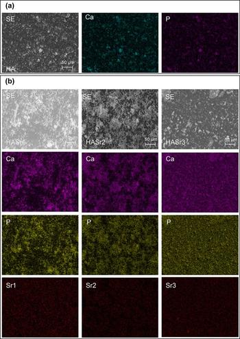
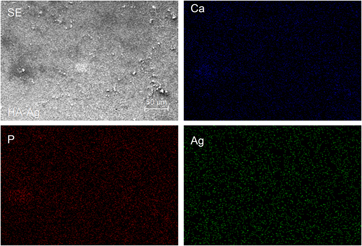
The distribution and homogeneity of Ca, P, Sr, and Ag in HA coatings were observed by elemental mapping method combined with EDAX analysis. Figures 3(a), 3(b), and 4 represent elemental mapping of HA, HASr, and HA-Ag coatings through selected regions, respectively. As shown in Fig. 3(a), Ca and P are homogenously distributed and clustered in the same regions. In the case of Sr-incorporated coatings, Sr is located on Ca/P nuclei, which carry HA aggregates, and has been found to participate in the structure through these regions homogenously [Fig. 3(b)]. However, Ag was observed to be not only homogenously distributed on Ca/P nuclei regions but also distributed uniformly throughout the structure (Fig. 4).
Elemental mapping of (a) HA and (b) HASr coatings and distributions of individual elements based on selected region of the sample.

Elemental mapping of HA-Ag coatings and distributions of individual elements based on selected region of the sample.

Raman spectroscopy
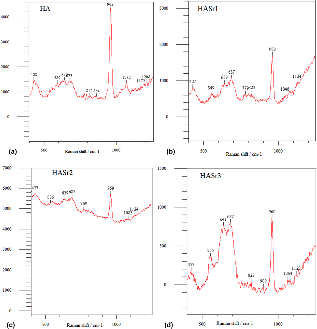
In Fig. 5, Raman spectra obtained from all types of Sr-incorporated HA coatings are compared with those of HA coatings according to the detected Raman spectroscopic bands and their assignments. In the spectrum of HA [Fig. 5(a)], an intense band situated around 962 cm−1 is assigned to P–O symmetric stretching mode (ν1) of the PO4 group, representing the most characteristic band of carbonated apatites [Reference Mandair and Morris40, Reference Quade, Schumacher, Bernhardt, Lode, Kampschulte, Voß, Simon, Uckermann, Kirsche and Gelinsky41]. The bands at 428 cm−1 and 589 cm−1 correspond to the ν2 bending vibration and ν4 mode of the PO4 group, respectively [Reference Tunçay, Demirtaş and Gümüşderelioğlu16]. A shift of stretching mode (ν1) of the PO4 group from 962 to 959 cm−1 is observed for HASr coatings [Figs. 5(b)–5(d)] with Sr incorporation to HA structure. This downshifting of the ν1 phosphate band to slightly lower values is due to the substitution of Ca by Sr in the apatite structure [Reference Quade, Schumacher, Bernhardt, Lode, Kampschulte, Voß, Simon, Uckermann, Kirsche and Gelinsky41, Reference Bigi, Boanini, Capuccini and Gazzano42]. The band shift correlates with the progressing Sr substitution, indicating increased Sr incorporation except in HASr3. O’Donnell et al. detected down-shifting of Raman bands of phosphate groups for modes ν1, ν2, and ν4, with increasing Sr and decreasing Ca content [Reference O’Donnell, Fredholm, de Rouffignac and Hill43], which is in a good correlation with our results.
Raman spectra of (a) HA, (b) HASr1, (c) HASr2, and (d) HASr3 coatings.

XRD and XRF results
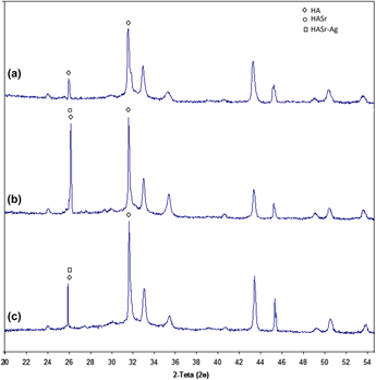
XRD patterns of HA, HASr, and HA-Ag coatings are given in Fig. 6 to determine phase purity and structure of HA coatings. The peaks observed at 2θ = 26° and 32° are the characteristic peaks of HA [Reference Demirtaş, Kaynak and Gümüşderelioğlu15]. The presence of wide peaks in the spectra indicates the amorphous HA structure, which is analogous to the crystallographic behavior of the biological apatite pattern [Reference Narasaraju and Phebe44]. As shown in Fig. 6, there are no differences in diffraction peaks between XRD patterns of HA and HASr, which indicates that there are no extraneous phases, such as dicalcium phosphate anhydrous, dicalcium phosphate dihydrate, and β-tricalcium phosphate, and only monolayer in HA structure. These results indicated that incorporation of Sr into HA structure did not affect the biomimetic HA, which conclude the suitability of biomimetic method for the production of HASr. However, the peak intensity is increased (peak at 26°) with Sr addition [Fig. 6(b)]. This result can be attributed to the fact that Sr is heavier and contains more electrons than Ca, which is consistent with the literature [Reference O’Donnell, Fredholm, de Rouffignac and Hill43]. Therefore, an increase in crystallinity of HA structure has occurred with Sr substitution. The XRD spectrum of HA-Ag coatings is similar to that of HA, but the peak intensities slightly increased and the 2θ values slightly shifted downward [Fig. 6(c)]. This shift can be explained by the change in the crystal sizes of the particles as a result of heat treatment after Ag coating [Reference Mirzaee, Vaezi and Palizdar30]. The XRD of HA-Ag also demonstrates that Ag coating as a second layer on biomimetic bone plates still exhibit the apatite characteristics with good crystal structure and no new phase or impurity.
XRD spectra of (a) HA, (b) HASr, and (c) HASr-Ag coatings.

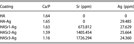
The amounts of strontium and silver within the biomimetic coatings on bone plates and Ca/P ratios were determined by XRF analysis and the results are demonstrated in Table I. Ca/P ratios of HA, HA-Ag, HASr1-Ag, HASr2-Ag, and HASr3-Ag coatings were found to be 1.64, 1.65, 1.63, 1.59, and 1.16, respectively. The results revealed that the amounts of measured Sr within HASr1-Ag, HASr2-Ag, and HASr3-Ag coatings were 0.4, 0.8, and 1.0 mM, respectively. It has been well studied that Sr substitution in HA has a superior effect both in vitro and in vivo, enhancing osteoblast proliferation and differentiation [Reference Canalis, Hott, Deloffre, Tsouderos and Marie45, Reference Chang, Tu, Chen, Komuves, Oda, Pratt, Miller and Shoback46, Reference Barbara, Delannoy, Denis and Marie47]. Moreover, Sr dose control over cells positively influences proliferation and osteogenic differentiation of cells in vitro, which is reported to be most effective between 0.001 and 1 mM [Reference Quade, Schumacher, Bernhardt, Lode, Kampschulte, Voß, Simon, Uckermann, Kirsche and Gelinsky41, Reference Braux, Velard, Guillaume, Bouthors, Jallot, Nedelec, Laurent-Maquin and Laquerrière48, Reference Schumacher, Lode, Helth and Gelinsky49]. Our results are in the range of the doses reported that positively influence proliferation and osteogenic differentiation of cells. Although, Sr has also been implicated in antimicrobial activity [Reference O’Sullivan, O’Hare, O’Leary, Crean, Ryan, Dobson and O’Neill50], it has been well known that silver exhibits good antibacterial activity toward mammalian cell [Reference Melaiye and Youngs26]. Previously, in vitro silver ion effects with the antibacterial activity against Staphylococcus epidermidis, which is the most common microorganism responsible for implant infections, have been investigated, and minimum inhibitory concentration (MIC) for silver ion was reported as 0.094 mM (∼10 ppm) [Reference Albers, Hofstetter, Siebenrock, Landmann and Klenke25]. The measured silver amounts determined from XRF results within HA coatings were in the range of 24–29 ppm (Table I), which confirms the previously reported effective doses against implant infections [Reference Albers, Hofstetter, Siebenrock, Landmann and Klenke25].
XRF analysis of biomimetic coatings showing Ca/P ratio and Sr and Ag amounts.

Release study of strontium and silver ions and mathematical modeling
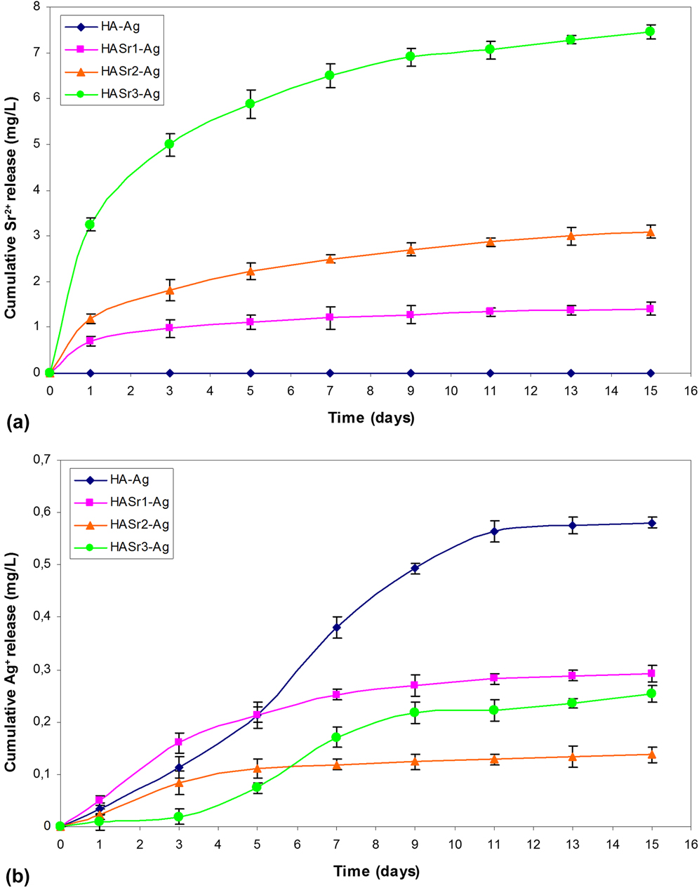
The release of Sr2+ and Ag+ ions into PBS medium was determined via ICP-OES by analyzing the supernatants of HA-Ag, HASr1-Ag, HASr2-Ag, and HASr3-Ag bone plates during 15 days of incubation (Fig. 7). Figure 7(a) represents cumulative in vitro release profiles of Sr2+ and Ag+ ions. Measurements revealed that the amount of Sr2+ ions released was 0.008 mM (HASr1-Ag), 0.014 mM (HASr2-Ag), and 0.037 mM (HASr3-Ag) at day 1, and 0.016 mM (HASr1-Ag), 0.035 mM (HASr2-Ag), and 0.085 mM (HASr3-Ag) at day 15. Although all bone plates exhibited similar controlled release profiles, a significant burst release of Sr2+ was determined for HASr3-Ag. Based on the incorporated strontium amount, according to the XRF data, the total amounts of strontium released from the HASr1-Ag, HASr2-Ag, and HASr3-Ag bone plates at the end of 15 days were determined to be 10.5%, 11.0%, and 21.6%, respectively. This study is highly motivated to adapt a defined Sr2+ concentration at the defect site. Thus, this would be a great interest for clinical applications to generate a sustained and controlled release of Sr2+ ions in therapeutically relevant concentrations. The mechanism behind osteogenic proliferation, differentiation, and bone matrix formation with the usage of strontium lies on that fact that Sr2+ ions activate the Wnt/β-catenin pathway via calcium-sensing receptors [Reference Yang, Yang, Tu, Zheng, Cai and Wang51]. The rationale behind this proposal is still unknown; however, the interaction of osteoblasts and Sr2+ ion plays an important role in the bone cell signaling with a concentration between 0.001 and 1 mM, which is reported as favorable for bone defect therapies [Reference Quade, Schumacher, Bernhardt, Lode, Kampschulte, Voß, Simon, Uckermann, Kirsche and Gelinsky41, Reference Braux, Velard, Guillaume, Bouthors, Jallot, Nedelec, Laurent-Maquin and Laquerrière48, Reference Schumacher, Lode, Helth and Gelinsky49]. The results obtained from release studies are in a good parallel with the previously reported doses (between 0.001 and 1 mM), which effects proliferation and osteogenic differentiation of cells [Reference Quade, Schumacher, Bernhardt, Lode, Kampschulte, Voß, Simon, Uckermann, Kirsche and Gelinsky41, Reference Braux, Velard, Guillaume, Bouthors, Jallot, Nedelec, Laurent-Maquin and Laquerrière48]. Moreover, after 15 days of incubation, the released doses of Sr2+, which were still in the range of the doses, positively influence cells.
Cumulative release profiles of (a) Sr2+ and (b) Ag+ from HA-Ag, HASr1-Ag, HASr2-Ag, and HASr3-Ag bone plates.

To reduce the risk of implant infections in orthopedic surgery, antimicrobial coatings, i.e., silver-coated implants, are attractive options for perioperative implant infections [Reference Gordon, Slenters, Brunetto, Villaruz, Sturdevant and Otto52]. The most common technique to incorporate silver within HA coatings is dip-coating method of HA coatings into AgNO3 for a period of time [Reference Feng, Kim, Wu, Park, Kim, Lim and Cui28]. However, this method has the limitation of quick depletion of silver in vivo/in vitro, which may be due to the fact that silver resides on the outer surface of the coating. Thus, for long-term bacterial effect, a sustained release of silver is suggested for implant systems. Although silver-coated implants have great interest in clinical practice, it has been known that Ag+ release may accumulate in the liver, kidney, and brain [Reference Gosheger, Hardes, Ahrens, Streitburger, Buerger and Erren53]. Moreover, the accumulation of ionic silver in the peri-implant tissue at unknown concentrations may affect the viability and differentiation of osteoblasts and osteoclasts [Reference Albers, Hofstetter, Siebenrock, Landmann and Klenke25]. Thus, in this study, the release of silver ions was determined by ICP-OES and the in vitro cumulative Ag+ ion release profiles are demonstrated in Fig. 7(b). Ag+ ion release was determined to be 0.099 mM (HA-Ag), 0.052 mM (HASr2-Ag), and 0.034 mM (HASr3-Ag) at day 5, and 0.269 mM (HA-Ag), 0.135 mM (HASr1-Ag), 0.064 mM (HASr2-Ag), and 0.118 mM (HASr3-Ag) at day 15. The results indicated that after 5 days, the released doses of Ag+ ions were in the range of previously described MIC (0.094 mM) for silver ion against S. epidermidis [Reference Albers, Hofstetter, Siebenrock, Landmann and Klenke25]. The total amounts of silver released from the HA-Ag, HASr1-Ag, HASr2-Ag, and HASr3-Ag bone plates at the end of 15 days were determined to be 98.5%, 52.8%, 26.7%, and 52.1%, respectively. The differences between the release profiles of silver from HASr2-Ag and HASr3-Ag bone plates possibly affect the amount of silver released. Although a similar controlled release profiles of silver from HASr1-Ag and HASr2-Ag bone plates is determined after a small burst release, long-term exposure of silver ions with lower concentrations revealed the sustained release from HASr2-Ag bone plates [Fig. 7(b)]. This result may be due to the amount of Sr incorporated in HA, which may change surface area and release mechanism. The release profiles of Ag+ form HA-Ag and HASr3-Ag bone plates are similar; however, the results indicate that high concentrations of silver are released from HA-Ag bone plates compared to all. In literature, the toxic effect of Ag+ ions on osteoblasts or mammalian cells were discussed [Reference Albers, Hofstetter, Siebenrock, Landmann and Klenke25, Reference Langanki, Ogle, Cameron, Lirtzman, Schroeder and Mirsch54]; nevertheless, these silver-coated HA bone plates need to be investigated further for systemic and long-term toxicity, osseointegration, and their related toxicity to osteoblasts and osteoclasts in vivo.
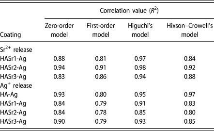
To evaluate the release mechanisms of Sr2+ and Ag+ from biomimetic coatings, release data were used by several mathematical models (zero order, first order, Higuchi, Hixson–Crowell), and correlation values (R 2) were determined (Table II) from the results of model fitting of the release profiles. As shown in Table II, according to correlation values, Sr2+ release data were well fitted to the Higuchi model for HASr1-Ag, HASr2-Ag, and HASr3-Ag coatings, which indicates that Sr2+ is released by diffusion [Reference Higuchi37], The results of model fitting of the Ag+ release profiles showed higher correlation values for Higuchi model for all the coatings (Table II). However, the best fitting release data of the Hixson–Crowell model for HA-Ag coatings (R 2 = 0.97) suggest that Ag+ is released from HA-Ag by dissolution, which may be due to the changes in surface area or changes in diameter of particles, and release is not occurred by diffusion [Reference Hixson and Crowell38]. Huang et al. indicated the fact that releasing Ag+ in the initial stage could lead to higher concentrations of Ag+ in the medium, which could have a bad effect on the osteoblasts [Reference Huang, Zhang, Zhang, Qiao, Zhang, Jia, Han, Gao, Xiao and Yang32]. According to their study, they speculated that dual release of Sr2+ and Ag+ might offset the potential cytotoxicity of Ag+ release. In this study, we concluded that dissolution of Ag+ from HA coatings could be avoided by the development of HA coating as multilayered biomimetic coatings.
Mathematical models and parameters based on Sr2+ and Ag+ release data.

Conclusions
Biomimetic coatings provided by 10× SBF-like solutions revealed an appropriate technique for the fabrication of HASr with a fine control of its strontium content and homogenous Ag coatings were achieved as a second layer on 316L SS bone plates. Results showed that strontium is successfully incorporated into the nano-assembled structure of bone-like apatite by a substitution of Ca2+ by Sr2+. Structural and surface properties can be controlled via changing the Sr2+ concentration in soaking medium. A controlled and sustained release of Sr2+ and Ag+ provided from HASr1-Ag, HASr2-Ag, and HASr3-Ag bone plates by a release mechanism of diffusion in therapeutically relevant concentrations, which is favorable to exploit dual effect of strontium and silver on bone bonding and antibacterial activity. However, further in vitro and in vivo studies should be carried out to characterize the biological properties of HASr-Ag bone plates and provide further evidence of their potential clinical utility.
Materials and methods
Treatment of bone plates before coating
The 316L SS fracture fixation bone plates were kindly provided by the Department of Orthopedics and Traumatology, Bülent Ecevit University. Before coating the substrates, which are 10 × 10 × 4 mm3 in size, an alkali treatment was performed as described before [Reference Lina, Hsub, Linb and Sun7]. Briefly, samples were soaked in 5 mL of 10 M NaOH aqueous solution at 60 °C for 24 h. Then, they were washed with distilled water and dried at 40 °C for 24 h in an air atmosphere. The alkali-treated substrates were then heated to 500–800 °C at a rate of 5 °C/min in Ni–Cr furnace and cooled to room temperature in the furnace [Reference Lina, Hsub, Linb and Sun7].
Coating of HA and HASr on bone plates from 10× SBF by biomimetic HA precipitation (first layer)
SBF solution used in this study has 10 times concentrated calcium and phosphate ions, and biomimetic HA precipitation was carried out using 10× SBF-like solution according to the method of Mavis et al., as described previously [Reference Maviş, Demirtaş, Gümüşderelioğlu, Gündüz and Çolak13]. Briefly, the reagent-grade chemicals, purchased from Merck (Darmstadt, Germany), NaCl (58.443 g), KCl (0.373 g), CaCl2·2H2O (3.675 g), MgCl2·6H2O (1.016 g), and NaH2PO4·H2O (0.250 g), were added into the ultrapure water in the given order to prepare the stock solution. Then, the samples were placed into a beaker of 100 mL of the stock solution. Precipitation was started with the addition of 10 mM NaHCO3 solution, and coating process was achieved at 37 °C for 24 h. Then, HA-coated bone plates were rinsed with distilled water and dried and stored at room temperature.
To prepare strontium-containing HA coatings, first, certain amounts of strontium nitrate [0.025% (w/v), 0.05% (w/v), 0.1% (w/v); Merck, Germany] were added into 100 mL stock solution. Then, the same coating process was performed as mentioned above. Strontium-containing HA-coated bone plates are denoted as HASr1, HASr2, and HASr3 based on their Sr inclusion concentrations of 0.4, 0.8, and 1.6 mM, respectively.
Coating of Ag on biomimetic HA- and HASr-coated bone plates (second layer)
Silver coating of bone plates was performed according to the dip-coating procedure described previously [Reference Hazer, Sakar, Dere, Altinkanat, Ziyal and Hazer34]. Polylactic acid (0.2 g; Cargill, Minnetonka, Minnesota) was dissolved in 10 mL of chloroform. About 0.01 mL of the AgNO3 (0.1 M) aqueous solution was added to this solution. Then, HA- and HASr-coated bone plates were dipped into this solution and dried in open air for 5 min. This procedure was repeated 20 times until a fine composite film layer was achieved on the surface of the plates. These film layered plates were dried under vacuum and then put in a furnace at 450 °C for 2 h to diminish the polymer. Thus, silver nanoparticles are adhered to the surface of the biomimetic HA- and HASr-coated bone plates and formed as a second layer on plates. Silver-coated bone plates are denoted as HA-Ag and HASr-Ag.
Characterization of coated bone plates
The structure and morphologies of HA, HASr, HA-Ag, and HASr-Ag coatings on bone plates were investigated by scanning electron microscope (SEM; FEI QUANTA FEG 450, Eindhoven, The Netherlands) at 15 kV under vacuum, and each sample was ion-sputtered with gold prior to examination. For the elemental analysis, the electron microscope was equipped with an energy dispersive X-ray attachment (EDAX/APOLLO X SSD device, Mahwah, NJ). The elemental maps of the prepared samples were investigated for Ca, P, Sr, and Ag. Structural changes and phase purity of the coatings were investigated by X-ray diffraction (XRD; PANalytical Empyrean diffractometer, Royston, United Kingdom; step size: 0.026°, time per step: 99.45 s, scan speed: 0.067°/s, 45 kV–40 mA Cu source). To support the XRD findings of HASr coatings and provide evidence of ionic substitution, Raman spectral characterization of the HASr-coated bone plates was performed using a Raman Spectrometer (RENİSHAW Invia, Edge Gloucestershire, United Kingdom) with an excitation wavelength of 532 nm. The composition of the coatings was quantitatively determined by X-ray fluorescence (XRF) on PANalytical equipment (Royston, United Kingdom) with a rhodium tube (4 kW). Therefore, the strontium and silver contents and also the Ca/P ratios could be verified.
In vitro release studies of Sr2+ and Ag+ from biomimetic bone plates
Sr2+ and Ag+ release studies from bone plates were performed by incubating bone plates in 10 mL phosphate buffered saline (PBS) solutions (pH = 7.4) at 37 °C. HA-Ag, HASr1-Ag, HASr2-Ag, and HASr3-Ag bone plates were immersed into tubes containing 10 mL PBS. Then, at predetermined time intervals (1, 3, 5, 7, 9, 11, 13, and 15 days), buffer solutions were changed with fresh ones. The amounts of Sr and Ag released from the bone plates were determined at selected time intervals through ICP-OES (PerkinElmer Optima 4300 DV, Wellesley, Massachusetts) analysis, and the cumulative release graphs were maintained.
Mathematical modeling and release kinetics
To investigate the mechanism of Sr2+ and Ag+ release from biomimetic coated bone plates, in vitro release data were analyzed using various kinetic models. The zero-order kinetic model [Eq. (1)] is described as follows [Reference Hadjiioannou, Christian, Koupparis and Macheras35]:
 $${Q_t} = \!\!{\rm{\ }}{Q_0} + {\rm{\ }}{K_0}t\quad ,$$
where Q 0 = initial amount of drug, Q t = cumulative amount of drug release at time “t,” and K 0 = zero-order release constant.
The first-order release [Eq. (2)] is defined as follows [Reference Bourne36]:
 $$\log {\rm{\ }}{Q_t} = \!\!\!{\rm{\ }}\log {\rm{\ }}{Q_0} + {\rm{\ }}{K_1}t\quad ,$$
where K 1 is the first-order release constant.
Higuchi described the release of drugs from insoluble matrix as a square root of time-dependent process based on the Fickian diffusion [Reference Higuchi37]:
 $${Q_t} = \!\!{\rm{\ }}{Q_0} + {\rm{\ }}{K_{\rm{H}}}{t^{{1 / 2}}}\quad ,$$
where K H is the Higuchi constant.
The Hixson–Crowell cube root law [Eq. (4)] is defined as follows [Reference Hixson and Crowell38]:
 $$\root 3 \of {{Q_t}} - \root 3 \of {{Q_0}} = {K_{{\rm{HC}}}}t\quad ,$$
where K HC is the Hixson–Crowell constant.
Modeling was performed using the parameters that provide the closest fit between experimental observations and the nonlinear function. The model that best fits the release data was selected based on the correlation coefficient (R 2) in the models described above. The model that gives the highest R 2 value is considered as the best fit of release data.
Statistical analysis
All data are expressed as means ± standard deviations of a representative of three similar experiments carried out in triplicate.
Acknowledgment
This study was financially supported by Bülent Ecevit University Scientific Research Projects, Project No. 2015-3997 1044-02.