Introduction
Gastro-oesophageal reflux disease is defined as the retrograde flow of gastric content into the oesophagus caused by transient lower oesophageal sphincter relaxation. In children, common clinical manifestations of gastro-oesophageal reflux disease include heartburn, vomiting and regurgitation, as well as irritability and feeding difficulty.Reference Vandenplas, Rudolph, DiLorenzo, Hassall, Liptak and Mazur1 Gastro-oesophageal reflux disease has been associated with a variety of extra-oesophageal manifestations relevant to Otolaryngologists, such as chronic cough, wheeze and stridor, a hoarse voice, frequent episodes of croup, difficult-to-treat asthma, recurrent chest infections, and dental erosion.Reference Tolia and Vandenplas2 These symptoms are collectively known as extra-oesophageal reflux disease.
Empirical treatment for gastro-oesophageal reflux disease is often instituted when extra-oesophageal reflux disease is the suspected aetiology. Treatment with proton pump inhibitors (PPIs) changes refluxate acidity, promotes oesophageal mucosa healing and provides symptomatic relief in over 90 per cent of gastro-oesophageal reflux disease patients.Reference Vandenplas, Rudolph, DiLorenzo, Hassall, Liptak and Mazur1 Although they are well tolerated, other studies suggest that PPIs benefit only 69 per cent of extra-oesophageal reflux disease patients.Reference Pina Dore, Pedroni, Pes, Maragkoudakis, Tadeu and Pirina3
Confirming a diagnosis of extra-oesophageal reflux disease is problematic owing to limitations of the currently available investigations. For example, pH probe monitoring, barium meals and gastroscopy only give information about the oesophageal manifestations of gastro-oesophageal reflux disease.Reference Vandenplas, Rudolph, DiLorenzo, Hassall, Liptak and Mazur1 Multichannel intraluminal impedance monitoring can detect both acid and non-acid reflux, show the degree of proximal migration of refluxate within the oesophagus and provide data on the association between reflux episodes and symptoms thought to be secondary to extra-oesophageal reflux disease.Reference Tutuian and Castell4 However, it is expensive, not widely available and involves time-consuming manual data analysis.Reference Samuels and Johnston5 There is also a lack of normative values for the paediatric population. The presence of lipid-laden macrophages in bronchoalveolar lavage fluid is reported to be neither a sensitive nor specific indicator of reflux aspiration.Reference Kitz, Boehles, Rosewich and Rose6, Reference Krishnan, Mitchell, Messina, Day and Bohane7 Bile acid is another marker, but it is only detected in the minority of cases involving duodenogastric reflux.
Diagnostic laryngobronchoscopy may be performed to exclude structural abnormality and identify macroscopic changes thought to be attributable to extra-oesophageal reflux disease or another pathology. Findings in the laryngopharynx can be scored using the reflux finding score.Reference Belafsky, Postma and Koufman8 Others have described signs of subglottic oedema and posterior commissure hypertrophy;Reference Wang, Liu, Liu, Zeng, Wu and Yang9 arytenoid and vocal fold oedema, enlarged lingual tonsils and cricotracheal cobblestoning;Reference Carr, Nguyen, Poje, Pizzuto, Nagy and Brodsky10 and ventricular obliteration, erythema and hyperaemia, and thick endolaryngeal mucus.Reference Simons, Rosen, Casselbrant, Chi, Schaitkin and Rubinstein11 Mucosal cobblestoning is a common finding in the larynx, posterior pharyngeal wall and trachea, although its aetiology remains uncertain.Reference Vakil, van Zanten, Kahrilas, Dent and Jones12 Despite correlating with gastro-oesophageal reflux disease findings in some studies, these changes are all non-specific, subjectively assessed and could have other inflammatory causes.
Pepsin, however, is produced when pepsinogen from the chief cells in the stomach comes into contact with acid produced by parietal cells deep within the gastric pits. Detection of this proteolytic enzyme at a site other than the stomach (such as the trachea) is thus an ideal method for confirming extra-oesophageal reflux disease. Pepsin is assayed in tracheal aspirates by either enzymatic methods or an enzyme-linked immunosorbent assay using monoclonal antibodies.Reference Tutuian and Castell4 These assays have a high sensitivity and specificity for reflux microaspiration. There is a strong association between a positive pepsin finding and underlying respiratory symptoms independent of having typical symptoms associated with gastro-oesophageal reflux disease.Reference Krishnan, Mitchell, Messina, Day and Bohane7 Rosen et al. also demonstrated that positive pepsin findings in lung samples is a strong predictor of reflux-related lung disease.Reference Rosen, Johnston, Hart, Khatwa and Nurko13
Pepsin in refluxate depletes the levels of two prominent laryngeal ‘protective’ proteins, carbonic anhydrase III and squamous epithelial stress protein 70, which predisposes to inflammatory changes.Reference Johnston, Dettmar, Bishwokarma, Lively and Koufman14 In addition, in animal studies, acid refluxate has been shown to induce major inflammation in respiratory epithelium.Reference Carr, Nguyen, Poje, Pizzuto, Nagy and Brodsky10
This study aimed first to investigate a possible association between the finding of macroscopic changes on laryngobronchoscopy and pepsin-positive tracheal aspirates, and second to investigate a possible correlation between a positive pepsin assay and the findings of standard tests for gastro-oesophageal reflux disease or specific clinical features, which may be useful for diagnosing extra-oesophageal reflux disease in the paediatric population.
Materials and methods
Data collection
A retrospective review was performed of the medical records of children who underwent laryngobronchoscopy and tracheal aspirate collection for pepsin analysis between January 2005 and February 2011 at a tertiary paediatric hospital. These patients presented with chronic respiratory and ENT symptoms, and extra-oesophageal reflux disease was suspected. There were no exclusion criteria. Data were collected on the presenting clinical features, medical history, the standard gastro-oesophageal reflux disease investigations performed and response to therapy. Laryngobronchoscopy results were specifically reviewed for macroscopic signs of extra-oesophageal reflux disease, focusing on cobblestoning, inflammatory changes and the site of abnormalities.
Tracheal aspirates were collected using a suction catheter and sputum trap during laryngobronchoscopy, with 2 ml of sterile normal saline to assist passage of the specimen from the suction tubing to the collection container. Aspirates were assayed for pepsin using the enzymatic fluorometric method.Reference Krishnan, Mitchell, Messina, Day and Bohane7 A pepsin assay value greater than 30 fluorometric units was considered a positive result, while a value less than 30 was deemed negative.Reference Krishnan, Mitchell, Messina, Day and Bohane7
A range of standard gastro-oesophageal reflux disease investigations (barium meal, gastroscopy and pH probe monitoring) had been performed in some, but not all, of the children with typical symptoms of gastro-oesophageal reflux disease as part of the investigation. A positive barium meal result was noted if a reflux event was detected in the proximal oesophagus during the procedure. Gastroscopy was considered positive if reflux-induced oesophagitis occurred upon biopsy, according to the Los Angeles classification system.Reference Lundell, Dent, Bennett, Blum, Armstrong and Galmiche15 A 24-hour pH probe investigation was considered positive (or abnormal) if the acid reflux index was more than 10 per cent in infants less than 1 year of age or more than 6 per cent in those more than 1 year of age.Reference Vandenplas, Rudolph, DiLorenzo, Hassall, Liptak and Mazur1
Ethics statement
Ethical approval was obtained from the Human Research Ethics Committee of the South Eastern Sydney Illawarra Area Health Service Network prior to commencement of the project.
Statistical analysis:
Data was entered into a Microsoft Excel 2007 spreadsheet (Microsoft; Washington, USA) and analysed using PRISM software (GraphPad; California, USA). The results of standard gastro-oesophageal reflux disease investigations were analysed for correlation with pepsin assay and laryngobronchoscopy findings.
The analysis consisted of a descriptive statistical review and an association study between relevant variables through calculating the odds ratio of having a positive result, with a 95 per cent confidence interval based on the sample size. Subgroup analysis of patients stratified by age, sex and recurrent croup, atopy and cystic fibrosis (CF) were also performed.
Results
Patient demographics
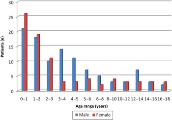
A total of 188 paediatric patients were included in this study, ranging in age from 1 month to 18 years: 84 (44.7 per cent) were female and 104 (55.3 per cent) were male (Figure 1). Of these, 44.7 per cent were under two years of age. The mean age was 3.99 (3.40–4.58) years and the median (interquartile range) was 2.46 (1.08–5.27) years. In all, 93 per cent of the children were receiving oral nutrition alone (i.e. without gastric or jejunal tube feeds or intravenous nutrition).
Graph showing patient age and sex distribution.

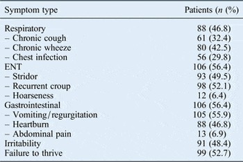
A total of 46.8 per cent of children presented with respiratory symptoms such as a cough, wheeze or recurrent chest infections and 56.4 per cent presented with an ENT symptom such as stridor, croup or hoarseness. A total of 56.4 per cent had gastrointestinal (GI) symptoms, including vomiting, regurgitation, heartburn, abdominal pain, irritability and/or failure to thrive (Table I). Medical histories included atopy (59.6 per cent), CF (39.9 per cent), developmental delay (10.1 per cent) and previous oesophageal atresia repair (5.8 per cent). There was overlap among the children's symptoms and their medical histories.
Presenting symptoms

Diagnostic tests
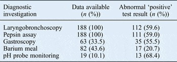
All 188 patients underwent laryngobronchoscopy and tracheal pepsin analysis. Results of these and the other standard investigations for gastro-oesophageal reflux disease are listed in Table II.
Results of diagnostic investigations

Laryngobronchoscopy findings
During laryngobronchoscopy for symptom investigation, macroscopic features such as erythema, oedema, ventricular obliteration, posterior commissure hypertrophy, increased secretions and cobblestoning were noted; these are suggestive of extra-oesophageal reflux disease. In all, 112 patients (59.6 per cent) had positive laryngobronchoscopy findings suggestive of extra-oesophageal reflux disease, including 55 (29.3 per cent) with cobblestoning. Sites of macroscopic abnormality included the larynx (58.9 per cent), trachea (41.1 per cent), cricoid region (34.8 per cent) and pharynx (16.1 per cent). Laryngobronchoscopy findings suggestive of extra-oesophageal reflux disease were significantly associated with the presence of respiratory (odds ratio 3.39, 95 per cent CI 1.84–6.25; p < 0.0001) and ENT (odds ratio 3.08, 95 per cent CI 1.68–5.65; p = 0.0003) symptoms, but not GI symptoms (odds ratio 0.77, 95 per cent CI 0.43–1.39; p = 0.46).
When laryngobronchoscopy and pepsin assay findings were compared, the presence of a macroscopic abnormality in the upper aerodigestive tract was significantly associated with a positive pepsin assay (p < 0.0001) (Figure 2). Cobblestoning (either the presence or absence) was mentioned in only 72 of the 188 laryngobronchoscopy reports analysed. The remaining 116 reports were therefore excluded from the analysis of this particular variable. Of those with positive laryngobronchoscopy findings, there was a significant association between the presence of cobblestoning and a positive pepsin assay (p < 0.0001). However, positive laryngobronchoscopy findings were not significantly associated with the results of other standard investigations for gastro-oesophageal reflux disease (Figure 2).
Forest plot showing the association of positive laryngobronchoscopy results with pepsin-positive tracheal aspirates and the standard investigations for gastro-oesophageal reflux disease.

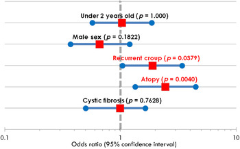
Several subgroups within our patient population were analysed separately. There was no significant association between positive laryngobronchoscopy findings and age under two years, male sex or CF (Figure 3). However, of the 112 (59.6 per cent) patients recorded as having a history of atopy, 77 (68.8 per cent) had positive laryngobronchoscopy findings, with cobblestoning identified in 35 (31.3 per cent) of these. There was a significant association between positive laryngobronchoscopy findings and a medical history of atopy (p = 0.004). Similarly, in the 98 (52.1 per cent) patients with recurrent croup, there was a significant association with macroscopic upper airway changes on laryngobronchoscopy (p = 0.0379).
Forest plot showing the subgroup analysis for patients with positive laryngobronchoscopy results.

Pepsin assay findings
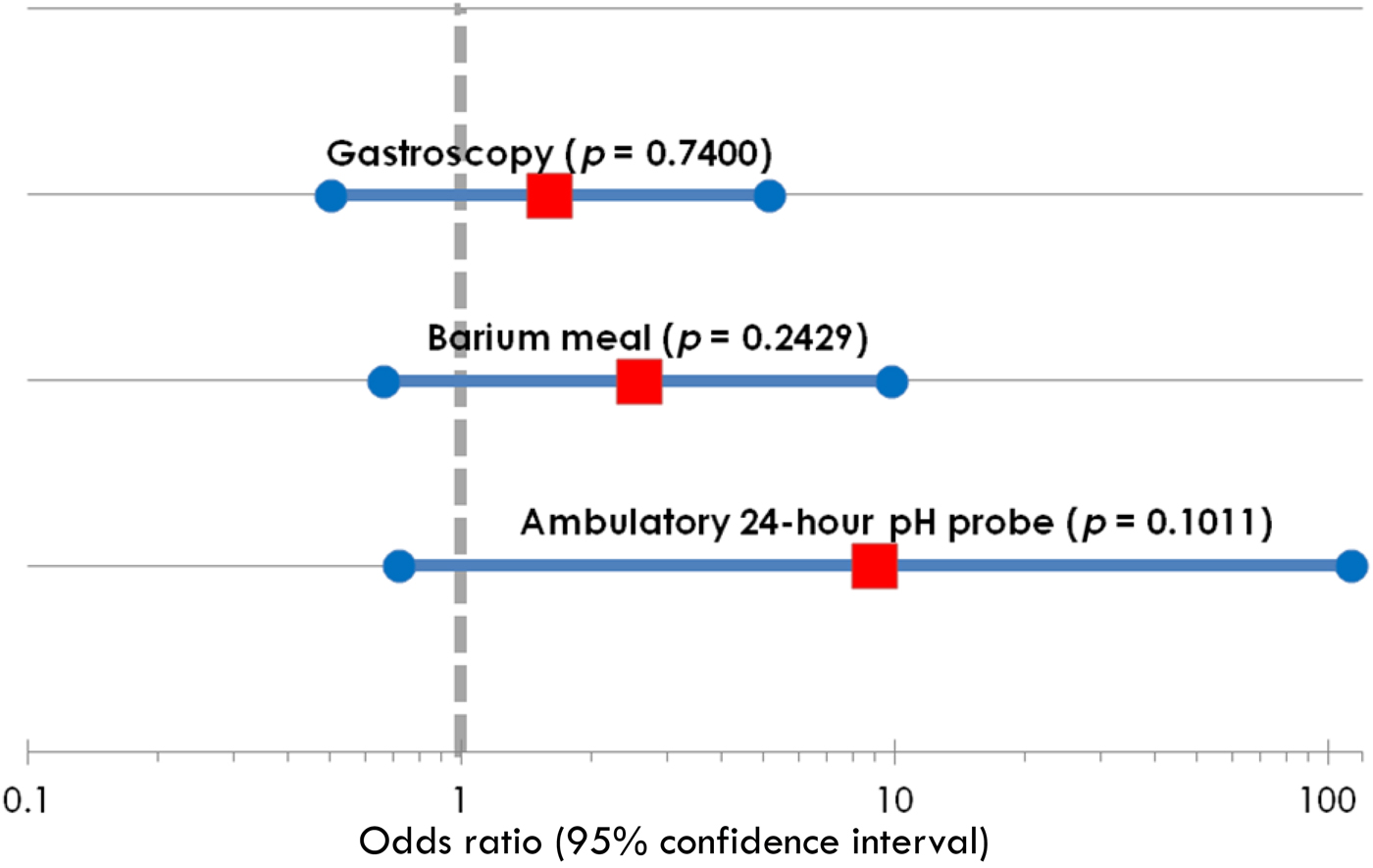
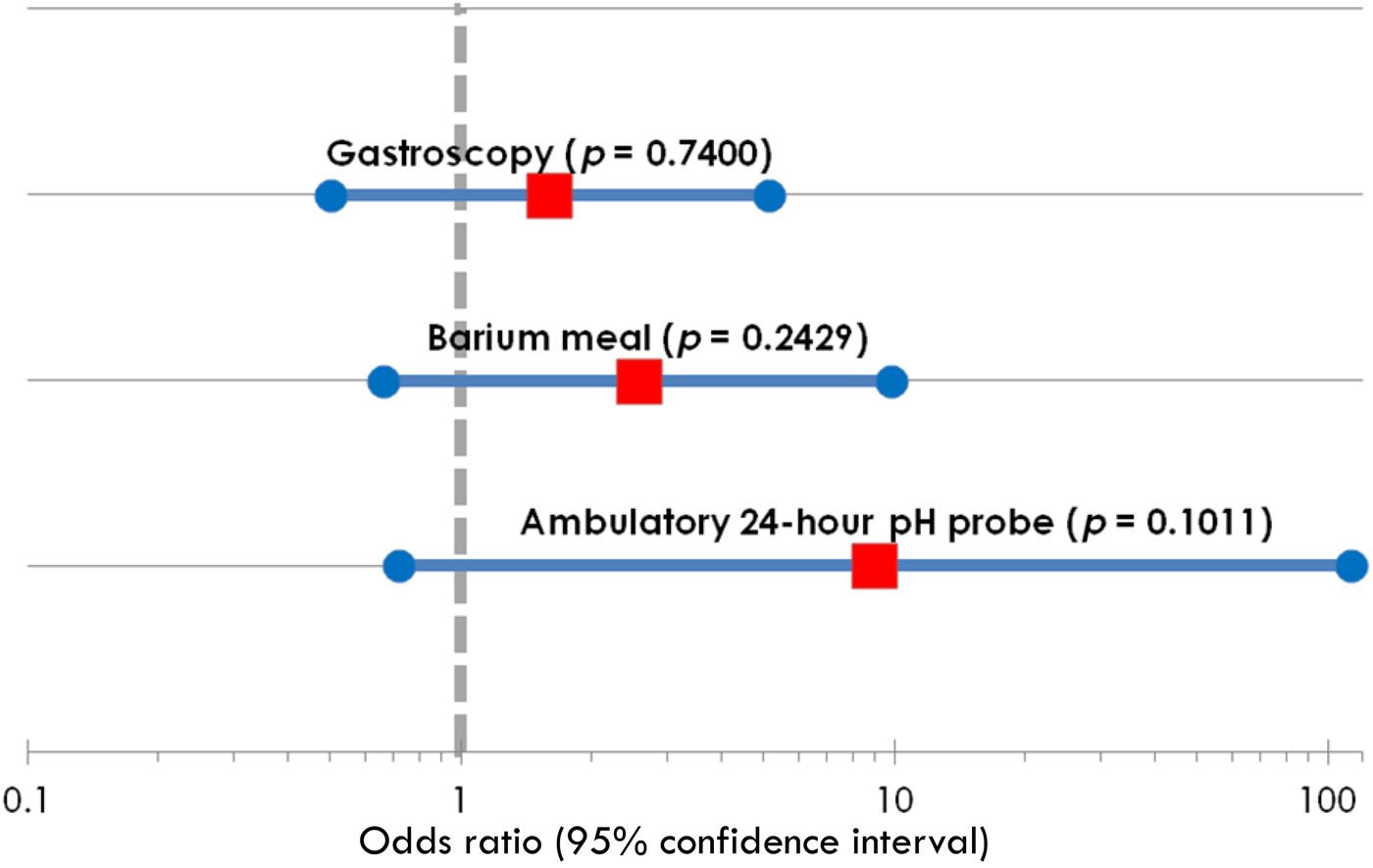
Of the 188 children who underwent tracheal aspirate analysis, there was a positive result for 111 (59.0 per cent) (Table II). However, there was no significant association between a positive pepsin result and the standard investigations for gastro-oesophageal reflux disease, including reflux oesophagitis on gastroscopy, reflux episodes seen during a barium meal and an abnormal pH probe result (Figure 4).
Forest plot showing the association of pepsin-positive tracheal aspirates with the results of standard gastro-oesophageal reflux disease investigations.
A positive pepsin result was significantly associated with the presence of respiratory (odds ratio 8.72, 95 per cent CI 4.48–16.98; p < 0.0001) and ENT (odds ratio 5.62, 95 per cent CI 2.98–10.60; p < 0.0001) symptoms. Surprisingly, a positive pepsin result was inversely related to the presence of GI symptoms (odds ratio 0.25 95 per cent CI 0.14–0.47; p < 0.0001).
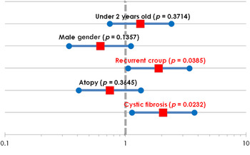
Subgroup analysis revealed no significant association between positive pepsin findings and age under two years, male sex or a medical history of atopy (Figure 5). However, positive pepsin assay findings were significantly associated with a history of CF (p = 0.0232) or recurrent croup (p = 0.0385).
Forest plot showing the subgroup analysis for patients with pepsin-positive tracheal aspirates.

Discussion
Children presenting with clinical symptoms thought to be secondary to extra-oesophageal reflux disease can be difficult to identify because of a dearth of readily available standardised investigations to delineate the aetiology. They often initially present to an ENT or respiratory medicine service and, depending on symptom type and severity, commence an empirical trial of anti-reflux medication such as an H2 antagonist or PPI on the presumption that acid reflux is a contributing factor. They may be referred to the Gastroenterology Department and undergo one or more of the standard gastro-oesophageal reflux disease investigations (barium meal, pH probe monitoring, gastroscopy or a multichannel intraluminal impedance study), even though these tests do not specifically examine extra-oesophageal sites. Laryngobronchoscopy visualises the airways and can detect macroscopic changes thought to be secondary to extra-oesophageal reflux disease;Reference Simons, Rosen, Casselbrant, Chi, Schaitkin and Rubinstein11 however, the findings may be subtle and non-specific. In contrast, the presence of pepsin (a substance foreign to respiratory and ENT tissue) in respiratory secretions helps to correlate changes on laryngobronchoscopy with reflux aspiration.Reference Tutuian and Castell4 Recent clinical practice guidelines by the North American Society for Pediatric Gastroenterology, Hepatology and Nutrition recommend the pepsin assay as one of the investigations that should be performed to diagnose extra-oesophageal reflux disease.Reference Vandenplas, Rudolph, DiLorenzo, Hassall, Liptak and Mazur1
In this study, changes on laryngobronchoscopy suggestive of extra-oesophageal reflux disease (including cobblestoning) were significantly associated with positive tracheal pepsin findings. In contrast, gastroscopy, pH probe monitoring and barium meal were shown to be poor indicators of extra-oesophageal reflux disease (Figures 2 and 4). This finding suggests that the inflammatory changes seen on laryngobronchoscopy in our study population were secondary to reflux aspiration occurring in the upper respiratory tract and pharyngeal regions due to the presence of pepsin and acid in the refluxate. This finding further supports the use of pepsin as a reliable diagnostic marker of extra-oesophageal reflux disease.Reference Tutuian and Castell4 The lack of a significant association between laryngobronchoscopy findings of extra-oesophageal reflux disease or pepsin positivity with positive results of standard investigations for gastro-oesophageal reflux disease is unsurprising because the latter mainly describe oesophageal pathology secondary to gastro-oesophageal reflux disease and not reflux aspiration.
We also found that changes on laryngobronchoscopy suggestive of extra-oesophageal reflux disease and a positive pepsin result were both more likely to be found in those children with chronic respiratory and ENT symptoms rather than those with typical GI symptoms of reflux. This finding is consistent with previous reports that extra-oesophageal reflux disease can exist despite the absence of typical gastro-oesophageal reflux disease symptoms such as vomiting, regurgitation and heartburn, as well as negative reflux findings on gastroscopy, barium meal and pH probe monitoring.Reference Farrokhi and Vaezi16 This phenomenon is particularly true for most paediatric cases of extra-oesophageal reflux disease. We acknowledge, however, that there can be a spectrum of severity and an overlap between gastro-oesophageal reflux disease and extra-oesophageal reflux disease in some patients.
The subgroup analysis found that a history of recurrent croup was significantly associated with positive changes on laryngobronchoscopy and positive pepsin assay findings (Figures 3 and 5). This result supports the hypothesis that extra-oesophageal reflux disease may be an aetiological factor in these patients. In contrast, in 112 out of 188 (59.6 per cent) children with an atopic history there was significant association with macroscopic changes on laryngobronchoscopy but not with positive pepsin findings. Some authors suggest that positive laryngobronchoscopy findings, including cobblestoning, may not exclusively result from laryngopharyngeal reflux and extra-oesophageal reflux disease.Reference Carr, Nguyen, Poje, Pizzuto, Nagy and Brodsky10 It is possible that allergic and infectious causes may contribute, such that some of the changes on laryngobronchoscopy identified in this group could be secondary effects of aero-allergens or other inflammatory agents.
However, a history of CF was significantly associated with positive pepsin findings, but not with changes on laryngobronchoscopy. This is consistent with reports of a high prevalence of gastro-oesophageal reflux disease and reflux aspiration in children with CF that contribute to lung disease severity, potential aspiration and bronchospasm.Reference Blondeau, Pauwels, Dupont, Mertens, Proesmans and Orel17, Reference Mousa and Woodley18
This study had several limitations inherent to its retrospective nature. There was selection bias because this study included only patients who had tracheal aspirates collected for pepsin assay by an ENT or respiratory specialist following changes seen on laryngobronchoscopy thought to be secondary to extra-oesophageal reflux disease, or a suspicion of this diagnosis in the clinical history. The study was performed at a tertiary referral centre with an unusually high proportion of patients coming from specialised clinics such as a CF clinic; hence, the study population was not representative of the general paediatric population. There was also no control group for comparison. Laryngobronchoscopy procedures were performed by a number of specialists, and there is likely to have been inter-observer variability in the identification of positive laryngobronchoscopy findings. The laryngobronchoscopy reports were usually completed by trainees and the level of recorded detail was not standardised. In addition, pepsin was assayed using an enzymatic fluorometric method, which is not as sensitive or specific as an enzyme-linked immunosorbent assay. However, the results of the enzymatic assay were subsequently found to correlate with those of an enzyme-linked immunosorbent assay using an anti-pepsin A monoclonal antibody.
This study's main strength is that it is the single largest paediatric study in which laryngobronchoscopy changes suggestive of extra-oesophageal reflux disease have been correlated with pepsin assays from respiratory secretions as well as clinical symptoms in a paediatric population. Others have primarily compared laryngobronchoscopy findings with positive standard gastro-oesophageal reflux disease investigations or correlated diagnostic laryngobronchoscopy findings with symptoms.Reference Wang, Liu, Liu, Zeng, Wu and Yang9, Reference Carr, Nguyen, Poje, Pizzuto, Nagy and Brodsky10 However, previous studies were limited in that they compared laryngobronchoscopy findings only with investigations that examined the oesophageal manifestations of gastro-oesophageal reflux disease but did not include a specific marker for reflux microaspiration resulting in extra-oesophageal reflux disease, such as pepsin.
• Gastro-oesophageal reflux is associated with extra-oesophageal manifestations in the upper aerodigestive tract
• There is no definitive diagnostic investigation for extra-oesophageal reflux disease
• The presence of pepsin in the trachea may be a useful marker of reflux aspiration
• Macroscopic changes correlate with tracheal pepsin in some affected children
• An association with tracheal pepsin suggests that extra-oesophageal reflux disease may contribute to recurrent croup and cystic fibrosis pathology
We hope that correlating laryngobronchoscopy findings with the presence of a sensitive and specific marker of reflux aspiration such as pepsin will provide objective evidence for the presence of extra-oesophageal reflux disease. This may avoid the need for an empirical trial with PPIs, which is sometimes unnecessary and can have side effects.Reference Pina Dore, Pedroni, Pes, Maragkoudakis, Tadeu and Pirina3 Conversely, a negative pepsin assay result despite positive laryngobronchoscopy findings could alter the diagnosis of extra-oesophageal reflux disease towards other conditions that warrant different further investigations such as microbiological analyses or allergen testing.
Conclusion
In this study, changes on laryngobronchoscopy, including cobblestoning, were significantly associated with positive pepsin findings in children presenting with chronic respiratory and ENT symptoms, and in those with a history of recurrent croup. This helps confirm the clinical diagnosis of extra-oesophageal reflux disease and can provide evidence for managing the suspected aetiology. Tracheal aspirate pepsin assays may become an essential objective investigation for reflux aspiration leading to extra-oesophageal reflux disease in the future.