Implications
Poor maternal nutrition alters muscle development and reduces postnatal muscle growth, but the mechanisms by which this occurs are unknown. Satellite cells are muscle progenitor cells that contribute to muscle growth in postnatal animals. Maternal nutrient restriction alters the expression pattern of essential transcription factors that regulate myogenesis, likely contributing to poor postnatal muscle growth through a reduced pool of myoblasts and reduced fusion into myotubes.
Introduction
Poor maternal nutrition during gestation can program the fetus, altering the development of several tissues and resulting in long lasting negative impacts on the offspring. Poor maternal nutrition during gestation results in reduced lean-to-fat ratio (Zhu et al., Reference Zhu, Ford, Means, Hess, Nathanielsz and Du2006), reduced muscle cross-sectional area (Reed et al., Reference Reed, Raja, Hoffman, Zinn and Govoni2014) and increased fat deposition (Bee, Reference Bee2004) in offspring. Insults to muscle progenitor cells, like poor maternal nutrition during prenatal development, likely contribute to these immediate and long-term negative outcomes. During development, muscle progenitor cells expressing paired box (Pax)-7 commit to the myogenic lineage and differentiate into mature myofibers in two waves of myogenesis. Myogenesis is governed by the appropriate temporal expression of the myogenic regulatory factors (MRF) Myf5, MyoD, myogenin and MRF4 (Yablonka-Reuveni and Rivera, Reference Yablonka-Reuveni and Rivera1994). Myf5 is expressed in quiescent and activated Pax7-positive progenitor cells. Activated satellite cells upregulate the expression of MyoD, which promotes extensive proliferation of the myoblast pool. MyoD activates transcription of myogenin, which directs cell cycle exit and the process of myoblast fusion into existing myofibers during periods of muscle growth. Finally, MRF4 expression is upregulated and the fiber matures into a functional contractile cell. By late gestation, myofiber hyperplasia slows and muscle growth occurs primarily through hypertrophy driven by increased protein synthesis and fusion of muscle progenitor cells with existing fibers. During this process, a portion of Pax7-expressing progenitor cells exit the cell cycle and are retained in the postnatal animal as quiescent satellite cells, which remain adjacent to myofibers and support postnatal growth (Hawke and Garry, Reference Hawke and Garry2001). Thus, maternal nutrition may affect both muscle development in the fetus and the establishment of satellite cells which allows for optimal postnatal muscle growth. For example, fetal offspring (day 75 of gestation) from obese ewes showed decreased abundance of MyoD and myogenin (Tong et al., Reference Tong, Yan, Zhu, Ford, Nathanielsz and Du2009), indicating that myogenesis is impaired during fetal development. Furthermore, satellite cells isolated from the offspring of undernourished rats failed to form myotubes comparable with those isolated from control offspring (de Melo et al., Reference de Melo, Aloulou, Duval, Vigneron, Bourgoin, Leandro, de Castro and Nagel2011). Therefore, poor maternal nutrition can program the nascent myofibers (affecting size and number) and the satellite cells (affecting functional capacity) leading to long-term negative effects on postnatal myogenesis. However, the effects of poor maternal nutrition on satellite cell differentiation are unknown. Thus, we hypothesized that restricted maternal nutrition during gestation would alter the temporal expression of myogenic regulatory factors in satellite cells of the offspring and decrease fusion into multinucleated myotubes in vitro.
Material and methods
Pregnant ewes were transitioned onto a control (100% NRC requirements for TDN) or restricted-fed (60% NRC requirements) diet at day 31±1.3 of gestation and remained on their respective diets until parturition as described in Reed et al. (Reference Reed, Raja, Hoffman, Zinn and Govoni2014). At birth, lambs from control-fed ewes (CON, n=12) and restricted-fed ewes (RES, n=12) were allowed to nurse to receive colostrum for up to 24 h. One-half of the lambs from each diet group were necropsied within 24 h of birth. The remaining lambs were maintained on the same control diet until necropsy at 3 months of age. At necropsy, muscle samples were collected from the left semitendinosus muscle (STN) from a subset of animals for satellite cell isolation (CON: birth, n=2; 3 months, n=3; RES: birth, n=3; 3 months, n=2). Satellite cells were isolated using a standard enzymatic digestion and differential centrifugation protocol and frozen until further use (Li et al., Reference Li, Reed and Johnson2009). In our hands, >95% of satellite cells isolated from sheep by this method are desmin positive, demonstrating purity of the population. To determine temporal expression of myogenic regulatory factors, satellite cells were cultured in growth media [Dulbecco’s modified Eagle medium (DMEM) containing 4 g/l d-glucose, 20% fetal bovine serum and 4 ng/ml fibroblast growth factor 2] and fixed and immunostained for Pax7, MyoD or myogenin after 24, 48 and 72 h in culture, while in the proliferative stage. The percent of cells positive for each antigen was calculated from 10 to 15 images per animal. To determine myogenic capacity for differentiation, satellite cells were cultured in growth media until confluent, at which time media was changed to differentiation media (DMEM containing 1 g/l d-glucose and 2% horse serum). After 96 h in culture, myotubes were fixed and immunostained for myosin heavy chain (MyHC). Hoescht 33 342 was used to visualize nuclei. Fusion index was calculated as the number of nuclei within multinucleated MyHC-positive myotubes divided by the number of total nuclei and multiplied by 100. Fusion index data were collected from 10 to 15 images per animal. Immediately after thawing, a Qiagen RNeasy Mini Kit (Qiagen, Valencia, CA, USA) was used to extract RNA from satellite cells isolated from lambs at birth. Expression of Pax7, MyoD and myogenin was measured by real-time RT-PCR using iTaq SybrGreen Master Mix (BioRad, Hercules, CA, USA) and the ABI 7900HT Fast Real-time PCR machine (Applied Biosystems, Foster City, CA, USA) as previously described (Reed et al., Reference Reed, Raja, Hoffman, Zinn and Govoni2014). Cell culture data were analyzed as a completely randomized design with treatments arranged as a 2×2 factorial with age of lamb and maternal diet as main effects. Analyses were performed using the MIXED procedure in SAS. Statistical models included all main effects and interactions. Image within animal was included as a random effect. Real-time data were analyzed as a completely randomized design with a main effect of maternal diet. When appropriate, means comparisons were made using a least square means statement and differences between least square means were separated using the PDIFF option. Significance was considered at P⩽0.05.
Results
After 24 h in culture, 74.0±3.5% of the cells were Pax7-positive [Pax7(+)] cells, demonstrating the myogenic nature of the isolated cells. There was no difference in the percent of Pax7(+) cells between CON and RES lambs at birth or 3 months of age (P=0.95), consistent with other work (de Melo et al., Reference de Melo, Aloulou, Duval, Vigneron, Bourgoin, Leandro, de Castro and Nagel2011). As expected, the percent of Pax7(+) cells decreased after 24 h in culture.
Myogenic regulatory factor expression in satellite cells from lambs at birth
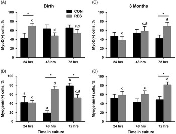
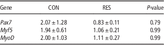
There was a significant interaction of diet×age of lamb×day in culture for both MyoD and myogenin (P<0.01). However, the relevant significant differences occurred within an age group, thus the data are presented according to the age of the animal. After 24 h in culture, there was a greater percent of MyoD(+) cells in cultures isolated at birth from RES lambs (69.5±6.5%; Figure 1A) compared with CON lambs (42.9±10.9%; P=0.03), suggesting earlier satellite cell activation. There were no differences in the percent of MyoD(+) satellite cells at 48 or 72 h in culture in CON and RES lambs. The percent of MyoD(+) satellite cells in RES lambs decreased after 24 h in culture (P=0.05; Figure 1A). The percent of cells that were myogenin(+) was greater at 48 h in RES lambs compared with CON lambs (RES: 70.9±7.0%; CON: 18.6±5.1%; P<0.001; Figure 1B), but less than CON after 72 h in culture (RES: 51.8±7.6%; CON: 78.4±6.2%; P<0.01; Figure 1B). The percent of myogenin(+) cells increased from 24 to 48 h in satellite cells from RES lambs (24 h: 40.8±5.8%; 48 h: 70.9±7.0%; P=0.002). This increase occurred later in cells from CON lambs, where the percent of myogenin(+) cells increased from 48 h to 72 h (48 h: 18.6±5.1%; 72 h: 78.4±6.2%; P<0.001). There were no significant differences observed between RES and CON lambs in the mRNA expression of Pax7, MyoD or Myf5 (Table 1) in the satellite cells of lambs necropsied at birth.
Restricted maternal nutrition during gestation alters MyoD and myogenin expression in offspring. Satellite cells isolated from lambs born to restricted-fed (RES) and control-fed (CON) ewes at birth (A, B) or 3 months of age (C, D) were cultured for 24, 48 and 72 h and immunostained for MyoD (A, C) and myogenin (B, D). Data presented are mean±SEM. *P<0.05 compared with CON at that time point. a, b Differences (P<0.05) over time in culture within CON. c, d Differences (P<0.05) over time in culture within RES.

Gene expressionFootnote 1 in satellite cells of 1-day-old lambs from control (CON) and restricted fed (RES) ewes

1 Mean fold change±SEM.
Myogenic regulatory factor expression in satellite cells from lambs at 3 months of age
After 72 h in culture, the percent of MyoD(+) cells was greater in cells isolated from 3 months old RES lambs compared with CON lambs (RES: 69.4±9.1%; CON: 42.2±7.6%; P=0.02; Figure 1C). The percent of MyoD(+) cells increased from 24 to 72 h in cells from RES lambs (P=0.01). There was no change in the percent of MyoD(+) cells over time in CON animals (P>0.28). The percent of myogenin(+) cells was greater in RES at 72 h of culture (RES: 81.2±6.4%; CON: 48.3±7.4%; P=0.003; Figure 1D) compared with CON. The percent of myogenin(+) cells increased from 48 to 72 h in cells from RES lambs (P=0.05). There was no change in the percent of myogenin(+) cells over time in cells from CON lambs (P>0.40). These data indicate that the satellite cell population from 3 months old RES lambs retains MyoD and myogenin, which would inhibit terminal differentiation into myofibers.
Maternal nutrient restriction impairs myoblast fusion index
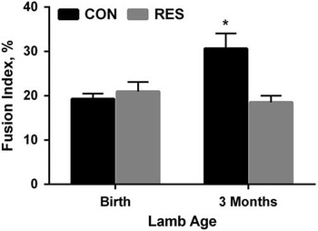
There was a significant age of lamb×diet effect on fusion index (P=0.0006). Fusion index was greater in satellite cells from CON lambs at 3 months of age compared with cells from RES lambs at 3 months of age and both CON and RES lambs at birth (P<0.01, Figure 2). There was no effect of maternal diet on fusion index in cells isolated from lambs at birth (P=0.5, Figure 2).
Restricted maternal nutrition during gestation decreases myoblast fusion in offspring at 3 months of age. Satellite cells isolated from lambs born to restricted-fed (RES) and control-fed (CON) ewes at birth or 3 months of age were allowed to differentiate for 96 h and immunostained for myosin heavy chain (MyHC). Fusion index was calculated as the number of nuclei in MyHC-expressing multi-nucleated myotubes divided by the total number of nuclei and multiplied by 100. Data presented are mean±SEM. *P<0.05.

Discussion
The process of myogenesis is governed by the myogenic regulatory factors, Myf5, MyoD, myogenin and MRF4. Myf5 and MyoD regulate progenitor cell proliferation and commitment to the myogenic lineage. Myogenin and MRF4 regulate cell cycle exit, terminal myoblast differentiation and fiber maturation. Importantly, these factors must be expressed in the appropriate temporal pattern to direct muscle fiber formation and growth during prenatal development and muscle growth postnatally. Disruption in the expression pattern or timing of expression of MRFs negatively affects muscle development and growth (reviewed in Mok and Sweetman (Reference Mok and Sweetman2011).
Nutrient restriction during gestation altered the temporal expression of MyoD and myogenin in satellite cells isolated from the offspring at birth, decreasing the time in the proliferative, MyoD(+) stage of myogenesis. Expression of MyoD and myogenin drives cell cycle exit, which may limit the number of myoblasts available for fusion into muscle. This, in turn, limits fetal myofiber formation, hypertrophy and early postnatal muscle growth. The rapid progression through these stages of myogenesis may also contribute to the decreased MyoD and myogenin expression in muscle of offspring of obese ewes (Tong et al., Reference Tong, Yan, Zhu, Ford, Nathanielsz and Du2009), impairing myogenesis during fetal development. Heat stress induced intrauterine growth restriction also resulted in decreased MyoD expression in satellite cells of the offspring after 3 days of culture (Yates et al., Reference Yates, Clarke, Macko, Anderson, Shelton, Nearing, Allen, Rhoads and Limesand2014). Together, these data demonstrate that the maternal environment has significant impacts on muscle development and satellite cell function, and importantly that in vivo treatments to the animal can alter function in vitro.
In contrast, satellite cells from RES lambs at 3 months of age retain expression of MyoD and myogenin compared with CON. Coupled with a reduced fusion index, this suggests that satellite cells from RES animals are deficient in their ability to fuse and contribute to muscle growth. Similarly, satellite cells isolated from the offspring of rats undernourished during gestation demonstrated formation of fewer muscle fibers (de Melo et al., Reference de Melo, Aloulou, Duval, Vigneron, Bourgoin, Leandro, de Castro and Nagel2011). Furthermore, RES offspring at 3 months of age have reduced muscle fiber cross sectional area compared with CON lambs (Reed et al., Reference Reed, Raja, Hoffman, Zinn and Govoni2014), supporting inhibited muscle growth as a result of restricted maternal nutrition during gestation.
In summary, restricted maternal nutrition during gestation alters the temporal expression of myogenic regulatory factors in satellite cells and decreases fusion index in cells isolated from 3 months old lambs. Altered satellite cell function likely contributes to the poor muscle growth in offspring of restricted-fed ewes, and may be programmed in utero with long lasting consequences for the offspring.
Acknowledgments
This work was supported in part by the Storrs Agricultural Experiment Station.