Introduction
The development of early chorionic villi is a complicated process involving multiple cell types (Knöfler et al., Reference Knöfler, Haider, Saleh, Pollheimer, Gamage and James2019). Among them, placental trophoblastic cells play an important role in embryo implantation and placental genesis (Knöfler et al., Reference Knöfler, Haider, Saleh, Pollheimer, Gamage and James2019). The differentiation of trophoblastic cells during placenta formation involves two routes, an invasion and migration route and a fusion route. In the first route, cytotrophoblast cells (CTBs) are differentiated into extravillous trophoblasts (EVTs) with invasion ability, which allows the embryo to implant in the uterus. Furthermore, this pathway is regulated by multiple factors, for instance, matrix metalloproteinases (Isaka et al., Reference Isaka, Usuda, Ito, Sagawa, Nakamura, Nishi, Suzuki, Li and Takayama2003), cytokines (Prabha et al., Reference Prabha, Molykutty, Swapna, Rajalekshmi and Gangadharan2001; Schäfer-Somi, Reference Schäfer-Somi2003; Lash et al., Reference Lash, Otun, Innes, Bulmer, Searle and Robson2005; Lockwood et al., Reference Lockwood, Oner, Uz, Kayisli, Huang, Buchwalder, Murk, Funai and Schatz2008), hormones (Shin et al., Reference Shin, Lee, Yang, Moon, Rha and Kim2003), and signalling pathways, i.e. the Wnt (Knöfler, Reference Knöfler2010) and phosphoinositide 3-kinase (PI3K) pathways. In the fusion route, CTBs fuse with syncytiotrophoblasts (STBs) to form the overlying syncytiotrophoblast layer, therefore promoting placental development and maturation (Kar et al., Reference Kar, Ghosh and SenGupta2007). Some CTBs are fused with STBs to expand and renew the syncytiotrophoblast, therefore promoting placental development and maturation, which is regulated by multiple factors, such as vascular endothelial growth factor (Crocker et al., Reference Crocker, Strachan, Lash, Cooper, Warren and Baker2001), human chorionic gonadotropin (hCG; Ullah et al., Reference Ullah, Dar, Ahmad, de Renty, Usman, DePamphilis, Faisal, Shahzad-Ul-Hussan and Ullah2018), and syncytin (syncytin 1 and syncytin 2; Langbein et al., Reference Langbein, Strick, Strissel, Vogt, Parsch, Beckmann and Schild2008; Ruebner et al., Reference Ruebner, Langbein, Strissel, Henke, Schmidt, Goecke, Faschingbauer, Schild, Beckmann and Strick2012). However, the mechanism of trophoblast cell syncytialization during placental development has not been fully elucidated.
Tspan5 is a member of the tetraspanin superfamily TspanC8 subgroup (Todd et al., Reference Todd, Doctor and Levy1998), which contains a common structure with four highly conserved hydrophobic transmembrane domains (García-Frigola et al., Reference García-Frigola, Burgaya, Calbet, de Lecea and Soriano2000). The members of this family can interact with other molecules, such as cholesterol (Huang et al., Reference Huang, Hays, Tomasek, Benyajati and Zhang2020), Fc receptor (Moseley, Reference Moseley2005), and tetraspanins (Berditchevski et al., Reference Berditchevski, Gilbert, Griffiths, Fitter, Ashman and Jenner2001; Israels and McMillan-Ward, Reference Israels and McMillan-Ward2010; Conley et al., Reference Conley, Stuck and Naash2012), and play multiple roles in cell adhesion (Reyes et al., Reference Reyes, Cardeñes, Machado-Pineda and Cabañas2018), inflammation (Brosseau et al., Reference Brosseau, Colas, Magnan and Brouard2018), cell motility and metastasis (Yaseen et al., Reference Yaseen, Monk and Partridge2017; Wang et al., Reference Wang, Liang, Yang, Lan, Han, Wang, Yin, Song, Zheng, Zhang, Pan, Liu, Zhu, Liu, Cui, Meng, Zhang, Liang, Guo, Liu, Hassan and Liu2018), cell growth (Li et al., Reference Li, Wang, Qiu, Da, Tiollais, Li and Zhao2012), signal transduction (Levy et al., Reference Levy, Todd and Maecker1998), and cell proliferation and differentiation (Berditchevski, Reference Berditchevski2001). The roles of Tspan5 in cerebral development (García-Frigola et al., Reference García-Frigola, Burgaya, Calbet, de Lecea and Soriano2000; García-Frigola et al., Reference García-Frigola, Burgaya, de Lecea and Soriano2001; Juenger et al., Reference Juenger, Holst, Duffe, Jankowski and Baader2005) and embryonic development (Gao et al., Reference Gao, Liang, Tang and Quan2018) have been reported, while the mechanism of Tspan5 in trophoblast cell syncytialization during placental development has not been shown.
Epithelial–mesenchymal transition (EMT) refers to a process in which epithelial cells lose their identity and acquire the ability to freely move to mesenchymal cells (Kokkinos et al., Reference Kokkinos, Murthi, Wafai, Thompson and Newgreen2010). This is regulated by multiple factors, including growth factors and transcription factors, which can directly or indirectly inhibit the expression of E-cadherin, therefore promoting EMT (Lamouille et al., Reference Lamouille, Xu and Derynck2014). Slug, also named Snail2, can cause the downregulation of E-cadherin and promote EMT. Twist attenuates intercellular adhesion by inhibiting the expression of E-cadherin (Kang and Massagué, Reference Kang and Massagué2004; Ng et al., Reference Ng, Zhu and Leung2011). Twist can promote the expression of N-cadherin and modulate the invasion of human trophoblastic cells (Ng et al., Reference Ng, Zhu and Leung2012). In addition, the EMT pathway in cultured trophoblasts can be collectively inhibited by maternal circulating miRNAs (HEamiRNAs; Tseng et al., Reference Tseng, Mahnke, Wells, Salem, Allan, Roberts, Newman, Walter, Kroenke, Grant, Akison, Moritz, Chambers and Miranda2019). Notch signalling is closely implicated in cell proliferation and differentiation, and precisely mediates information communication between cells (Reichrath and Reichrath, Reference Reichrath and Reichrath2020b). We hypothesized that Notch signalling could be involved in the fusion of trophoblastic cells. Coincidently, Tspan5 can activate Notch signalling via A disintegrin and metalloprotease 10 (ADAM10), which is essential for embryonic development (Dornier et al., Reference Dornier, Coumailleau, Ottavi, Moretti, Boucheix, Mauduit, Schweisguth and Rubinstein2012; Jouannet et al., Reference Jouannet, Saint-Pol, Fernandez, Nguyen, Charrin, Boucheix, Brou, Milhiet and Rubinstein2016). Loss of Tspan5 in osteoclast lineage cells results in the attenuation of Notch activation (Zhou et al., Reference Zhou, Fujiwara, Ye, Li and Zhao2014). However, whether Tspan5 can mediate trophoblast cell syncytialization during placental development by activating Notch signalling still needs to be explored.
In this study, BeWo cells derived from human chorionic cancer cells were selected as the cell model to study trophoblastic cell fusion. The effects of Tspan5 on trophoblast syncytialization and the involvement of the Notch pathway were explored.
Materials and Methods
Sample collection and treatment
Seminal fluid was collected from healthy male volunteers and isolated by density gradient centrifugation (Irvine, America; Phillips et al., Reference Phillips, Dhaliwal, Verstegen-Onclin and Verstegen2012). Then, semen liquid was decanted and the cells were treated with IVF G-Series culture medium (Vitrolife, Sweden) according to the manufacturer’s instructions. Ova were collected from healthy female volunteers and added to IVF G-Series culture medium for 2–5 h. Then, granulosa cells were removed by enzymatic digestion. External fertilization was performed, and the fertilization conditions were observed under a microscope. Healthy embryos were collected and cultured in a CO2 incubator (6.0% CO2, 37°C, 100% humidity). The embryo culture medium was collected on the third and sixth days after fertilization. The study was approved by the Meizhou People’s Hospital (Huangtang Hospital). Informed consent was obtained from all participants before sample collection.
Cell culture
BeWo cells were obtained from the American Type Culture Collection (ATCC) and stored in a −80°C freezer. BeWo cells were cultured in Dulbecco modified Eagle’s medium (DMEM; Invitrogen, Carlsbad, CA, USA) with 10% fetal bovine serum (FBS; Invitrogen), 100 μg/ml streptomycin and 100 U/ml penicillin (Invitrogen). BeWo cells were fused under treatment with 10 μM forskolin (FSK; Sigma-Aldrich, USA) for 48 h. All cells were cultured at 37°C in a humidified 5% CO2 incubator.
Immunofluorescence
The BeWo cells were fixed with methanol for 5 min at room temperature, followed by washing three times with phosphate-buffered saline (PBS). The BeWo cells were blocked in 5% bovine serum albumin (BSA; Sigma-Aldrich, USA) for 1 h, followed by washing three times with PBS. After that, the BeWo cells were incubated with primary anti-E-cadherin antibodies (Abcam, England, No. ab40772) and anti-Tspan5 antibodies (Abcam, No. ab236881) at 4°C overnight, followed by washing three times with PBS. Then, they were incubated with anti-rabbit IgG antibodies (Invitrogen) at room temperature for 2 h, washed three times, stained with 4′,6-diamidino-2-phenylindole (DAPI) for 10 min and washed three times with PBS. After blocking with glass slides, they were observed under a fluorescence microscope.
Quantitative real-time polymerase chain reaction (qRT-PCR)
Total RNA from BeWo cells was extracted using TRIzol reagent following the manufacturer’s instructions (TaKaRa Bio Inc., Otsu, Shiga, Japan). Then, RNA concentration and purity were detected by spectrophotometry. Subsequently, 1 μg of total RNA was reverse transcribed into cDNA using a PrimeScriptTM RT Reagent Kit (TaKaRa). The expression of genes was detected using the SYBR premix Ex Taq II kit (TaKaRa) on an ABI Prism 7500 HT Sequence Detection System (Applied Biosystems, CA, USA). The reaction procedures were designed as follows: predenaturation at 95°C for 10 min, followed by 35 cycles of denaturation at 95°C for 30 s, annealing at 60°C for 30 s, and extension for 60 s at 72°C. The relative expression levels of targeted genes were analyzed by the 2−ΔΔCt method. The primer sequences are listed in Table 1.
Oligonucleotide primer sets for real-time qPCR

Western blotting
Total proteins were extracted using RIPA lysis buffer supplemented with protease inhibitor and phosphatase inhibitors (Selleck, China). The protein samples were separated by 12% sodium dodecyl sulfate-polyacrylamide gel electrophoresis (SDS-PAGE) and transferred onto polyvinylidene difluoride (PVDF) membranes (Millipore, MA, USA). Soon afterwards, the PVDF membranes were blocked with 5% BSA for 1 h. After washing, the PVDF membranes were incubated with primary antibodies diluted at 1:1000 at 4°C overnight: Tspan5 (cat no. ab236881, Abcam), β-hCG (cat no. ab53087, Abcam), syncytin 1 (cat no. ab179693, Abcam), syncytin 2 (cat no. ab230235, Abcam), E-cadherin (cat no. 14472, Cell Signalling Technology, USA), N-cadherin (cat no. 13116, Cell Signalling Technology), vimentin (cat no. 5741, Cell Signalling Technology), Snail (cat no. 3879, Cell Signalling Technology), Slug (cat no. 9585, Cell Signalling Technology), Twist (cat no. 69366, Cell Signalling Technology), NOTCH-1 (cat no. 3608, Cell Signalling Technology), Jagged-1 (cat no. 70109, Cell Signalling Technology), β-actin (cat no. 4970, Cell Signalling Technology). After washing, the membranes were then incubated with horseradish peroxidase (HRP)-conjugated secondary antibodies diluted at 1:3000 (Cell Signalling Technology) at room temperature for 2 h. The protein bands were exposed using an enhanced chemiluminescence (ECL) kit (Millipore, USA) and analyzed using ImageJ software (NIH, Bethesda, MD, USA).
ELISA
Healthy embryos were cultured, and the embryo culture medium was collected on the third and sixth days after fertilization. The total protein content in the culture supernatant on the sixth day was quantified and adjusted to an equal level on the third day. The levels of Tspan5 in the culture supernatant were examined using ELISA kits (sensitivity: <0.095 ng/ml; detection range: 0.156–10 ng/ml; inter-assay coefficients of variations: <15%; intra-assay coefficients of variations: <10%) according to the manufacturer’s instructions (RunYu, Shanghai, China).
Construction of expression vectors
The sh-Tspan5 vectors, Tspan5 overexpression vectors and matching negative control vector pcDNA3.1 (GenePharma, China) were constructed. Afterwards, the recombinant plasmids were transformed into DH5α cells, which were cultured at 37°C overnight. After extraction and identification, the indicated vector-only or combined vectors were incubated with isopycnic Lipofectamine 2000 (Invitrogen, USA) in DMEM for 10 min, after which the mixtures were transfected into BeWo cells following the product’s instructions.
Cell transfection
BeWo cells were transfected with sh-Tspan5 (sh-NC as a control) or oe-Tspan5 (oe-NC as a control) according to the instructions for Lipofectamine 2000 (Invitrogen). Briefly, 1 μg of plasmid was mixed in 200 μl DMEM without serum, while 2 μl of Lipofectamine 2000 was mixed with another 200 μl DMEM without serum. The 200 μl DNA system was added to 200 μl of Lipofectamine 2000 for 10 min to form the transfection complexes at room temperature. The cells were incubated with the transfection complexes in a 12-well plate for 6 h, after which the medium was replaced. Afterwards, the cells were cultured for another 48 h at 37°C. Then, the expression levels of related genes in transfected cells were determined by qRT-PCR and western blotting.
Statistical analysis
For statistical analyses, one-way analysis of variance (ANOVA) followed by Tukey’s post-hoc test and Student’s t-test were performed using SPSS software (v.12.0; SPSS Inc., Chicago, IL, USA). GraphPad Prism 8 software was used for data analysis. The results are presented as the mean ± standard deviation (SD) of three independent experiments. A P-value < 0.05 was considered statistically significant.
Results
Tspan5 was upregulated in embryo culture medium, and the fusion of BeWo cells was induced by FSK
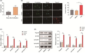
On the third and sixth days after fertilization, the embryo culture medium was collected and detected by ELISA. The expression of Tspan5 significantly increased in the embryo culture medium on the sixth day compared with the third day after fertilization (Figure 1A), indicating that Tspan5 could be involved in embryonic development. To further study the role of Tspan5, BeWo cells were selected as the study model. The fusion of BeWo cells was detected by immunofluorescence after treatment with FSK for 48 h. The results showed that BeWo cells fused after treatment with FSK compared with the control group, whereas dimethyl sulphoxide (DMSO) used by itself in cell culture could not cause BeWo cell fusion. In addition, the E-cadherin expression level significantly decreased after treatment with FSK compared with the control and DMSO groups. In contrast, Tspan5 significantly increased after induction by FSK (Figure 1B). hCG and syncytin promoted CTB fusion with STBs to expand and renew syncytiotrophoblasts and promoted placental development and maturation in the fusion route. Therefore, we also detected the expression levels of Tspan5, β-hCG and two syncytin family members (syncytin 1 and syncytin 2) by qRT-PCR and western blotting. The results showed that the levels of Tspan5, β-hCG, and syncytin 1/2 were significantly upregulated after treatment with FSK compared with the control group and the DMSO group (Figure 1C,D).
Cell fusion and Tspan5 expression were detected after treatment with FSK. (A). Tspan5 was detected in embryo culture medium by ELISA. (B) Syncytium formation and expression of Tspan5 were detected by immunofluorescence. (C) The expression levels of Tspan5, β-hCG, and syncytin 1/2 were detected by qRT-PCR. (D) The expression levels of Tspan5, β-hCG, and syncytin 1/2 were detected by western blotting. The experiments were carried out in triplicate. **P < 0.01; ***P < 0.001.

EMT occurred during the BeWo cell fusion process
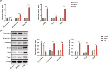
We detected the involvement of EMT-related factors during BeWo cell fusion. The expression levels of E-cadherin, N-cadherin, vimentin, Snail, Slug and Twist were measured after treatment with FSK for 48 h by qRT-PCR and western blotting. The results showed that E-cadherin expression significantly decreased in the FSK group compared with the control and DMSO groups, whereas the level of N-cadherin was significantly upregulated in the FSK group (Figure 2A,B), which was in accordance with the typical characteristics of EMT. Furthermore, the expression of Vimentin, Snail, Slug and Twist were significantly upregulated in the FSK group (Figure 2A,B). These results indicated that these factors Vimentin, Snail, Slug and Twist were upregulated, therefore promoting EMT during BeWo cell fusion.
Detection of EMT-related factors in BeWo cells after treatment with FSK. (A) The expression levels of E-cadherin, N-cadherin, Vimentin, Snail, Slug and Twist were detected by qRT-PCR. (B) The expression levels of E-cadherin, N-cadherin, Vimentin, Snail, Slug and Twist were detected by western blotting. The experiments were carried out in triplicate. *P < 0.05; **P < 0.01; ***P < 0.001.

Downregulation of Tspan5 inhibited EMT and BeWo cell fusion
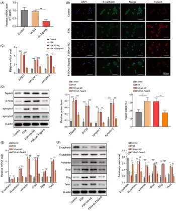
To further explore the regulatory mechanisms of Tspan5 in cell fusion, we knocked down Tspan5 in BeWo cells by cell transfection. The Tspan5 level was significantly downregulated after transfection with sh-Tspan5 compared with the control and sh-NC groups (Figure 3A), which indicated that the expression-blocking expression vector had been successfully constructed. The immunofluorescence results showed that BeWo cells could undergo cell fusion after treatment with FSK for 48 h. However, cell fusion was obstructed after transfection with sh-Tspan5 (Figure 3B). Also, FSK treatment upregulated the expression of Tspan5, while the knockdown of Tspan5 exerted an opposite effect (Figure 3B). However, compared with sh-NC group, downregulation of Tspan5 resulted in a slight decrease in cell fusion, but there was no significant difference (Figure S1). Furthermore, the knockdown of Tspan5 increased the expression of E-cadherin and reduced Tspan5 expression (Figure S1). In addition, the expression levels of the cell fusion-related factors β-hCG and syncytin 1/2 were significantly downregulated after transfection with sh-Tspan5 compared with the sh-NC group in FSK-induced cells (Figure 3C,D). The expression levels of N-cadherin, Vimentin, Snail, Slug and Twist were significantly decreased in the sh-Tspan5 group compared with the sh-NC group in FSK-induced cells, whereas E-cadherin had the opposite expression (Figure 3E,F), indicating that the EMT process was blocked by the downregulation of Tspan5.
Fusion of BeWo cells and EMT-related factors were detected after knockdown of Tspan5. (A) The expression level of Tspan5 was detected by qRT-PCR after transfection with sh-Tspan5. (B) Syncytium formation, and the expression of Tspan5 were detected after transfection with sh-Tspan5. (C, D) The expression levels of Tspan5, β-hCG, and syncytin 1/2 were detected by qRT-PCR and western blotting, respectively. (E, F) The expression levels of E-cadherin, N-cadherin, Vimentin, Snail, Slug and Twist were detected by qRT-PCR amd western blotting, respectively. The experiments were carried out in triplicate. **P < 0.01; ***P < 0.001.

Tspan5 promoted the EMT process by activating Notch signalling
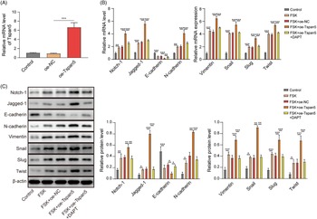
The above findings showed that the downregulation of Tspan5 inhibited the EMT process, but the mechanism needed to be further explored. An overexpression vector was constructed and transfected into BeWo cells. The expression of Tspan5 significantly increased after transfection with oe-Tspan5 compared with the control and oe-NC groups (Figure 4A). Furthermore, to define the effect of Notch signalling on the EMT process, we detected the Notch signalling-related factors Notch-1 and Jagged-1 by qRT-PCR and western blotting. DAPT, a repressor of Notch signalling, was used as a control. The results showed that the Notch-1 and Jagged-1 expression levels were upregulated after treatment with FSK for 48 h compared with the control group, indicating that Notch signalling was activated during cell fusion (Figure 4B,C). As expected, the Notch-1 and Jagged-1 expression levels further significantly increased after overexpression of Tspan5 compared with the oe-NC group in FSK-induced cells, which could be rolled back using the repressor DAPT. These results indicated that Tspan5 could activate Notch signalling. Furthermore, the upstream and downstream relationship between Notch signalling and EMT was explored by blocking Notch signalling with DAPT. EMT-related factors E-cadherin, N-cadherin, vimentin, Snail, Slug and Twist were detected by qRT-PCR and western blotting. As expected, the overexpression of Tspan5 resulted in the significant downregulation of E-cadherin and the upregulation of N-cadherin, Vimentin, Snail, Slug and Twist (Figure 4D,E), indicating that Tspan5 could promote the EMT process, which was in line with our previous results. Interestingly, after blocking Notch signalling with DAPT, E-cadherin expression significantly rose to normal levels in the oe-NC group in FSK-induced cells, whereas the expression levels of N-cadherin, vimentin, Snail, Slug and Twist significantly dropped to normal levels in the FSK and oe-NC groups (Figure 4D,E). These results indicated that blocking Notch signalling inhibited the EMT process and that Tspan5 could regulate EMT progression via the Notch protein, therefore playing an important role in the fusion of BeWo cells.
Detection of Notch signalling and EMT-related factors after overexpression of Tspan5. (A) The expression level of Tspan5 was detected by qRT-PCR after overexpression of Tspan5. (B, C) Detection of Notch-1 and Jagged-1 with or without the repressor DAPT by qRT-PCR and western blotting after overexpression of Tspan5, respectively. The expression levels of E-cadherin, N-cadherin, Vimentin, Snail, Slug and Twist were detected with or without the repressor DAPT by qRT-PCR and western blotting after overexpression of Tspan5, respectively. The experiments were carried out in triplicate. *P < 0.05; **P < 0.01, ***P < 0.001.

Discussion
Cell fusion plays a vital important role in fetal development (Crocker et al., Reference Crocker, Strachan, Lash, Cooper, Warren and Baker2001; Langbein et al., Reference Langbein, Strick, Strissel, Vogt, Parsch, Beckmann and Schild2008; Ruebner et al., Reference Ruebner, Langbein, Strissel, Henke, Schmidt, Goecke, Faschingbauer, Schild, Beckmann and Strick2012; Ullah et al., Reference Ullah, Dar, Ahmad, de Renty, Usman, DePamphilis, Faisal, Shahzad-Ul-Hussan and Ullah2018). In this study, we observed that Tspan5 was significantly upregulated and that the involvement of EMT-related factors was enhanced in FSK-induced cell fusion. Moreover, the downregulation of Tspan5 in BeWo cells inhibited the EMT process. Tspan5 promoted the EMT process by activating Notch signalling. This study, for the first time, revealed Tspan5 expression in embryo culture medium and the regulatory role of Tspan5 on the EMT process in BeWo by FSK treatment, which is of great significance for a better understanding of placental development.
Tspan5 belongs to the tetraspanin family TspanC8 subgroup and participates in the regulation of cell adhesion, migration and fusion (Hemler, Reference Hemler2001). Tspan5 is primarily located in the cytoplasm and on the surfaces of extravillous trophoblasts (EVCTs) and cytotrophoblasts (CTs) in human villi (Gao et al., Reference Gao, Liang, Tang and Quan2018) and is critical to placental development. The progression of EMT gives cells the ability to move freely and lays a foundation for cell fusion and the development of tissues and organs. In this study, we found that Tspan5 was upregulated, and typical EMT progression occurred in BeWo cells induced by FSK. A significant decrease in E-cadherin expression and an increase in N-cadherin expression are typical features of EMT progression (Kokkinos et al., Reference Kokkinos, Murthi, Wafai, Thompson and Newgreen2010), which can be directly or indirectly regulated by Snail, Slug, ZEB1/2 and Twist (Lamouille et al., Reference Lamouille, Xu and Derynck2014). A previous study reported that Twist could decrease E-cadherin expression (Ng et al., Reference Ng, Zhu and Leung2011) and increase N-cadherin expression (Ng et al., Reference Ng, Zhu and Leung2012). Consistently, we found that upregulation of Tspan5 resulted in a reduction in E-cadherin and an increase in N-cadherin, vimentin, Snail, Slug and Twist expression, indicating that Tspan5 promoted EMT progression.
Notch signalling, which was first discovered in fruit flies, is evolutionarily highly conserved and governs many important processes, such as cell fate decisions during embryonic development. Many scientific studies have convincingly demonstrated that Notch signalling represents one of the most important pathways in regulating the embryogenesis of invertebrates and vertebrate animals (Bahrampour and Thor, Reference Bahrampour and Thor2020; Reichrath and Reichrath, Reference Reichrath and Reichrath2020a). ADAM10 is essential for embryonic development through the activation of Notch proteins (Dornier et al., Reference Dornier, Coumailleau, Ottavi, Moretti, Boucheix, Mauduit, Schweisguth and Rubinstein2012; Jouannet et al., Reference Jouannet, Saint-Pol, Fernandez, Nguyen, Charrin, Boucheix, Brou, Milhiet and Rubinstein2016; Matthews et al., Reference Matthews, Szyroka, Collier, Noy and Tomlinson2017). ADAM10 is a ubiquitous transmembrane metalloprotease and can cleave the extracellular regions of as many as 40 transmembrane target proteins as a ‘molecular scissor’, including Notch signal-related proteins (Dornier et al., Reference Dornier, Coumailleau, Ottavi, Moretti, Boucheix, Mauduit, Schweisguth and Rubinstein2012; Jouannet et al., Reference Jouannet, Saint-Pol, Fernandez, Nguyen, Charrin, Boucheix, Brou, Milhiet and Rubinstein2016; Noy et al., Reference Noy, Yang, Reyat, Matthews, Charlton, Furmston, Rogers, Rainger and Tomlinson2016; Matthews et al., Reference Matthews, Szyroka, Collier, Noy and Tomlinson2017). Tspan5 has been shown to interact with ADAM10 and promote the maturation of ADAM10 (Haining et al., Reference Haining, Yang, Bailey, Khan, Collier, Tsai, Watson, Frampton, Garcia and Tomlinson2012), therefore promoting the activation of Notch signalling. In this study, we found that Tspan5 activated Notch signalling, which was consistent with previous reports. However, whether ADAM10 is involved in this process and the possible mechanisms of action in embryonic development need to be further studied.
In short, we demonstrated that Tspan5 could activate Notch signalling, which promoted the EMT process of BeWo cells. These findings could provide a better understanding of the syncytialization of trophoblast cells and embryonic development. The promotion of placental trophoblast fusion by Tspan5 involves some factors, and aberrant expression of these factors may lead to abnormal placentation and a variety of diseases, such as miscarriage, stillbirth, preterm labour, intrauterine growth restriction (IUGR) and preeclampsia (Knöfler et al., Reference Knöfler, Haider, Saleh, Pollheimer, Gamage and James2019). Therefore, these molecules can be used as indicators of normal development of the placenta and therapeutic targets of diseases.
Supplementary material
The supplementary material for this article can be found at https://doi.org/10.1017/S0967199423000369
Acknowledgements
Thanks to Guangdong Medical Research Fund Projects (B2020129) and the Scientific Research and Cultivation Project of Meizhou People’s Hospital (PY-C2019017) for their financial support.
Ethics approval
The study was approved by the Meizhou People’s Hospital (Huangtang Hospital). Informed consent has been obtained from all participants before sample collection.
Competing interests
The authors declare that they have no conflict of interest.