Implications
This study provides evidence that bovine lactoferrin (bLF), injected into layer breeder eggs, improves tibia bone strength, tibia bone weight and egg weight of laying hens. Therefore, bLF has a positive role in the laying hens, and can be considered as an anabolic agent for bone disorder, such as osteoporosis. The present results also suggest that in ovo injection of bLF is an effective strategy to enhance the integrity and health of bones in the laying hens.
Introduction
Lactoferrin is a non-heme iron binding glycoprotein of the transferrin family found in blood, milk and exocrine secretions including nasal, pancreatic juice, lachrymal, vaginal, seminal and specific granules of neutrophils (Baker et al., Reference Baker, Anderson, Baker, Haridas, Jameson, Norris, Rumball and Smith1991). The role of this glycoprotein as a pleiotropic factor on cell growth and differentiation, embryonic development and potent antimicrobial and immuno-modulatory activities is well known, and other investigations have demonstrated that as a potent anabolic factor promotes bone growth (Brock, Reference Brock2002; Malet et al., Reference Malet, Bournaud, Lan, Mikogami, Tome and Blais2011). This glycoprotein stimulates osteoblast proliferation, reduces apoptosis of osteoblasts by 50% to 70%, and enhances thymidine incorporation into osteocytes. A similar trend is also reported in chondrocytes (Cornish et al., Reference Cornish, Callon, Naot, Palmano, Banovic, Bava, Watson, Lin, Tong, Chen, Chan, Reid, Fazzalari, Baker, Baker, Haggarty, Grey and Reid2004). In contrast, lactoferrin reduces or even inhibits osteoclastogenesis in a concentration-dependent fashion (Lorget et al., Reference Lorget, Clough, Oliveira, Daury, Sabokbar and Offord2002). Therefore, lactoferrin contributes to the stabilization of the osseous tissue. Most research on lactoferrin is undertaken in the human studies. In other animal species, information regarding lactoferrin is also reported. In a study reported by Blais et al. (Reference Blais, Malet, Mikogami, Martin-Rouas and Tome2009), bovine lactoferrin (bLF) was added to diets of ovariectonmized mice at a level of 1, 5, 10, 15 and 20 g/kg for 27 weeks, and it was shown that bLF improved bone mineral density (BMD) and femoral failure load in a dose-dependent manner. These researchers also reported that addition of bLF to the culture medium at a level of 5 to 100 and 50 to 1000 μg/ml could improve growth and alkalin phosphatase activity of osteoblasts, respectively. Each egg contains over 2.3 g of calcium in the form of calcium carbonate, which is roughly equivalent to 10% of a hen’s total calcium stores (Etches, Reference Etches1987) and because bone of the laying hen contributes ∼35% of the calcium in each eggshell (Mueller et al., Reference Mueller, Schraer and Schraer1964), therefore the modern hybrid laying hen is highly susceptible to osteoporosis. Osteoporosis, which is caused by loss of structural bone following sexual maturity, results in bone fragility and susceptibility to fracture, causing severe pain (Whitehead and Fleming, Reference Whitehead and Fleming2000). Therefore, skeletal problems related to structural bone loss, such as osteoporosis, clearly influence egg production and shell quality in laying hens (Parkinson and Cransberg, Reference Parkinson and Cransberg2002). The bone loss results from imbalance between the activities of osteoclasts and osteoblasts that leads to uncoupling of bone resorption and bone formation.
Studies have shown that several of the major diseases of later life, originate in impaired intrauterine growth and development (Langley and Jackson, Reference Langley and Jackson1994; Godfrey and Barker, Reference Godfrey and Barker2000). Studies have also shown that thermal manipulation during embryogenesis had long-lasting effects on body composition and tissue development in poultry (Loyau et al., Reference Loyau, Bedrani, Berri, Métayer-Coustard, Praud, Coustham, Mignon-Grasteau, Duclos, Tesseraud, Rideau, Hennequet-Antier, Everaert, Yahav and Collin2015). Therefore, we hypothesized that improvement in maternal effects by in ovo injection of bLF may have long-term effects on the bird. In other words, these improvements in the embryo may with a positive correlation transfer to adult birds and their products. To date, no information about the use of lactoferrin on bone growth in poultry studies is reported. Hence, many studies still need to be carried out to obtain a better understanding of its activity to enable the full and safe utilization of this glycoprotein. Therefore, the aim of the present study was to evaluate the effects of in ovo injection of bLF using tibia measurements and performance.
Material and methods
All procedures used during this study were approved by the University of Bu-Ali Sina Animal Care committee.
Eggs, injection procedure and incubation
A total of 1080 fertile eggs of White Leghorn layer breeder (Bovans White strain, 88 weeks old) were obtained from a commercial hatchery and were randomly divided into four groups with three replicates per each group, each comprising 270 eggs; (1) control group (group including 100 μl of 0.9% saline normal as a vehicle, injectable solution, sterile with osmolarity of 380 mOsm/l, electrolyte concentration was 154 and 154 mEq/l sodium and chloride, respectively); (2, 3 and 4) groups including 22.5 (low), 45 (medium) and 67.5 μg (high) of bovine lactoferrin (Sigma Chemical, St. Louis, MO, USA) in normal saline. Concentrations of bLF were chosen based on the in vitro part of Blais et al.’s (Reference Blais, Malet, Mikogami, Martin-Rouas and Tome2009) study. Before injection, bLF was dissolved in normal saline and was stored at 6°C. Then, the injection site of each egg was sterilized with 75% ethanol. Finally, a single hole (slightly above of egg equator towards the blunt end) was made with a dental drill bit and injection was implemented into the albumen with a 22-gauge needle. Then the injection site was sealed with sterile paraffin and eggs were set in an incubator under routine conditions (37.8°C and 56% RH) and automatically turned every hour. On day 18, eggs were transferred to the hatcher, which was set at 36.5°C and 65% RH until 21 days of incubation.
Pullet rearing management
On day 21 of incubation, chicks were examined for abnormalities. Healthy birds were feather-sexed, and total 251 female chicks vaccinated for Marek’s disease. Hatchability of eggs set was calculated for the control and treated (injected) groups. A sample of 12 chicks corresponding to each group was euthanized to determine bone characteristics. The rest of the chicks were weighed and placed into floor pens. Each treatment group had three replicates of 15 to 18 birds depending upon the hatch size. Mortality was recorded as it occurred. Birds were fed a starter diet from zero to 3 weeks, a grower diet from 4 to 9 weeks, a pullet diet from 10 to 15 weeks, and a prelay diet from 16 to 19 weeks of age (Supplementary Table S1). Feed allocation and all other management conditions were based on Centurion Poultry Inc. (2011) as a reference. Birds were individually weighed and feed consumption for each replicate was recorded weekly. Feed consumption was divided by the number of birds in each replicate to determine average feed consumption.
Production period
At 15 weeks of age, 201 pullets were housed in colony cages (40×50 cm) at three birds per cage. For the calculation of performance values, each group of six birds in two adjacent cages was considered as a replicate. BW was recorded individually at 20 and 28 weeks of age. All replicates were managed according to the Centurion Poultry Inc. (2011). A diet formulated to meet nutrient requirements throughout the experiment. The diet (Supplementary Table S1) and water were available ad libitum. The temperature of housing was in the range of 18°C to 22°C; relative air humidity ranged from 65% to 70%. The experiment lasted 9 weeks. Egg production and egg weight for each replicate was obtained daily throughout the experiment. Egg mass was calculated as, weekly egg number in replicate×average egg weight.
Tibia measurements
For the bone measurements, eight birds from each treatment were selected at random, weighed and euthanized in this particular case. The right tibia from each bird was removed, cleaned of adhering tissues without boiling. Tibia length, from the longitudinal epicondyle to the distal end, and the diameter at the center of diaphysis were determined, by measuring the widest and narrowest sites using calipers (mm). Tibia volume was also obtained according to water method. All bones were weighed in the presence of air, then reweighed while suspended in water at room temperature. The weighing accuracy was within 0.001 g. Bone volume was calculated assuming that the specific gravity of water is 1.0 g/cm3 at room temperature (Zhang and Coon, Reference Zhang and Coon1997). Bone density was obtained by dividing the tibia weight by its volume. All samples were kept on ice at the time of sample collection. After measurements were obtained, the right tibias were wrapped in saline-soaked towels, individually sealed in plastic bags, to minimize moisture loss, and frozen (−80°C) until the day of determination of breaking strength, ashing and collagen analysis (Baird et al., Reference Baird, Eggett and Fullmer2008). The left tibias from each treatment were assigned to histological measurements.
Reproductive measurements
The ovarian weight, yellow-yolky ovarian follicle weight and number; number of post-ovulatory follicle, and magnum weight of the euthanized birds were also obtained.
Bone biomechanical properties
The three-point bending test was performed using a mechanical testing device (BT1-FR0.5TH.D14, Zwick, Ulm, Germany) to acquire the biomechanical properties of the tibia bone. Specimens were thawed at room temperature in plastic bags and kept moist during testing. Span length was 62.33 mm throughout the experiment. Loading rate was 0.5 mm/s. A load cell (Zwick/Roell Co., Ulm, Germany) with a maximum capacity of 500 N was used. The load was applied at the midpoint, on the anterior surface, of the tibia until the occurrence of bone fracture. Fracture or ultimate stress was recorded as the maximum stress value on the stress v. strain curve and ultimate strain as the maximum strain before the stress starts decreasing from its ultimate value. The three-point bending test was performed to test the posterior surface in tension and the anterior surface in compression. Stress (σ) v. strain data were obtained for each tibia. At the fracture site using a digital vernier caliper, cortical thickness as a mean value of measurements at four locations of mid-shaft of the tested tibia, was obtained with the following equation: [(a−a′)+(b−b′)]/4 (American Society of Agricultural Engineers, 2001). Cross-sectional area of the tibia cortex (A) and moment of inertia (I xx ) were calculated as structural parameters of resistance (Martin, Reference Martin1991 and Reference Martin1993) using the following equations (Gere and Timoshenko, Reference Gere and Timoshenko1990):
 $$A=\pi /4[ab{\minus}(a{\minus}2t)(b{\minus}2t)]\quad \quad I_{{xx}} =\pi /4[ab^{3} {\minus}(a{\minus}2t)(b{\minus}2t)^{3} ]$$
where a is the outside major diameter (mm); a′ is the inside major diameter (mm); b is the outside minor diameter (mm); b′ is the inside minor diameter (mm) and t is cortical thickness (mm).
With the stress v. strain curve, elastic modulus (slope) and area under the curve (a measure of toughness or the amount of energy needed to cause a fracture) were also calculated. Elastic modulus was calculated as the slope of the linear portion of the stress v. strain curve between 25% and 75% of ultimate failure. Area under the stress–strain curve was calculated by numerically integrating the stress v. strain curve up to failure (Turner, Reference Turner2006).
With the calculations given below; ultimate force (F) was also determined (Gere and Timoshenko, Reference Gere and Timoshenko1990),
 $$\eqalignno{ \sigma=\,& M(b/2)/I_{{xx}} \cr M=\,& FL/4 $$
where σ is stress; M is the maximum moment; b is the outside minor diameter; F is ultimate force and L is the span length.
Following mechanical testing, tibia samples were cut into 0.5 to 1 cm strips. Then samples were powdered and 4 to 6 mg of each duplicate powder sample was oven-dried (OSK 95000; electric oven, Ogawa Seiki Co. Ltd., Tokyo, Japan) at 110°C for 2 h. Ash was obtained in a muffle furnace (OSK 1488; Ogawa Seiki Co., Ltd.) at 600°C for 14 h in tarred ceramic crucibles. Tibia ash was expressed as a percentage of dry bone weight (not fat-free; Kim et al., Reference Kim, Donalson, Herrera, Kubena, Nisbet and Ricke2005). Bone mineral content (BMC) in grams for each tibia, and BMD in grams per square centimeter, were also determined (Mazzuco and Hester, Reference Mazzuco and Hester2005). Hydroxyproline was measured by the Monnier et al.’s (Reference Monnier, Vishwanath, Frank, Elmets, Auchot and Kohn1986) method as an indicator of collagen content.
Statistical analysis
All experiments were set up as completely randomized designs. All data were analyzed by one-way ANOVA using the GLM procedure (SAS Institute, 2004). Variables with significant F-tests (P<0.05) were compared using Duncan’s multiple range test. Variation within treatment was expressed as the standard error of the treatment mean.
Results
No significant differences were observed in weight and hatchability of eggs set, and chick BW at hatch (P>0.05). Percentage hatchability in the control, 22.5, 45 and 67.5 μg of bLF per egg was 48.92%, 47.39%, 44.31% and 44.92%, respectively. Tibia measurements at hatch also did not differ among treatments (P>0.05; data not shown).
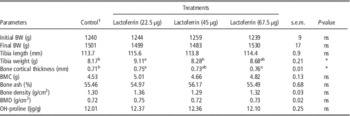
Tibia weight at hatch was 0.194, 0.197, 0.200 and 0.205 g in the control, 22.5, 45 and 67.5 μg of bLF per egg, respectively. No statistically significant differences (P>0.05) were found among all the treatments in this respect. Effects of in ovo injection of bLF on BW and tibia characteristics of 28-week-old laying hens are presented in Table 1. Tibia weight of laying hens was affected by bLF treatments and was significantly greater (P<0.05) for the treatment having 22.5 μg of bLF per egg than the control. Tibia cortical thickness was also increased by bLF treatments but these differences were significant (P<0.05) only for 22.5 and 67.5 μg of bLF per egg. No significant differences were obtained in other tibia characteristics (P>0.05).
Effects of in ovo injection of bLF on BW and tibial bone characteristics of 28-week-old laying hens

BMC=bone mineral content; BMD=bone mineral density.
a,bMeans in a row with common superscript are not significantly different (P>0.05); ns = non-significant; *P<0.05.
1 Vehicle-injected control.
The effects of in ovo injection of bLF on reproductive-system measurements of laying hens are shown in Table 2. As can be seen, no statistically significant differences (P>0.05) were found among all the treatments in this particular case.
Effects of in ovo injection of bLF on reproductive system of 28-week-old laying hens

ns=non-significant.
1 Vehicle-injected control.
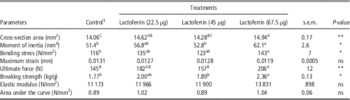
The results of bone biomechanical properties are shown in Table 3. Cross-sectional area of the tibia cortex is increased by 3.98% and 6.25% in treatments having 22.5 and 67.5 μg of bLF per egg, respectively, in comparison with the control (P<0.01). Maximum strain, elastic modulus and area under the curve were not affected (P>0.05) by in ovo injection of bLF, while moment of inertia, bending stress, ultimate force and breaking strength were significantly increased (P<0.05) in treatment having 67.5 μg of bLF per egg in comparison with the control.
Effects of in ovo injection of bLF on tibia biomechanical properties of 28-week-old laying hens

A,B,CMeans in a row with common superscript are not significantly different (P>0.01).
a,b,cMeans in a row with common superscript are not significantly different (P>0.05).
ns = non-significant; *P<0.05; **P<0.01.
1 Vehicle-injected control.
Performance trait values of 28-week-old laying hens are listed in Table 4. With the exception of egg weight (P<0.05), other performance traits were not affected by bLF treatment. The treatments having 45 and 67.5 μg of bLF per egg were significantly higher (P<0.05) in egg weight than the control.
Effects of in ovo injection of bLF on performance of laying hens from 20 to 28 weeks of age

EW=egg weight; HDP=hen-day egg production; EM=egg mass; FI=feed intake; FCR=feed conversion ratio.
a,bMeans in a row with common superscript are not significantly different (P>0.05); ns = non-significant; *P < 0.05.
1 Vehicle-injected control.
Discussion
At least until now, there is no study regarding the role of in ovo injection of lactoferrin in poultry. This study describes the effects of in ovo injection of bLF on laying-hen bone strength. Tibia measurements were not affected by in ovo injection of bLF at hatch that may be explained by the short period of embryonic stage. No significant effect of bLT treatments was observed on hen BW at 28 weeks of age. This result is in agreement with the literature reports of Svoboda et al. (Reference Svoboda, Drabek and Ficek2005), Uchida et al. (Reference Uchida, Oda, Sato and Kawakami2006) and Blais et al. (Reference Blais, Malet, Mikogami, Martin-Rouas and Tome2009), in piglet, rat and mice. As expected bLF treatments, especially in low concentration, caused higher tibia weight compared with the control, indicating the success of using bLF in the laying-hen bone. Higher tibia weight in the current study is in agreement with previously reported data by Cornish et al. (Reference Cornish, Callon, Naot, Palmano, Banovic, Bava, Watson, Lin, Tong, Chen, Chan, Reid, Fazzalari, Baker, Baker, Haggarty, Grey and Reid2004), Grey et al. (Reference Grey, Banovic, Zhu, Watson, Callon, Palmano, Ross, Naot, Reid and Cornish2004) and Hou et al. (Reference Hou, Xue and Lin2012), who reported that lactoferrin promotes bone growth. These studies established lactoferrin as an anabolic factor in osteoblasts, which also reduces osteoclast differentiation, causing an overall effect of increase in bone growth through the promotion of bone formation and the inhibition of bone resorption. In fact, lactoferrin plays an important immunomodulatory function, decreasing the secretion of a number of osteolytic cytokines such as TNFα and IL-1β and stabilizing mast cells. Thus, its direct effects on the activity and development of bone cells may be complemented by these cytokine-mediated effects (Cornish et al., Reference Cornish, Callon, Naot, Palmano, Banovic, Bava, Watson, Lin, Tong, Chen, Chan, Reid, Fazzalari, Baker, Baker, Haggarty, Grey and Reid2004). Tibia cortical thickness was notably increased in bLF treatments, especially in low and high concentrations. Cortical bone is the compact outer layer of bone and the predominant bone in the shafts of long bones, which contributes to bone strength (Nilas, Reference Nilas1993). Bone strength is related to bone biomechanical properties (e.g. force, energy and stiffness; Zernicke et al., Reference Zernicke, Salem, Barnard and Schraamm1995). In the present study, cortical tibia thickness supports and further explains the biomechanical results in which in ovo injection of bLF can improve laying-hen bone strength. The results of other tibia characteristics for evaluating bone, namely BMC, bone ash, bone density, BMD and hydroxyproline content, showed that bLF failed to alter the bone status of laying hen. Yet, nearly all of these variables in bLF treatments were numerically higher than the control. Between significant tibia characteristics, the coefficient of variation (CV) of tibia cortical thickness (3.89%) was smaller than the CV of tibia weight (6.12%), which may indicate that the tibia cortical thickness is potentially a better indicator of bone status than the tibia weight.
No detectable effects were observed on reproductive measurements among treatments, indicating that use of bLF during the embryonic stage did not affect the reproductive system of the adult laying hen. Guo et al. (Reference Guo, Jiang, Ibrahim, Zhang, Zhang, Zhang and Zheng Ren2009) and Blais et al. (Reference Blais, Malet, Mikogami, Martin-Rouas and Tome2009) also reported that lactoferrin supplementation did not significantly modify the uterine weight in rat and mouse. It is well demonstrated that estrogen can promote bone growth. This function is also documented for lactoferrin (Blais et al., Reference Blais, Malet, Mikogami, Martin-Rouas and Tome2009; Malet et al., Reference Malet, Bournaud, Lan, Mikogami, Tome and Blais2011). Because there are no significant differences in reproductive system among treatments in this study, therefore it seems likely that improvement in the bone status of bLF treatments is only related to in ovo injection of bLF that with a positive correlation is transferred to adult birds.
There are a number of biomechanical properties that can be used to characterize the bone integrity. Each of these properties reflects a characteristic of the bone. Cross-sectional area of the tibia cortex and moment of inertia were greater in treatment having 67.5 μg of bLF per egg than the control. These structural criteria are generally positively correlated with bone strength (Martin, Reference Martin1991 and Reference Martin1993; Turner, Reference Turner2006). Therefore, the increased ultimate force and bending stress (force per unit area) can be attributed to their increased sizes, as indicated by significant changes in the cross-sectional area and the moment of inertia of the tibiae. The biomechanical status of bone may be poorly described by just one of these properties. For instance, maximum strain is inversely related to the brittleness and stiffness of the bone (Turner, Reference Turner2006). In the present study, therefore according to the maximum strain (0.0119 mm) and stiffness (13 831 N/mm2), tibias with high concentration of bLF should tend to be brittle, but also should tend to be stiff, resulting in reduced work to failure and increased risk of bone fracture. But in fact, area under the stress–strain curve showed that the high concentration of bLF has improved bone status, resulting in increased work to failure and decreased risk of bone fracture. Collagen’s elastic properties can make bone less brittle and stiff, allowing it to sustain greater force without fracturing (Wang et al., Reference Wang, Shen, Li and Agrawal2002). But in the current study, the amount of collagen in bones did not increase by high concentration of bLF, which may indicate that while bLF does not affect the amount of collagen in developing bone, it could affect properties of the collagen matrix itself by affecting collagen fiber length, fiber orientation or crosslink concentration. Overall, the results indicate that bone status was improved with high concentration of bLF, as measured by the tibia weight, cortical thickness, moment of inertia, ultimate force and bending stress. Egg weight from this treatment was significantly greater than other treatments. Taylor et al. (Reference Taylor, Horvat-Gordon, Moore and Bartell2013) also found that improvements in bone status by melatonin could significantly improve egg weight in laying hens. In Ebeid study (Reference Ebeid2011) also no improvement between tibial bone measurements and consequently in egg weight was reported. Hens are able to maintain their performance by mobilizing bone reserves to meet the demands of egg formation and utilize dietary sources to replenish these reserves (Knowles and Wilkins, Reference Knowles and Wilkins1998; Ekmay and Coon, Reference Ekmay and Coon2010). Therefore, it seems that under identical conditions of the rearing period, the increase in egg weight caused by high concentration of bLF is due to the further capacity of bones in absorption and metabolism of dietary minerals, which may play an important role in improving eggshell weight and consequently in egg weight.
In this study, the improvement in bone status is evidenced by increasing tibia bending stress at 67.5 μg of bLF per egg. In conclusion, bLF caused dramatic improvement in bone biomechanical properties, even with small changes in cortical thickness. The present study, therefore, suggests that cortical tibia thickness is a suitable variable for evaluating bone status reflecting bone integrity and strength. Finally, the present study shows that bLF (67.5 μg of bLF per egg) injected into layer breeder eggs before incubation can be used to improve bone status and egg weight of laying hens at 28 week of age, while having no detrimental effect on embryo hatchability.
Acknowledgments
The authors thank Bu-Ali Sina University for providing facilities and financial support for this study. The authors also wish to thank to the staff of the Department of Animal Science of this university for their excellent scientific collaboration.
Supplementary material
To view supplementary material for this article, please visit http://dx.doi.org/10.1017/S1751731115001093