Introduction
Cells spend large amounts of energy on anabolism of lipids in the form of fatty acids and sterols. Subsequently, these lipids are stored in a biologically inert manner, either as triacylglycerols or stearyl esters, to prevent them from reacting with other cellular substances and to eliminate intrinsic cellular toxicity (Ruggles et al., Reference Ruggles, Garbarino, Liu, Moon, Schneider, Henneberry, Billheimer, Millar, Marchadier, Valasek, Joblin-Mills, Gulati, Munkacsi, Repa, Rader and Sturley2014). Thus, regulation of cellular lipid levels is crucial to ensure the smooth functioning of biochemical processes in lipid metabolism (Tong et al., Reference Tong, Billheimer, Shechtman, Liu, Crooke, Graham, Cohen, Sturley and Rader2010).
The ‘ARE2 required for viability 1’ (ARV1) gene was discovered from Saccharomyces cerevisiae as an important gene involved in intracellular sterol trafficking and sterol uptake mechanisms in eukaryotic cells, and required in yeast for viability at high temperatures (Tinkelenberg et al., Reference Tinkelenberg, Liu, Alcantara, Khan, Guo, Bard and Sturley2000). Since accumulation of sterols within cells is cytotoxic, several molecular mechanisms exist in eukaryotic cells to regulate intracellular sterol levels. Cholesterol, an important molecule in this class, is tightly regulated. Elevated intracellular cholesterol levels trigger the conversion of cholesterol into cholesteryl esters by the enzyme acyl-CoA cholesterol acyltransferase (ACAT), leading to storage of cholesterol, down-regulation of its biosynthesis, and diminished transcription of the gene encoding the low-density lipoprotein (LDL) receptor, all of which reduces the cellular uptake of cholesterol. ACAT-related enzymes 1 and 2 (ARE1 and ARE2) catalyse the esterification of free sterols. Of these isoforms, ARE2 plays a major role in cellular sterol esterification (Mullner & Daum, Reference Mullner and Daum2004). Furthermore, it has been shown that none of these isoforms are required for viability and normal growth, using yeast cells deficient in ARE1 and ARE2 (Tinkelenberg et al., Reference Tinkelenberg, Liu, Alcantara, Khan, Guo, Bard and Sturley2000). Therefore in the absence of the function of the latter two genes, ARV1 was identified as an essential gene for the viability of yeast cells, complementing the function of ARE1 and ARE2 (Tinkelenberg et al., Reference Tinkelenberg, Liu, Alcantara, Khan, Guo, Bard and Sturley2000).
Although many ARV1s have been detected in eukaryotes, only yeast and human ARV1s have been significantly studied to date. Yeast ARV1 is a protein of 321 amino acids (Villasmil et al., Reference Villasmil, Ansbach and Nickels2011), with three transmembrane domains localized in both the endoplasmic reticulum (ER) and Golgi bodies (Villasmil & Nickels, Reference Villasmil and Nickels2011). The N-terminal region of the ARV1 protein consists of a block of amino acid residues and a putative zinc-binding motif, the ARV1 homology domain (AHD), which is conserved among many multicellular eukaryotes (human, worm and plants). The spacing between cysteine residues found in the AHD of the N-terminal domain resembles that of zinc ribbons. This is thus considered to be a potential zinc-binding motif. The C-terminus of ARV1 is ER luminal, while the N-terminal region is localized in the cytosol. This protein is thought to be a multimeric protein in yeast. The human ARV1 is located on chromosome 1 and is presumed to encode at least 11 alternatively spliced variants and an unspliced form. Altogether ten different isoforms of the protein are produced, and the isoform that has 271 amino acids is considered to be the human ARV1. This protein also consists of three putative transmembrane domains and shares 44% homology with yeast (Tinkelenberg et al., Reference Tinkelenberg, Liu, Alcantara, Khan, Guo, Bard and Sturley2000).
Previous studies conducted with yeast have indicated that ARV1 plays a vital role in cellular lipid homeostasis, by participating in cellular sterol uptake, intracellular sterol trafficking, phospholipid metabolism, sphingolipid metabolism and other important functions unrelated to lipid metabolism (Villasmil et al., Reference Villasmil, Ansbach and Nickels2011). Moreover, it has also been reported that yeast lacking functional ARV1 can be functionally complemented by the human ARV1 gene, demonstrating the functional conservation among eukaryotic classes (Villasmil et al., Reference Villasmil, Ansbach and Nickels2011).
To date, only a few ARV1s have been characterized, and the role of ARV1 in nematodes is yet to be addressed (Tong et al., Reference Tong, Billheimer, Shechtman, Liu, Crooke, Graham, Cohen, Sturley and Rader2010). Therefore, the main objective of this study was to functionally characterize a putative ARV1 identified from Setaria digitata by carrying out: (1) homologous property and phylogenetic relationship analyses using identified homologous counterparts of SdARV1; (2) gene expression analysis using reverse transcription-polymerase chain reaction (RT-PCR) in different developmental stages of S. digitata; and (3) yeast complementation assay using SdARV1 to analyse temperature sensitivity, growth and sterol content in complemented yeast strains, with respect to wild and mutant yeast strains.
Materials and methods
Identification of cDNA sequence corresponding to SdARV1
A cDNA library of S. digitata was constructed in the vector λ Zap according to the manufacturer's instructions (Stratagene, San Diego, California, USA). The library was excised in vivo in Escherichia coli, XL1-Blue MRF, to give phagemid colonies, which were randomly picked and their plasmid DNA was extracted using the alkaline lysis method. Bidirectional sequencing of randomly picked plasmid DNA was carried out with vector-derived primers using a Thermo Sequenase™ CY5 Dye Terminator kit and ALF Express™ DNA Sequencer (Amershan–Pharmacia, Amersham, Bucks, UK). A clone, designated pSDC14, containing S. digitata ARV1 (SdARV1) was completely sequenced (GenBank accession number KY884304), after which a homology search showed it to have sequence similarity with ARV1 in the National Center for Biotechnology Information (NCBI) database.
Sequence characterization and phylogenetic analysis
Prediction of physicochemical properties of the putative SdARV1 was conducted using the program DNAsist 2.2 (https://dnassist.en.softonic.com/) and homologous protein sequences for SdARV1 were identified by executing BLASTP at NCBI using SdARV1 as the query sequence. Secondary structural features of SdARV1 were predicted using the PSIPRED protein sequence analysis workbench (http://bioinf.cs.ucl.ac.uk/psipred/). Structural analysis of SdARV1 for potential transmembrane domains was performed using TMHMM21 v 2.0 (http://www.cbs.dtu.dk/services/TMHMM/) and HMMTOP22 v 2.0 (http://www.enzim.hu/hmmtop/) servers. SdARV1 was compared at the pairwise level using the EMBOSS needle server (https://www.ebi.ac.uk/Tools/psa/emboss_needle/) and multiple sequence alignment was carried out using the program ClustalW2 (Larkin et al., Reference Larkin, Blackshields, Brown, Chenna, McGettigan, McWilliam, Valentin, Wallace, Wilm, Lopez, Thompson, Gibson and Higgins2007). The phylogenetic analysis was carried out using MEGA software version 6 (Tamura et al., Reference Tamura, Stecher, Peterson, Filipski and Kumar2013), by implementing the neighbour-joining method supported by bootstrapping values taken from 5000 replicates.
RNA isolation, cDNA preparation and RT-PCR analysis of SdARV1
Microfilariae and adult males and females of S. digitata were collected from the peritoneal cavity of cattle slaughtered at Wellisara abattoir in Sri Lanka, and were washed with phosphate-buffered saline (pH 7.4). RNA extraction was carried out using a Genospin-R™ total RNA extraction kit (CeyGen, Colombo, Sri Lanka) according to manufacturer's instructions. Briefly, about 50 mg of microfilariae, adult male or female worms, were separately ground to a fine powder in liquid nitrogen, were transferred into nuclease-free 1.5-ml microcentrifuge tubes, each containing 200 μl of tissue lysis buffer and 40 μl of proteinase K, and incubated at 56°C overnight until the tissue was completely digested. Then, 400 μl of binding buffer and 350 μl of absolute ethanol were added to each tube and mixed well, prior to letting them stand for 2 min at room temperature. Each mixture was then transferred to a Genospin-R™ column (CeyGen) placed in a collection tube and washed separately with 500 μl of wash buffer-1 and wash buffer-2, prior to eluting the RNA with 30 μl of elution buffer. Following determination of total RNA concentration using an QubitR 2.0 Fluorometer (Invitrogen, California, USA), the reverse transcription reaction was carried out in 20-μl reaction mixtures, each containing 200 ng of total RNA, 8.8 μl of RNase-free water, 2.0 μl of 10× M-MuLV buffer (New England BioLabs, Ipswich, Massachusetts, USA), 1.00 μl of deoxyribonucleoside triphosphates (dNTPs; 10 mm), 0.2 μl of RNase inhibitor (40 U/μl), 2.0 μl of oligo-dT primers (50 μm) and 1.0 μl of reverse transcriptase (200 U/μl; New England Bio-labs) at 42°C for 1 h. The reverse transcription was carried out simultaneously for all the RNA samples isolated.
The primers designed for RT-PCR to determine the expression of SdARV1 are given in table 1. The cDNAs prepared from adult male and female worms and microfilariae were used separately as the templates for RT-PCR. The reaction was carried out in 25 μl of master mix containing 5.0 μl of cDNA, 0.6 μl of ThermoRead™ Polymerase (5 U/μl; CeyGen), 10 μl of master mixture (10 mm dNTPs, 10× Taq buffer, 25 mm MgCl2) and 1.0 μl of each primer (5.0 μm). The thermal cycle parameters were: initial denaturation at 95.0°C for 5 min; followed by 40 cycles of denaturation at 95.0°C for 30 s, annealing at 59.5°C for 30 s and extension at 72.0°C; with a final extension step of 72.0°C for 7 min. In the same reaction, 5.0 μl of PCR water and 1.0 μl of previously extracted pSDC12 plasmid DNA (10 ng/μl) having the SdARV1 cDNA sequence were used as the templates for negative and positive controls, respectively. After the completion of PCR cycles, a portion of 3 μl of each PCR mixture was run on a 1.0% agarose gel with a 100 bp ladder marker (Biobasic, Ontario, Canada) and observed under UV light.
Primer name, the purpose of usage and its corresponding sequences.

Yeast complementation and functional assays
PCR amplification and cloning of SdARV1 into the yeast vector pNEV
The coding region of SdARV1 was PCR amplified, using the pSDC14 clone containing SdARV1, previously identified and sequence confirmed, using forward and reverse PCR primers (table 1), engineered with the restriction enzyme site of NotI using initial denaturation at 94°C for 3 min; followed by 35 cycles of denaturation at 94°C for 1 min, annealing at 57°C for 30 s and extension at 72°C for 1 min; and a final extension step of 72°C for 3 min. PCR was carried out in 25 μl of master mix containing 13.5 μl of PCR water, 7.5 μl of master mixture (10 mm dNTPs, 10× Taq buffer, 25 mm MgCl2), 1.0 μl of forward primer (5 μm), 1.0 μl of reverse primer (5.0 μm), 1.0 μl of template (pSDC12 plasmid DNA (10 ng/μl) and 0.125 μl of ThermoRead™ Polymerase (5 U/μl)). The resultant PCR products and pNES expression vector (containing the PMA-1 promoter) were simultaneously digested with NotI restriction enzymes (200 U/μl, New England Bio Labs) following the manufacturer's protocol and resolved on a 1% agarose gel. Subsequently, the corresponding DNA bands were detached and purified using the CEYGEN SpinClean™ gel band and product purification kit. Following the addition of the Ligation Kit Mighty Mix (Clonetech, Mountain View, California, USA), purified PCR products and vector were ligated by incubating overnight at 4°C. Thereafter, the ligated product was transformed into E. coli DH5α cells and recombinant colonies were identified by PCR screening, and plasmid DNA was extracted. Finally, the sequence-confirmed recombinant plasmid (pNEV–SdARV1, containing a single copy of SdARV1 under the control of the PMA-1 promoter) was used for further complementation studies.
Transformation of SdARV1 into the ARV1 mutant yeast strain
The yeast competence cells prepared from the ARV1 mutant strain (Y35151, MATa/MATα; ura3Δ0/ura3Δ0; leu2Δ0/leu2Δ0; his3Δ1/his3Δ1; met15Δ0/MET15; LYS2/lys2Δ0; YLR242c::kanMX4/YLR242c::kanMX4; http://www.euroscarf.de) were gently thawed at room temperature, and 40 μl of these cells were mixed with 5 μl of the ligation mixture in a 0.2-cm electroporation cuvette and subjected to an electric pulse using an electroporator (25 μF and 2.5 kV) (BioRad, Hercules, California, USA). The electroporated cells were pelleted and resuspended in 1 ml of TE buffer, and a 200 μl aliquot of the resuspension was spread on Complete Minimal and Drop-out plates (Sigma-Aldrich, St. Louis, Missouri, USA). The Ura+ transformants were initially isolated at 37°C in YPAD replica plating and then minimum medium containing leucine and histidine (40 μg/ml). They were confirmed by obtaining a PCR amplicon corresponding to SdARV1 using primers specific to SdARV1. Total RNA was extracted from confirmed recombinant yeast colonies (SdARV1 cDNA sequence-transformed mutant yeast, SdARV1-yeast), the wild-type yeast strain and the ARV1 mutant yeast strain, using the Genospin-R™ total RNA extraction kit. Semi-quantitative RT-PCR was carried out for all extracted RNA samples following the protocol described above.
Functional assessment
Serial blotting assay
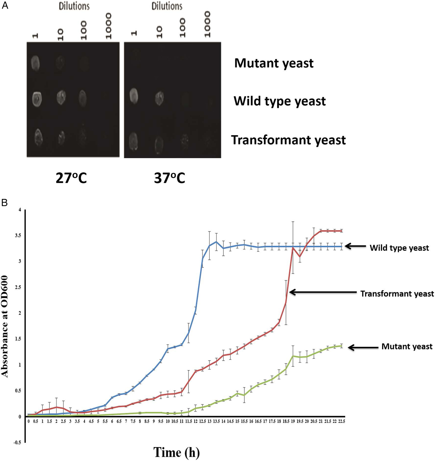
Serial dilutions of cultures containing the yeast ARV1 mutant strain, the pNEV–SdARV1 yeast strain and the wild type were spotted on to plates, such that each row represented a strain while each column represented a dilution, to compare growth behaviours of yeast strains at permissive (27°C) and restrictive (37°C) temperatures. Briefly, single colonies from previously prepared YPAD plates of the yeast strains mentioned above were picked and inoculated separately into 5-ml aliquots of YPD broth, and incubated while shaking at 27°C until the OD600 reached 1.0 A. Then 10 μl was taken from each culture and diluted with 90 μl of sterile deionized water and, using these, six tenfold dilutions were prepared, and 10 μl of these dilutions were spotted in rows representing a particular yeast strain (mentioned above) across the plate with 1-cm distance between two spots. This procedure was performed in two YPAD plates simultaneously, incubating one at 37°C and other at 27°C for 3 days. The plates were observed after 3 days.
Construction of growth curves
Single colonies of the ARV1 mutant strain, wild type and pNEV–SdARV1 yeast strain from the streaked plates were picked and inoculated separately into 5-ml aliquots of YPD broth, and grown at 30°C with horizontal shaking until the OD600 reached 1.0 A. A 1-ml portion from each culture was transferred to 200 ml of YPD broth and incubated at 30°C with continuous shaking, and the absorbance measured at 600 nm, taking samples of all three cultures at 30-min intervals for 24 h. These readings were used to plot growth curves.
Microscopic observation of yeast cells
A drop of cell suspension culture was spread over a microscope glass slide and was fixed by exposing to a flame. A drop of ammonium crystal violet was added, allowing the dye to bind with the film for 30 s. Then excess stain was removed by washing with tap water. Next, iodine acetone was added to the film and left for 30 s. This was followed by the immediate addition of a drop of diluted carbolfucsine at room temperature for another 30 min. Finally, the stained yeast cells were fixed on to the glass slide, by exposing to a flame, for morphological analysis under the microscope.
Total sterol extraction and quantification from yeast cells
A single colony from each of the previously prepared ARV1 mutant strain, wild type and pNEV–SdARV1 yeast strain were picked and inoculated separately into 200-ml batches of YPD broth, and incubated while shaking at 27°C. After the OD600 reached approximately 1.0 A in all the cultures, cells were harvested by centrifugation at 3500 rpm for 15 min. Then, to each 100 mg harvested air-dried cell pellet in a Falcon tube, 3 ml of methanol, 2 ml of 0.5% pyrogallol, 2 ml of 60% KOH and 50 μl of 1 mg/ml cholesterol were added, vortexed gently and the contents were then transferred to round-bottom flasks and placed in a water bath equilibrated to 90°C for 2 h incubation. Samples were allowed to cool in the round-bottom flasks and removed to new Falcon tubes, to which 5 ml of heptane was added, and centrifuged at 5000 rpm for 5 min to facilitate phase separation. Extracted heptane was transferred to a new Falcon tube while repeating extraction for two more times. The extracted heptane then was evaporated in a water bath set at 55°C under a stream of nitrogen before storing the samples at −20°C. The above procedure was repeated in triplicate with three separate colonies from each plate.
The extracted sterol was quantified using the Cholesterol LiquiColor Test (Allain et al., Reference Allain, Poon, Chan, Richmond and Fu1974; MacLachlan et al., Reference MacLachlan, Wotherspoon, Ansell and Brooks2000) according to manufacturer's instructions (Interchem, California, USA). In brief, the spectrophotometer (Jenway 6300, Cole-Parmer, Staffordshire, UK) was calibrated with standard cholesterol. Each extracted sample was dissolved in 50 μl of saline to which 500 μl of enzyme reagent was added and mixed well, then incubated at 37°C for 5 min. Readings were obtained from the samples at a wavelength of 540 nm.
Results and discussion
Sequence characterization, functional prediction and evolutionary relationship of S. digitata ARV1
The SdARV1 cDNA sequence of 761 bp, with 5′ and 3′ untranslated regions (UTRs) of 34 bp and 73 bp, respectively, and with an open reading frame (ORF) of 651 bp that codes for a protein of 217 amino acids, was identified. The protein has a predicted molecular weight and isoelectric point of 25 kDa and 9.0, respectively. In secondary structure prediction using PISPRED (http://bioinf.cs.ucl.ac.uk/psipred/), four sheets and seven helixes were detected for SdARV1 (fig. 1A). With respect to transmembrane domain prediction using the TMHMM program (http://www.cbs.dtu.dk/services/TMHMM-2.0/), two such domains were identified with high probability in the regions of amino acid residues 112–134 and 144–166, with the N-terminal ER luminal region (0.8) and C-terminal ER region (0.6) also of high probability. However, HMMTOP (http://www.enzim.hu/hmmtop/index.php) predicted a cytoplasmically localized N-terminal region and three transmembrane domains in the regions of amino acid residues 120–137, 144–163 and 194–213 (fig. 1B). The first two transmembrane predictions had a consensus in both servers, whereas the third did not. However, collectively these results further support the hypothesis that SdARV1 could be a transmembrane protein, as in yeast.
The prediction of SdARV1 secondary structure and membrane topology. (A) Predicted SdARV1 secondary structural features from PSIPRED. The sheets are indicated in arrows and the helices are indicated in cylinders. The letters ‘E’ and arrows, ‘H’ and cylinders and ‘C’ indicate potential areas for beta-strands, alpha-helices and coil regions of SdARV1, respectively. (B) Predicted membrane topology of SdARV1 via TMHMM21 v 2.0 and HMMTOP22 v 2.0 servers. The ARV1 homology domain (AHD) is indicated as a box. HMMTOP predicted a cytoplasmically localized N-terminal region and three transmembrane helices for SdARV1 at amino acid residues 120–137, 144–163 and 194–213 (cylinders).

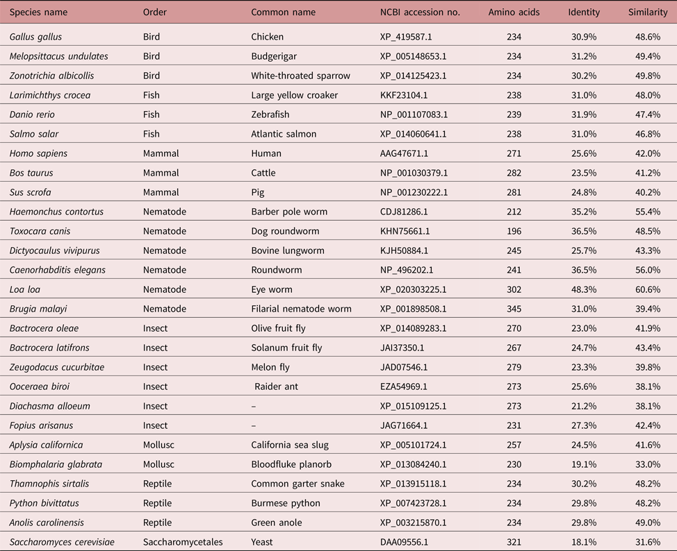
To gain insights into primary protein structural relationships, the putative SdARV1 amino acid sequence was compared in a pairwise manner with ARV1 sequences identified from different organisms. It was found that ARV1 of Loa loa has the highest sequence identity (48.3%, table 2). The ARV1 sequences derived from mammals showed the fewest sequence identities and similarities with SdARV1. However, through overall pairwise sequence comparison, SdARV1 seems to share substantial levels of sequence identity and similarities with all the other species.
Pairwise sequence comparison.

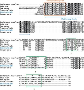
As revealed by the generated multiple sequence alignment (MSA) (fig. 2), amino acid positions C11, C14, L21, C35, V39, D40, Y42, E44, D46, V48, I52, L56, A61, R64, H65 and N69 in the ARV1 homology domain (AHD) were identified to be highly conserved over the range of taxonomic groups compared. Appropriate multicellular eukaryotes were chosen so that they included organisms from widely varying taxa, to identify residues that are highly conserved through the evolutionary timeline. Identical amino acids at analogous positions are present to carry out a vital function and are likely to be involved in the formation of the active site or binding site for specific molecules.
ClustalW multiple sequence alignment of the deduced amino acid sequence of SdARV1 with other known ARV1 proteins. As revealed by the generated multiple sequence alignment (MSA), amino acid positions C11, C14, L21, C35, V39, D40, Y42, E44, D46, V48, I52, L56, A61, R64, H65 and N69 in the ARV1 homology domain (AHD) were identified to be highly conserved over the range of taxonomic groups compared (black shading). The putative zinc-binding motif is indicated with a brown box. The ARV1 homology domain is shown with the blue double-headed arrow. The locations of putative transmembrane domains are indicated with green boxes 1, 2 and 3.

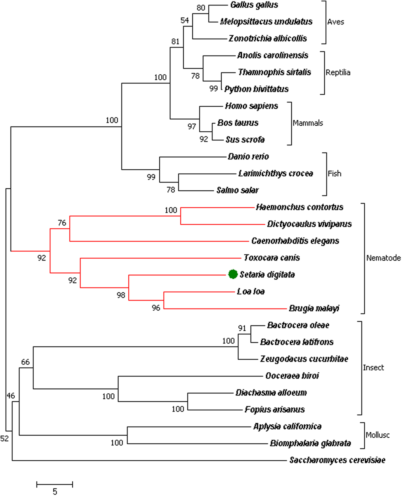
To clarify phylogenetic relationships, SdARV1 identified in this study and ARV1 members retrieved from the database were analysed by generating a phylogenetic tree. As reflected by the formulated tree, SdARV1 was grouped with other nematode ARV1 counterparts, while members from mammals, reptiles, birds, fish, insects and molluscs were clustered in different groups in an evolutionarily distant branch from nematodes (fig. 3). As compared to the insect and mollusc clade, the nematode clade showed closer phylogenetic relationships to the vertebrate clade where fish, mammals, reptiles and birds were included. This observation hints at an evolutionary advancement of nematode ARV1s over those of other invertebrates such as insects and molluscs. As far as the nematode phylum in the phylogenetic tree is concerned, SdARV1 showed the closest phylogenetic relationships with the ARV1 counterparts from Loa loa (eye worm) and Brugia malayi (filarial nematode worm). It also exhibited a close phylogenetic relationship with Haemonchus contortus (barber pole worm), Caenorhabditis elegans (roundworm) and Dictyocaulus vivipurus (bovine lungworm), along with Toxocara canis (dog roundworm). This clear-cut clustering of nematodes in the phylogeny hints of a distinct biological role of ARV1 in the phylum Nematoda. Taken together, these results suggest the conserved nature of the ARV1 counterpart among different animal groups, perhaps performing group-specific functions in intracellular sterol homeostasis.
Phylogenetic analysis of SdARV1 with selected, full-length ARV1 amino acid sequences of several other species. The numbers at the nodes represent the bootstrap values for 5000 replicates. The Setaria digitata homologue is indicated with a green-coloured dot and the nematode phylum is indicated in black parentheses and red branch lines. Animal groups: Aves, Reptilia, Mammals, Fish, Insects and Molluscs are also indicated using black parentheses. The NCBI-GenBank accession numbers of the amino acid sequences of the organisms used in the comparison are mentioned in table 1, with the exception of Setaria digitata: KY884304

Expression of SdARV1 in different life stages of S. digitata
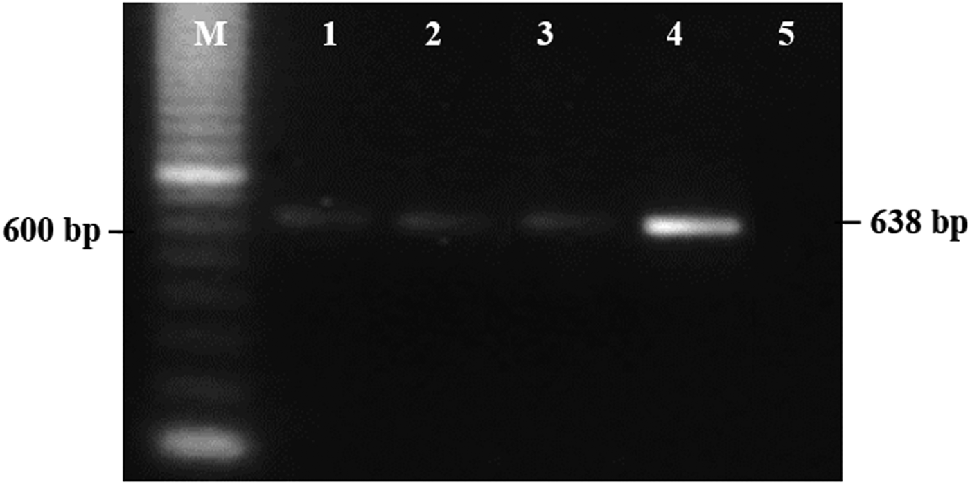
To analyse SdARV1 expression, RT-PCR was carried out and the expression was detected in microfilariae, and adult male and female worms, showing the expressional importance of SdARV1 in these stages (fig. 4). ARV1 has been validated as a crucial component of sterol metabolism and subcellular localization, and sterols are an important structural component of membranes and organelles (Shechtman et al., Reference Shechtman, Henneberry, Seimon, Tinkelenberg, Wilcox, Lee, Fazlollahi, Munkacsi, Bussemaker, Tabas and Sturley2011). Thus, it can be assumed that SdARV1 plays similar functional roles in early developmental stages as well as in the adult stage of S. digitata.
RT-PCR-based SdARV1 expression analyses in development stages of S. digitata using agarose gel electrophoresis. Lanes: 1, microfilariae; 2, adult male worm; 3, adult female worm; 4, positive control; 5, negative control; M, the 100 bp Biobasic ladder marker.

Functional assessment of SdARV1 via yeast complementation assays
Two distinguishable phenotypes have been reported, when ARV1 is deleted or mutated, i.e. abnormal growth attributes and altered state of the sterol metabolism pathway (Shechtman et al., Reference Shechtman, Henneberry, Seimon, Tinkelenberg, Wilcox, Lee, Fazlollahi, Munkacsi, Bussemaker, Tabas and Sturley2011). In the current study, evidence of complementation of these two major attributes was gathered by transforming the recombinant SdARV1 into ARV1 mutant yeast strains, and the success of transformation was confirmed by showing mRNA expression in the yeast transformant (see supplementary fig. S1).
Determination of the temperature sensitivity and monitoring of the growth rates of yeast strains
Previous work reported that the functional involvement of ARV1 was mandatory for cell survival at restrictive temperatures such as 37°C (Tinkelenberg et al., Reference Tinkelenberg, Liu, Alcantara, Khan, Guo, Bard and Sturley2000). Therefore, the temperature sensitivities of mutant, wild-type and pNEV–SdARV1-transformed yeast were determined at 27°C (permissive temperature) and 37°C (restrictive temperature) via a serial blotting assay (fig. 5A) by duplicating the assay in YPAD plates.
Growth analysis of ARV1 mutant yeast strain (Y35151), ARV1 mutant strain transformed with pNEV–SdARV1 and wild-type yeast. (A) The growth of serially diluted yeast strains (three tenfold dilutions) in YDPA plates at permissive temperature (27°C) and non-permissive temperature (37°C). (B) The comparison of growth curves of yeast strains in YPD broth over the period of time. The Y-axis indicates the absorbance measured at OD600 and the X-axis indicates the time in hours. The cells were grown at 27°C. Error bars represent the SD (n = 3).

A high level of growth was observed in wild-type cells at both 27°C and 37°C, indicating that the growth of the wild type is independent of temperature. However, the yeast mutant strain, which was deemed to show hardly any growth at the restrictive temperature, exhibited significantly higher growth when transformed, compared to its original strain, in almost every cell dilution tested in the blotting assay, indicating that the temperature-sensitive and growth-defective phenotype of the mutant yeast cells can be restored back to the original phenotype by introducing SdARV1. Similar approaches have been used in previous studies to restore growth and temperature defects due to ARV1 mutation of yeast by complementing through the introduction of the ARV1 gene isolated from humans (Tinkelenberg et al., Reference Tinkelenberg, Liu, Alcantara, Khan, Guo, Bard and Sturley2000).
To further clarify the effect of SdARV1 on yeast cell viability and growth levels, cells grown in YPD cultures at a non-permissive temperature, 37°C, were analysed using the spectrophotometer. The growth curve was generated by plotting the absorbance (A) of the culture against time, and the growth rates were compared (fig. 5B). According to the growth curves, the growth rate of mutant yeast was significantly lower than that of wild-type and transformed yeast. When comparing the growth curve of transformant with the growth curve of wild type, the growth rate of transformant cells was slightly lower, but still significantly higher than that of the yeast mutant. Collectively, it may be postulated that the reason for the lowest growth rate in the yeast mutant was the lack of ARV1 function, because the wild-type yeast carrying the wild yeast ARV1 exhibited the highest growth rate. Meantime, the yeast cells transformed with SdARV1 showed moderate growth rates, indicating that SdARV1 complements the functions of the yeast ARV1 gene to a significant extent.
A distinguishable morphological difference was observed in transformed and mutant yeast strains when they were observed under the microscope following Gram staining (see supplementary fig. S2B, C), and typical wild-type yeast cells and mutant yeast cells were seen as oval and abnormally round shapes, respectively (see supplementary fig. S2A, C). Since the ARV1 gene is involved in sterol redistribution in cell membranes, it can be hypothesized that lack of ARV1 function alters the physical appearance of yeast cells and this can be regained by transformation following complementation of the yeast mutant phenotype by SdARV1 (see supplementary fig. S2), as seen in this study.
Sterol extraction from yeast and quantification of sterol levels
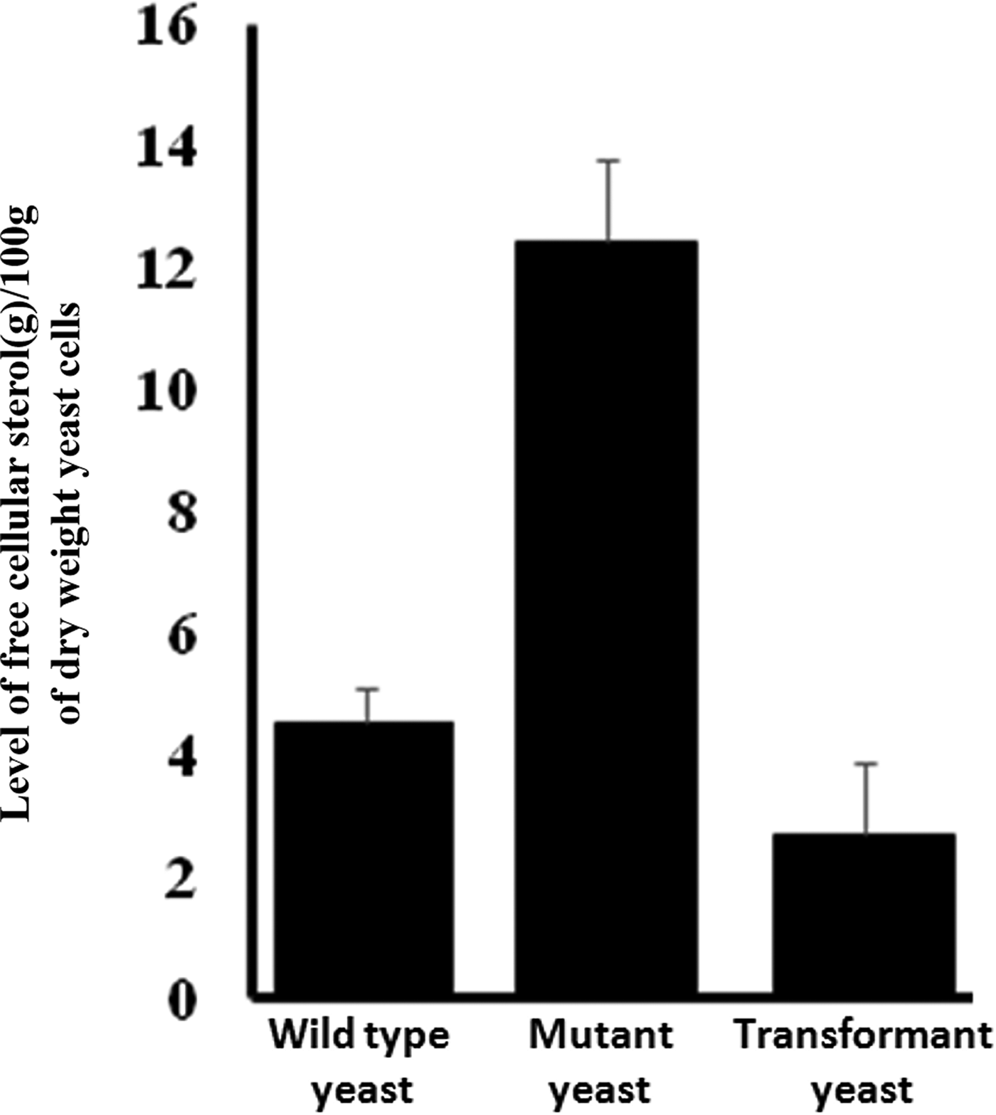
To investigate the effect of complementation further, an enzyme-based detection of free sterol levels of wild-type, mutant and transformant strains was carried out. The free sterol level of the mutant strain was significantly higher, compared to the wild-type and transformants, which had approximately similar levels of sterols. The effect of complementation on sterol levels was significant, and seems reversible when introducing SdARV1 into the yeast mutant (fig. 6). Thus, the results of sterol-level detection further strengthen the hypothesis that the SdARV1 gene has complemented the ARV1 function of yeast, and that SdARV1 is a true homologue of yeast and human ARV1.
Free sterol levels of wild-type yeast, mutant yeast and transformant yeast strains. The Y-axis indicates the level of free cellular sterol (g) in 100 g dry weight of yeast cells and the X-axis indicates the type of yeast strains used in this study. Error bars represent the SD (n = 3).

In conclusion, ARV1 of S. digitata and its homologues identified in this study have crucial information for phylogenetic analysis that follows classical evolutionary trends. Through complementation of general phenotypic defects of ARV1 mutant yeast strains, such as growth rates, temperature sensitivity and sterol levels, it can be confirmed that SdARV1 is a true homologue of ARV1 of yeasts and humans. Finally, the findings of the current study enhance our interest in nematode ARV1, encouraging us to conduct further studies that would detail the role of ARV1 in sterol metabolism and other biological functions.
Supplementary material
To view supplementary material for this article, please visit https://doi.org/10.1017/S0022149X17001109
Financial support
This research received no specific grant from any funding agency, commercial or not-for-profit sectors.
Conflict of interest
None.