Introduction
The frontal-subcortical pattern of cognitive deficits associated with Parkinson’s disease, which is most often diagnosed after age 50, is well-documented. In contrast, the cognitive profile of early-onset Parkinson’s disease (EOPD), defined by age at onset of 50 or younger (Hedrich et al., Reference Hedrich, Kann, Lanthaler, Dalski, Eskelson, Landt and Klein2001; Lucking et al., Reference Lucking, Durr, Bonifati, Vaughan, De Michele, Gasser and Brice2000) and affecting up to 10% of all PD patients (Schrag & Schott, Reference Schrag and Schott2006) is not well defined. Parkin mutations are a common cause of parkinsonism and are the most common mutations associated with EOPD, particularly among those ≤30 years of age (Hedrich et al., Reference Hedrich, Kann, Lanthaler, Dalski, Eskelson, Landt and Klein2001; Lucking et al., Reference Lucking, Durr, Bonifati, Vaughan, De Michele, Gasser and Brice2000; Schrag & Schott Reference Schrag and Schott2006; Wang et al., Reference Wang, Clark, Louis, Mejia-Santana, Harris, Cote and Marder2008), although mutations and variants in other genes have also been found in these cases including leucine-rich repeat kinase-2 (LRRK2) and glucocerebrosidase (GBA).
Mutations in the parkin gene have been identified in homozygous states, that is, when the same mutation is inherited from each parent; compound heterozygous states, that is, when different mutations in the same gene are inherited from each parent; and heterozygous states, that is, when a single mutation is inherited from one parent. The likelihood that one parkin mutation may predispose to later disease onset is supported by studies that have identified heterozygous mutations in patients with Parkinson’s disease (Clark et al., Reference Clark, Afridi, Karlins, Wang, Mejia-Santana, Harris and Marder2006; Foroud et al., Reference Foroud, Uniacke, Liu, Pankratz, Rudolph, Halter and Nichols2003; Lesage et al. Reference Lesage, Lohmann, Tison, Durit, Durr and Brice2008; Moro et al., Reference Moro, Volkmann, Konig, Winkler, Hiller, Hassin-Baer and Klein2008; Pramstaller et al., Reference Pramstaller, Schlossmacher, Jacques, Scaravilli, Eskelson, Pepivani and Klein2005) and PD onset is earlier in carriers of two mutations (homozygous or compound heterozygous) when compared to carriers of one mutation (heterozygous), suggesting that a dosage effect may exist. However, the pathogenicity of heterozygous mutations is debated (Lincoln et al., Reference Lincoln, Wiley, Lynch, Langston, Chen, Lang and Farrer2003; Oliveira et al., Reference Oliveira, Scott, Martin, Nance, Watts, Hobble and Vance2003).
Studies of the cognitive profile of EOPD and the role of parkin on cognitive function are limited. Early animal models of parkin deficiency reported decreased exploratory behavior in parkin-deficient mice (Zhu et al., Reference Zhu, Maskri, Herold, Bader, Stichel, Gunturkun and Lubbert2007) but another study did not observe any behavioral differences (Perez & Palmiter, Reference Perez and Palmiter2005). In a family based study, no differences were found on Mini-Mental State Examination (MMSE; Folstein, Folstein, & McHugh, Reference Folstein, Folstein and McHugh1975) performance between 101 parkin carriers with one or two mutations identified and 85 noncarriers (Lucking et al., Reference Lucking, Durr, Bonifati, Vaughan, De Michele, Gasser and Brice2000). Khan et al. (Reference Khan, Graham, Critchley, Schrag, Wood, Lees and Quinn2003) reported a mean MMSE score of 28/30 in their study of 24 EOPD patients with parkin mutations. Given that two patients from their sample had at least 45 years of parkin-related PD, these normal scores on the MMSE were interpreted as suggesting that parkin mutations do not affect brain areas implicated in cognitive dysfunction. We did not detect any difference in MMSE performance when 43 parkin carriers were compared to 596 controls (Alcalay et al., Reference Alcalay, Mejia-Santana, Tang, Rakitin, Rosado, Ross and Caccappolo2010).
To date, only one study has systematically compared the cognitive performance of EOPD parkin carriers and noncarriers with an extensive neuropsychological battery (Lohmann et al., Reference Lohmann, Thobois, Lesage, Broussolle, du Montcel, Ribeiro and Brice2009). In that study EOPD patients who were parkin homozygotes or compound heterozygotes (n = 21) did not differ in cognitive performance from parkin noncarrier (n = 23) s, nor from unaffected siblings with parkin mutations (n = 9), on any of the neuropsychological measures assessed. Four patients with parkin mutations and one healthy heterozygous parkin carrier scored within the abnormal range on a general measure used as part of the neuropsychological evaluation (i.e., Mattis Dementia Rating Scale) but comparisons of the other tests scores to normative data were not presented, making it difficult to assume normal cognitive performance for these patients or even for their unaffected siblings.
Here, we examined the possible effects of parkin mutations on cognitive functioning using the largest sample of EOPD patients assembled to date who were recruited based on an age at onset of motor signs ≤50 years, independently of family history of PD. We compared the neuropsychological performance of EOPD parkin carriers to EOPD noncarriers in this way, among EOPD patients, parkin carriers represented cases while noncarriers were used as controls. We hypothesized that EOPD patients who were carriers of parkin mutations would exhibit more cognitive impairment compared to EOPD noncarriers and that the pattern of impairment would be consistent with that observed in Parkinson’s disease; specifically, we expected to find subtle frontal-subcortical deficits. In addition, to evaluate the role of parkin mutation status on cognitive function independently of PD diagnosis, we compared the neuropsychological profile of family members who were noncarriers of parkin mutations to that of family members who carried mutations but were not diagnosed with PD. We hypothesized that mutation carriers would have subtle cognitive impairment when compared to noncarriers; therefore examining the cognitive profile of unaffected family members may assist in identifying individuals at risk for the development of PD and provide information about whether cognitive changes represent premotor manifestations of EOPD.
A secondary aim of the present study was to examine the cognitive profile of a large series of EOPD patients given that the literature on the neuropsychological performance of this group is limited. The few previous studies that have reported on the neuropsychological performance of EOPD patients have been constrained by limited test batteries or small sample sizes.
Methods
Subjects
Study design and statistical analysis
Probands, that is, cases with EOPD defined by an age at onset of PD ≤50 years, were recruited at Columbia University from 1998 to 2003 through the Genetic Epidemiology of PD study (GEPD; n = 247), using methods previously described (Marder et al., Reference Marder, Levy, Louis, Mejia-Santana, Cote, Andrews and Ottman2003a). Additional probands with EOPD were recruited from 13 centers participating in the Consortium of Risk for Early-onset PD (CORE-PD) study from 2004 through 2009 (n = 709; Marder et al., Reference Marder, Tang, Mejia-Santana, Rosado, Louis, Comella and Clark2009). Institutional review boards at all participating sites approved the protocols and consent procedures. To ensure that an accurate history could be obtained from each subject, only patients scoring ≥ 24 on the MMSE were included. Inclusion criteria in both studies required the diagnosis of PD based on the UK brain bank criteria (Hughes, Daniel, Kilford, & Lees, Reference Hughes, Daniel, Kilford and Lees1992), and an age at onset < 50 years. In addition to the MMSE, Part I of the CORE-PD assessment included demographic information, a Unified Parkinson’s Disease Rating Scale (UPDRS; Fahn & Elton, Reference Fahn1987), a validated family history interview (Marder et al., Reference Marder, Levy, Louis, Mejia-Santana, Cote, Andrews and Ottman2003b) and a blood sample for DNA. Part II of the CORE-PD study focused on probands with parkin mutations and their first-degree family members (parents, siblings, and children) who were 18 years and older. Families were further expanded by collecting the same information on first-degree relatives of each newly discovered family member found to have PD or carry a parkin mutation (Marder et al., Reference Marder, Tang, Mejia-Santana, Rosado, Louis, Comella and Clark2009). Cutoff scores for MMSE were not applied to family members. These participants were further evaluated with a detailed examination including a neuropsychological battery and a psychiatric evaluation, including the Beck Depression Inventory-II (BDI II; Beck, Ward, Mendelson, Mock, & Erbaugh, Reference Beck, Ward, Mendelson, Mock and Erbaugh1961). In addition to an in-person evaluation, a videotaped assessment of the UPDRS was evaluated by a movement disorders specialist (E.D.L.).
Molecular genetic analyses
We have previously described the methods used to detect mutations in the parkin gene in the 247 cases recruited in GEPD (Wang et al., Reference Wang, Clark, Louis, Mejia-Santana, Harris, Cote and Marder2008). In CORE-PD, we screened 709 samples obtained from PD probands for point mutations using denaturing high performance liquid chromatography and a parkin genotyping array (Clark et al., Reference Clark, Haamer, Mejia-Santana, Harris, Lesage, Durr and Marder2007) and for copy number variation (exon deletions and duplications) using semiquantitative multiplex polymerase chain reaction (Clark et al., Reference Clark, Afridi, Karlins, Wang, Mejia-Santana, Harris and Marder2006). The genotyping array was used to analyze amplicons in DNA samples with abnormal elution profiles and has excellent sensitivity and specificity for detection of sequence variants when compared to the gold standard of sequencing (Clark et al., Reference Clark, Haamer, Mejia-Santana, Harris, Lesage, Durr and Marder2007). In the present study, parkin mutations were assessed by sequencing in 26 probands initially recruited in GEPD. Mutations in the other 69 probands in CORE-PD who had Part II evaluations were assessed using denaturing high performance liquid chromatography and the genotyping array and multiplex polymerase chain reaction. DNA samples from all 204 relatives were analyzed using the genotyping array and semi-quantitative multiplex polymerase chain reaction. The genotyping array also included mutations in other genes associated with PD, including the LRRK2 G2019S mutation and the glucocerebrosidase (GBA) N370S and L444P mutations (Clark et al., Reference Clark, Haamer, Mejia-Santana, Harris, Lesage, Durr and Marder2007). Participants with mutations in these genes were excluded, as described below.
Neuropsychological evaluation
The neuropsychological battery was composed of measures corresponding to five cognitive domains selected based on previous research assessing predictors of cognitive decline in PD (Muslimovic, Post, Speelman, DeHaan, & Schmand, Reference Muslimovic, Post, Speelman, DeHaan and Schmand2005; Muslimovic, Post, Speelman, DeHaan, & Schmand, Reference Muslimovic, Post, Speelman, DeHaan and Schmand2009). These domains included psychomotor speed, attention, memory, visuospatial function, and executive function. With the exception of verbal fluency tests, additional tests of language function were not included in the battery because language dysfunction is rarely reported in nondemented PD patients (Levin, Llabre, & Weiner, Reference Levin, Llabre and Weiner1989; Lewis, Lapointe, & Murdoch., Reference Lewis, Lapointe and Murdoch1998).
Psychomotor speed was assessed with the Trail Making Test, Part A (TMT-A; Reitan & Wolfson, Reference Reitan and Wolfson1992; Spreen & Strauss, Reference Spreen and Strauss1991) and Stroop Color Word Test Part A (word reading) and Part B (color naming) trials (Golden, Reference Golden1978). Attention was examined with the Trail Making Test Part B (TMT-B; Reitan & Wolfson, Reference Reitan and Wolfson1992) and Stroop Color Word Test Part C (color word trial; Golden, Reference Golden1978). Memory was assessed with the California Verbal Learning Test-II (CVLT-II; Woods, Delis, Scott, Kramer, & Holdnack, Reference Woods, Delis, Scott, Kramer and Holdnack2006; Artioli i Fortuny, Romo, Heaton, & Pardee, Reference Artioli i Fortuny, Romo, Heaton and Pardee1999), Wechsler Memory Scale-R Visual Reproduction subtest (WMS-R Visual Reproduction; Wechsler, Reference Wechsler1987), and Benton Visual Retention Test-Revised Delayed Recall trial (BVRT; Benton, Reference Benton1974). Visuospatial function was examined with the Benton Visual Retention Test-Revised, Matching Trial (BVRT; Benton, Reference Benton1974) and the Benton Facial Recognition Test (BFRT; Benton, Sivan, Hamsher, Varney, & Spreen, Reference Benton, Sivan, Hamsher, Varney and Spreen1994). Executive function was assessed with the Controlled Oral Word Association Test (COWAT; Benton et al., Reference Benton, Sivan, Hamsher, Varney and Spreen1994), Category fluency (animals; Luteijn & Barelds, Reference Luteijn and Barelds2004; Spreen & Strauss Reference Spreen and Strauss1991), and CVLT-II Recognition Errors (False Positives). MMSE scores were obtained on all participants as part of the screening process for proband recruitment and these scores were included in data collection and analyses as well. The Digit Span subtest from the Wechsler Adult Intelligence Scale, 3rd Edition (WAIS-III) was added to the battery after data collection had begun; therefore, limited data are available for this test and it was subsequently not included in the domain scores.
Practical considerations demanded that the test battery be able to be administered in participants’ homes. Therefore, testing was designed to be as brief as possible and able to be completed by patients with motor impairment. The battery was comprised of measures that could be administered in English or Spanish. For the purposes of Spanish test administration, all items and instructions for the tests were translated into Spanish and then translated back again to ensure accuracy. Tests were administered in person, either in English or Spanish, by a bilingual psychometrician (L.R.) who received training on test administration and scoring by a neuropsychologist (E.C.). The psychometrician was not aware of the genetic status of the participants. All participants were maintained on their medications for neuropsychological testing. All test results were scored by the psychometrician and double-scored by a second psychometrician.
Statistical Analysis
General
Analyses were performed separately for the PD proband group and for first-degree relatives who were not diagnosed with PD. Twenty-three participants who were also carriers of LRRK2 or GBA mutations irrespective of whether they had PD were excluded from the analyses to examine the effect of parkin mutation status independently. Individual neuropsychological test scores for all probands and their family members were transformed to create Z-scores using means and standard deviations of the entire sample which included all cases and their family members. Composite scores for each domain were computed by averaging the mean Z-scores from the individual tests comprising each domain.
To examine the cognitive functioning of subjects from a more clinically relevant perspective, raw scores from individual neuropsychological tests were standardized using age- and education-based norms (Appendix 1). Impaired performance on a test was defined as a score that fell at least 1.5 standard deviations below the mean of the normative sample (i.e., from published norms).
Probands
Demographic and disease characteristics, individual test scores and domain scores of 43 EOPD probands who carried a parkin mutation (heterozygote, homozygote, compound heterozygote) and a sample of EOPD noncarriers (n = 52) were compared using χ2 tests with Kruskal-Wallis post hoc tests for categorical variables and Student’s t test for continuous variables. Mean domain scores were also compared by gender. Logistic regression models were constructed to predict group membership (dichotomous outcome = parkin carriers vs. noncarriers), with domain scores, age, education, gender, BDI-II score, side of disease onset, age at onset and initial PD symptom used as covariates. Educational level was truncated at 20 years. Given the high variability in the initial symptom presentation of PD, we dichotomized this variable to either the most common presenting symptom (rest tremor) versus others (e.g., gait instability, micrographia, etc.). Any covariates associated with group membership at p < .10 in bivariate analyses were included in subsequent multivariate regression models. In separate analyses, proband parkin-carriers were further divided into two groups: those with a single mutation (heterozygotes) and those with two mutations (compound heterozygotes and homozygotes). The domain scores and individual test scores (both raw and standardized) of these two groups were compared to those of noncarrier probands with one-way analysis of variance (ANOVA) and Tukey post hoc tests to assess the gene-dosage effect (one vs. two mutations) on cognitive function. These analyses were conducted with and without Spanish speakers (n = 14) to examine the possible effect of the language testing was administered in.
First-degree relatives
The demographics and cognitive performance of first-degree relatives were compared similarly to that of probands; that is, two analyses were conducted comparing the performance (i.e., domain scores) of parkin carriers to that of noncarriers and also comparing the performance of family members who were either carriers of one mutation, two mutations (compound heterozygotes only, as there were no homozygote family members) or noncarriers. A backward-stepwise regression with Generalized Estimating Equations (GEE; Zeger & Liang, Reference Zeger and Liang1986) was used to evaluate the effect of family relationship on test (domain) performance. This approach takes into account the possibility that characteristics among family members are correlated. Finally, this same GEE approach was used to compare the cognitive performance of probands with PD (cases) and their non-affected siblings (controls).
Results
Probands
Demographics
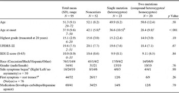
After excluding probands with other known mutations including GBA (n = 8) or LRRK2 (n = 2), 95 EOPD patients (54 men, 41 women) were evaluated, including 52 noncarriers, 23 parkin heterozygotes, and 20 cases with two parkin mutations (16 compound heterozygotes and 4 homozygotes). Demographic and clinical characteristics of the proband group are shown in Table 1.
Clinical and demographic characteristics of probands by parkin carrier status

Note. Values are means +/− standard deviations. Analyses reflect differences at the .001 level for comparison of adjacent groups. Values with different superscript letters reflect significant group differences on post-hoc testing, i.e. age at onset was significantly different in each group. UPDRS = Unified Parkinson’s Disease Rating Scale; BDI = Beck Depression Inventory.
aThese data were not collected within the GEPD study, resulting in a reduced n.
When the sample was dichotomized (parkin carriers vs. noncarriers) the groups were similar with regard to demographics and disease characteristics, except for age at onset which was younger in the parkin carrier group. When the proband group was divided into three groups, noncarriers, heterozygotes, and carriers of two mutations, again, only age-at-onset was significantly different among the groups (Table 1).
Neuropsychological performance
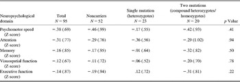
Performance on cognitive domains was similar among the mutation groups (Table 2). No significant differences were found on either raw or standardized test scores, whether two or three groups were compared. In the logistic regression model where parkin status was the dependent variable, no cognitive domain was associated with mutation status, either in a univariate model nor when data was stratified by demographic or disease-related variables including age, education, gender, side of disease onset, age at onset, initial PD symptom, or BDI-II score. The language used for test administration (English vs. Spanish) did not change these results.
Composite domain scores of probands (reported as Z-scores) by parkin carrier status
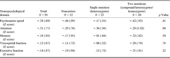
Note. Values are means +/− standard deviations. Analyses reflect comparisons across three groups on post hoc testing.
Individual test scores in both raw and standardized form are provided in table form (supplementary Table 1, which is available online) for descriptive purposes and provide a reference for the pattern of mean scores seen on this comprehensive battery. Supplementary Table 1 can be found as an online supplement. Probands were not impaired, that is, did not score ≥1.5 SD on any measure when mean standardized scores calculated from normative data were examined for noncarriers and carrier groups.
First-Degree Relatives
Demographics
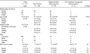
We studied 217 family members (39 parents, 101 siblings, and 77 children) including 146 noncarriers (22 parents, 77 siblings, 47 children), 65 heterozygotes (16 parents, 21 siblings, 28 children), and 6 compound heterozygous (1 parent, 3 siblings, 2 children; in four families), after excluding family members with PD diagnoses (n = 8, all of whom were siblings) or carriers of other genetic mutations (3 LRRK2 and 10 GBA mutation carriers; Table 3).
Demographic characteristics of unaffected first-degree relatives by parkin carrier status
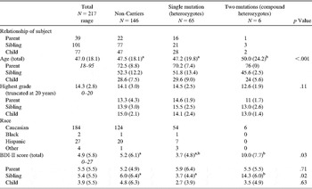
Note. Values are means +/− standard deviations. Values with different superscript letters reflect significant differences across groups on post-hoc testing; a = highest score, b = lowest score. Analyses reflect differences at the .001 level for comparison of adjacent groups. BDI = Beck Depression Inventory.
Neuropsychological performance
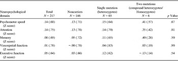
Whether two (parkin mutation carrier vs. noncarrier) or three groups (heterozygotes, compound heterozygotes, wild-type) were compared using one way ANOVA and Tukey post hoc tests, no significant differences were found for the five cognitive domains (Table 4).
Composite domain scores of unaffected first-degree relatives (reported as Z-scores) by parkin carrier status
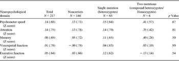
Note. Values are means +/− standard deviations. Analyses reflect comparisons across three groups on post hoc testing.
No covariates emerged in the backward step-wise regression approach and domain scores were not associated with carrier status outcome. Individual test scores including raw and standardized scores are provided for descriptive purposes in supplementary Table 2, which is available as an online supplement. None of the mean standardized scores for individual tests fell 1.5 SDs below the normative mean for relatives.
Given our negative findings we performed a power analysis using PS2 software (version 3.0.2) to determine the detectable difference in domain score for each genetic group (heterozygotes, compound heterozygotes/homozygotes) compared to noncarriers. This study had 80% power to detect a large difference (.76 SD) among proband groups (compound heterozygotes/homozygotes, heterozygotes, and noncarriers) and a medium effect size among family members (heterozygotes vs. noncarriers).
We further tested whether our battery was sensitive for PD-related cognitive changes. When the mean domain scores of probands with PD were compared to those of their siblings without PD (n = 101), probands were found to perform lower within the domains of speed (p < .001) and attention (p < .001), using a backward-stepwise regression with GEE after adjusting for sex, age, and education.
Discussion
Mutations in the parkin gene were recognized over a decade ago as a genetic cause of EOPD (Kitada et al., Reference Kitada, Asakawa, Hattori, Matsumine, Yamamura, Minoshima and Shimizu1998; Periquet et al., Reference Periquet, Latouche, Lohmann, Rawal, DeMichele, Ricard and Brice2003). Using the largest systematically collected sample of EOPD cases to date (Marder et al., Reference Marder, Tang, Mejia-Santana, Rosado, Louis, Comella and Clark2009) this study examined the possible association between parkin mutation status and cognitive function by assessing the neuropsychological performance of EOPD patients as well as that of their unaffected first-degree relatives. Consistent with previous studies reporting normal cognitive function in parkin-carrier EOPD patients (Khan et al., Reference Khan, Graham, Critchley, Schrag, Wood, Lees and Quinn2003, Reference Khan, Horta, Eunson, Graham, Johnson, Chang and Lees2005; van Nuenen et al. Reference van Nuenen, Weiss, Bloem, Reetz, van Eimeren, Lohmann and Siebner2009) we found no significant differences in neuropsychological test performance between parkin carrier and noncarrier probands, even when examining the possible effects of demographic and disease-related factors including age, education, gender, side of disease onset, age at onset, initial PD symptom, and BDI-II score. We did not find that parkin mutation status confers cognitive vulnerability on any subgroup, including gender, side of onset and initial presenting symptom; however, our study was not powered for these subanalyses. Furthermore, performance between noncarriers and carrier subgroups, (i.e., heterozygotes, compound heterozygotes/homozygotes) did not differ, suggesting that there is no gene-dosage effect on cognitive functioning. It is important to note that our sample included only non-demented probands, a limitation which may have affected our ability to detect cognitive differences among probands with and without mutations.
When we tested our hypothesis that neuropsychological assessment may detect populations at risk for PD among family members with parkin mutations, we found no differences in cognitive functioning in unaffected first-degree relatives across genotypes, suggesting that cognitive changes may not represent premotor manifestations in parkin carriers at risk for the development of PD. The fact that unaffected relatives with parkin mutations did not exhibit impaired performance on any mean test score suggests that their genetic status does not affect their cognitive functioning on tests used in the present battery.
Our null findings, that is, that parkin carrier and noncarrier probands did not differ in neuropsychological test performance and that unaffected parkin carriers did not exhibit preclinical cognitive changes or impairment that precedes motor impairment, are strengthened by our use of a comprehensive neuropsychological battery as well as our use of a very large sample of EOPD patients who were recruited based on of motor signs as opposed to family history of PD status. The negative findings in this study suggest that either there is no phenotypic difference among the mutation groups or that differences in cognitive function were too subtle to be detected in our sample using this battery.
This study had limitations. First, despite its large sample size of EOPD cases, the smaller number of mutation carriers found allowed us to detect only a large effect in probands and a moderate effect in family members. Second, the neuropsychological battery used may not have been sensitive enough to capture subtle differences. As previously described, we were limited by our need for a brief test battery that could be administered to a diverse population (e.g., patients with motor impairments, Spanish speakers) in their homes. Neuropsychological test data was analyzed as domain scores so as to reduce the data, and as individual test scores to avoid the loss of data that may occur with the use of pre-established domains. The categorization of tests within specific cognitive domains often appears arbitrary, however in devising our domain scores we attempted to replicate the domains used by Muslimovic et al. (2005, Reference Muslimovic, Post, Speelman, DeHaan and Schmand2009) in their previous studies that assessed predictors of cognitive decline in PD. In this way, data from the Trail Making Test and Stroop were categorized as tests of psychomotor speed (TMT Part A, Stroop color and word reading trials) and attention (TMT Part B, Stroop color-word trial). The authors also included fluency tasks within the domain of executive functioning (as opposed to language measures) and so this methodology was used in the present study. Additional support for the use of fluency tasks as executive measures is provided by the Dujardin et al. (Reference Dujardin, Duhamel, Becquet, Grunberg, Defebvre and Destee1999) study that found that the number of perseverative responses on phonemic fluency tasks differentiated first-degree relatives of PD patients from healthy controls.
Restricting our proband sample to those with MMSE scores > 23 may have limited the likelihood of identifying cognitive deficits; however, this limitation does not apply to the family members’ data, since we did not restrict recruitment of unaffected relatives based on MMSE performance. Finally, our cross-sectional design precludes follow-up information that would allow us to examine cognitive change over time. Our use of normative data was reported so as to provide a clinically relevant illustration of the cognitive performance of EOPD patients. When we compared probands’ mean scores on individual test measures with normative data, none of the genetic groups exhibited impaired performance, defined as a mean score more than 1.5 standard deviations below the mean of age and education norms, on any measure. In fact, mean test scores did not fall below even one standard deviation, reflecting performance within normal limits for the proband group. As such, we can assume in terms of the actual observed values that this group is cognitively normal. Since cognitive impairment is frequent in PD, long-term follow-up of EOPD patients will assist in determining whether the rate of cognitive impairment differs between mutation groups.
Acknowledgements
Elise Caccappolo, Department of Neurology and Taub Institute for Research on Alzheimer’s Disease and the Aging Brain, College of Physicians and Surgeons, Columbia University; Roy N. Alcalay, Department of Neurology, College of Physicians and Surgeons, Columbia University; Helen Mejia-Santana, Department of Neurology, College of Physicians and Surgeons, Columbia University; Ming-X. Tang, Department of Neurology and Taub Institute for Research on Alzheimer’s Disease and the Aging Brain, College of Physicians and Surgeons, Columbia University; Brian Rakitin, Department of Neurology and Taub Institute for Research on Alzheimer’s Disease and the Aging Brain, College of Physicians and Surgeons, Columbia University; Llency Rosado, Department of Neurology, College of Physicians and Surgeons, Columbia University; Elan D. Louis, Department of Neurology, Taub Institute for Research on Alzheimer’s Disease and the Aging Brain, Gertrude H. Sergievsky Center, College of Physicians and Surgeons, Department of Epidemiology, Mailman School of Public Health, Columbia University; Cynthia L. Comella, Department of Neurology/Movement Disorder Section, Rush University; Amy Colcher, Parkinson’s Disease and Movement Disorders Center, Pennsylvania Hospital; Danna Jennings, The Institute for Neurodegenerative Disorders; Martha A. Nance, Struthers Parkinson’s Center, Park Nicollet Clinic; Susan Bressman, The Alan and Barbara Mirken Department of Neurology, Beth Israel Medical Center, Department of Neurology, Albert Einstein College of Medicine; William K. Scott, Dr. John T Macdonald Foundation Department of Human Genetics and John P. Hussman Institute for Human Genomics, Miller School of Medicine, University of Miami; Caroline M. Tanner, Parkinson’s Institute; Susan F. Mickel, Marshfield Clinic, Department of Neurology; Howard F. Andrews, New York State Psychiatric Institute, Data Coordinating Center; Cheryl Waters, Department of Neurology, College of Physicians and Surgeons, Columbia University; Stanley Fahn, Department of Neurology, College of Physicians and Surgeons, Columbia University; Lucien J. Cote, Gertrude H. Sergievsky Center, College of Physicians and Surgeons, Columbia University; Steven Frucht, Department of Neurology, College of Physicians and Surgeons, Columbia University; Blair Ford, Department of Neurology, College of Physicians and Surgeons, Columbia University; Michael Rezak, Department of Neurology, at North Shore University Health System, Department of Neurology, Northwestern University, Feinberg School of Medicine; Kevin Novak, Department of Neurology, at North Shore University Health System, Department of Neurology, Northwestern University, Feinberg School of Medicine; Joseph H. Friedman, Parkinson’s Disease and Movement Disorders Center of NeuroHealth, Department of Clinical Neurosciences, The Warren Alpert School of Medicine of Brown University; Ronald F. Pfeiffer, Department of Neurology, College of Medicine, University of Tennessee Health Science Center; Laura Marsh, Morris K. Udall Parkinson’s Disease Research Center of Excellence, Department of Psychiatry and Behavioral Sciences, Department of Neurology and Neurological Sciences Johns Hopkins University School of Medicine; Brad Hiner, Medical College of Wisconsin; Andrew D. Siderowf, Department of Neurology, University of Pennsylvania Health System; Barbara M. Ross, Taub Institute for Research on Alzheimer’s Disease and the Aging Brain, College of Physicians and Surgeons, Columbia University; Miguel Verbitsky, Taub Institute for Research on Alzheimer’s Disease and the Aging Brain, College of Physicians and Surgeons, Columbia University; Sergey Kisselev, Taub Institute for Research on Alzheimer’s Disease and the Aging Brain, College of Physicians and Surgeons, Columbia University; Ruth Ottman, Department of Neurology, College of Physicians and Surgeons, Columbia University, Gertrude H. Sergievsky Center, College of Physicians and Surgeons, Department of Epidemiology, Mailman School of Public Health, Columbia University, Division of Epidemiology, New York State Psychiatric Institute; Lorraine N. Clark, Taub Institute for Research on Alzheimer’s Disease and the Aging Brain, College of Physicians and Surgeons, Columbia University, Department of Pathology and Cell Biology, College of Physicians and Surgeons, Columbia University; and Karen S. Marder, Department of Neurology, and Taub Institute for Research on Alzheimer’s Disease and the Aging Brain, College of Physicians and Surgeons, Gertrude H. Sergievsky Center, College of Physicians and Surgeons, Columbia University, Department of Psychiatry, Columbia University Medical Center.
This study was funded by NIH NS36630, UL1 RR024156 (KM), NS050487, NS060113 (LNC), and the Parkinson’s Disease Foundation (K.M., S.F., and L.N.C.). The authors thank Denise Gonzalez and Diana Ruiz for their assistance.
Supplementary Material
To review these additional data and analyses, please access the online-only supplementary Tables 1 and 2. Please visit journals.cambridge.org/INS, then click on the link “Supplementary Materials” at this article.