Introduction
Fish of the perciform suborder Notothenioidei dominate the diversity, abundance, and biomass in the subzero waters of the Antarctic continental shelf. Although they are a benthic group and lack a swim bladder, diversification in buoyancy is the hallmark of the radiation at the organismal level (Eastman Reference Eastman1993). Habitation of the water column is best exemplified by the neutrally buoyant clade of the family Nototheniidae. This clade, identified by morphological (Balushkin Reference Balushkin2000) and molecular (Near et al. Reference Near, Pesavento and Cheng2004, Sanchez et al. Reference Sanchez, Dettaï, Bonillo, Ozouf-Costaz, Detrich and Lecointre2007) datasets, includes Dissostichus mawsoni Norman 1937, D. eleginoides Smitt 1898, Pleuragramma antarcticum Boulenger 1902, Aethotaxis mitopteryx DeWitt 1962, and Gvozdarus svetovidovi Balushkin 1989. Adult specimens of Dissostichus mawsoni (Eastman & DeVries Reference Eastman and DeVries1981), Pleuragramma (DeVries & Eastman Reference DeVries and Eastman1978) and Aethotaxis (Near et al. Reference Near, Kendrick, Detrich and Jones2007) have been determined to be neutrally buoyant by measuring weight in seawater; neutral buoyancy has not been documented in D. eleginoides. Gvozdarus is known from only two specimens (DeWitt et al. Reference DeWitt, Heemstra, Gon, Gon and Heemstra1990) and its buoyancy has not been measured. The evolution of neutral buoyancy has enabled these species to utilize unfilled niches in the water column, especially those involving pelagic piscivory and zooplanktivory.
Neutral buoyancy in this clade is achieved through a combination of static mechanisms including reduced skeletal ossification, sometimes involving marked paedomorphy, and substantial accumulation of neutral lipids within the white axial musculature or in subcutaneous tissue (DeVries & Eastman Reference DeVries and Eastman1978, Eastman & DeVries Reference Eastman and DeVries1981, Reference Eastman and DeVries1982, Eastman Reference Eastman1993). The lipid, primarily triacylglycerols (Eastman & DeVries Reference Eastman and DeVries1981, Clarke et al. Reference Clarke, Doherty, DeVries and Eastman1984, Nachman Reference Nachman1985, Friedrich & Hagen Reference Friedrich and Hagen1994, Hagen et al. Reference Hagen, Kattner and Friedrich2000), is stored in either typical adipocytes or large sacs consisting of complexes of adipocytes (Eastman & DeVries Reference Eastman and DeVries1989). There is little hydrodynamic contribution to lift since the pectoral fins and associated red musculature are employed in labriform locomotion and activity levels are low.
In the skin and subcutaneous tissue of D. mawsoni from McMurdo Sound, lipid composes 74% of the wet weight and > 95% of the total lipid is triacylglycerols (Clarke et al. Reference Clarke, Doherty, DeVries and Eastman1984). The lipid content of white muscle is 7–23% of wet weight (Lin et al. Reference Lin, Dobbs and DeVries1974, Eastman & DeVries Reference Eastman and DeVries1981, Clarke et al. Reference Clarke, Doherty, DeVries and Eastman1984), depending on where the sample is taken, and lipid class composition in most specimens is primarily (60–69%) triacylglycerols (Eastman & DeVries Reference Eastman and DeVries1981, Clarke et al. Reference Clarke, Doherty, DeVries and Eastman1984, Nachman Reference Nachman1985). There are no remarkable features in the distributions of free and triacylglycerol fatty acids and monounsaturated oleic acid (18:1) is the dominant component (Nachman Reference Nachman1985). Such monounsaturated fatty acids are preferentially catabolized in the tissues of several species of non-neutrally buoyant notothenioids (Lund & Sidell Reference Lund and Sidell1992, Crockett & Sidell Reference Crockett and Sidell1993, Sidell et al. Reference Sidell, Crockett and Driedzic1995). On the basis of substrate competition assays, it has been hypothesized that saturated fatty acids are metabolized less well than monounsaturates and polyunsaturates and remain in adipose tissue, constituting the lipid component used for buoyancy in well nourished specimens (Hazel & Sidell Reference Hazel and Sidell2004). It is not known, however, whether the extensive lipid deposits in the white muscle and subcutaneous tissue of D. mawsoni and other neutrally buoyant species relinquish their triacylglycerols as a caloric resource for metabolism, thus serving a role in both energy storage and buoyancy.
The Antarctic toothfish, Dissostichus mawsoni, is one of the largest notothenioids, reaching lengths of 2 m and weights of > 100 kg (Eastman & DeVries Reference Eastman and DeVries2000). Although they are not neutrally buoyant as subadults (Eastman & Barry Reference Eastman and Barry2002, Eastman & Sidell Reference Eastman and Sidell2002, Near et al. Reference Near, Russo, Jones and DeVries2003), Ross Sea specimens attain neutral buoyancy at 80–100 cm standard length (Near et al. Reference Near, Russo, Jones and DeVries2003), approximately the same size (100 cm total length) at which they reach sexual maturity (Yukov 1982, Fenaughty Reference Fenaughty2006). Many aspects of its biology remain unknown although the developing fishery for D. mawsoni in Commission for the Conservation of Antarctic Marine Living Resources (CCAMLR) Subarea 88.1 has led to the capture of specimens throughout the Ross Sea and appreciably north of the shelf break. More specifically, large individuals in poor condition (so called “axe handles”) have been discovered near isolated seamounts north of the shelf break (Fenaughty Reference Fenaughty2006). Although tissue samples are not available, the existence of specimens in this condition provides insight into aspects of the lipid storage, lipid utilization and life history of this enigmatic species.
In this paper we will: 1) describe the axe handle morphology (emaciated specimens of low condition factor) captured near seamounts north of the Ross Sea shelf, 2) document the frequency, Fulton's condition factor (K), sex ratio, gonadal maturity and geographic distribution of axe handle specimens, 3) describe the location and method of storage of lipid deposits in typical specimens of D. mawsoni from the southern Ross Sea shelf, and 4) discuss, from an evolutionary perspective, lipid and protein metabolism as related to migration, feeding and reproduction of D. mawsoni in the Ross Sea.
Materials and methods
Specimens and localities
We obtained our data from specimens captured at two localities within CCAMLR Subarea 88.1. They were caught from mid-December to early April during 2001–2007 by two longlining vessels owned and operated by Sanford Limited, a major New Zealand seafood company. Scientific observers and contract science staff aboard these vessels collected the relevant data.
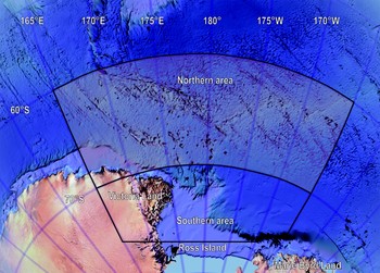
We calculated separate analyses of Fulton's condition factor (K) for specimens collected in Subarea 88.1 north and south of latitude 70°S which approximates to the continental slope. We used this latitude as the delineator as it effectively divides Subarea 88.1 into the contiguous Antarctic shelf to the south and an oceanic area to the north, consisting of isolated banks, ridges and seamounts. These areas are referred to in this analysis as “north” and “south”. The northern area, part of the Pacific-Antarctic Ridge, extends north to 60°S and the southern area approaches ≈ 78°S near the Ross Ice Shelf, encompassing most of the western and central Ross Sea (Fig. 1).
Features of the Ross Sea floor and general sampling locations for specimens of Dissostichus mawsoni collected from northern oceanic and southern shelf areas delineated by 70°S.

Measurements
For each specimen we obtained total length (L T) in cm rounded down and body weight (W) in kg. We made measurements of L T by compressing the dorsal and ventral lobes of the caudal fin towards the midline of the fish and measuring the full extent between the snout and the posterior margin of the compressed fin. We took body weights with hanging scales accurate to ± 0.5 kg. We converted data to mm and g and calculated Fulton's condition factor as an index (K) of weight per unit length:

where W = body weight in grams and L T is the total length in millimetres. This index assumes an isometric length-weight regression with an exponent (b) of ≈ 3, an assumption that is valid for D. mawsoni (Fenaughty Reference Fenaughty2006 where modal values of b = 2.9–3.1, R 2 = 0.92–0.99), hence larger values of K indicate greater weight per unit length associated with a more rotund body. Since nutritional state is one of the biological factors reflected in K (Busacker et al. Reference Busacker, Adelman, Goolish, Schreck and Moyle1990, p. 372), thinner/undernourished fish have values of K < 1 whereas in fatter/adequately nourished specimens K > 1. Thus values of K are effectively utilized to monitor the gross nutritional balance in populations (Bolger & Connolly Reference Bolger and Connolly1989), especially as reflected by lipid content, and fluctuations in feeding activity or food supply over an extended period (Weatherley & Gill Reference Weatherley and Gill1987, p. 18).
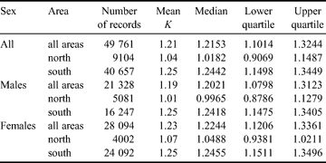
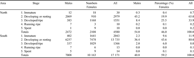
After we groomed the data by removing records lacking a recorded weight and obvious erroneous outliers, the working dataset for analysis of K consisted of 49 761 records (Table I). We analysed the data in Microsoft Excel which uses the method described by Freund & Perles (Reference Freund and Perles1987) to calculate quartile values. We used quartiles to circumvent the non-normality of the dataset and therefore avoided transforming it and possibly obscuring the intuitive biological value of the condition factor. Quartiles are also well suited to and easily interpreted in the graphical comparisons that are a key component in our analysis.
Summary of data on condition factor (K) with reference to sex and region of CCAMLR Subarea 88.1.
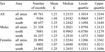
Number of records for “All” does not reflect the sum of “Males” and “Females” because sex was not determined for 339 specimens in the dataset.
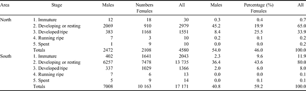
We also determined the sex of each specimen as well as the macroscopic gonadal maturity stage based on the five-point scale slightly modified from that of Kock & Kellermann (Reference Kock and Kellermann1991). In addition, there were records where the biological sampling data included gonad weights, measured with motion compensated scales to an accuracy of ± 10 g. These 21 751 records included 2472 males and 2108 females in the north, and 7008 males and 10 163 females in the south. We used these values to calculate a gonadosomatic index (GSI). The GSI is used to provide useful information on changes in reproductive maturity using gonad weight as a proxy for the progression through maturity stages. The GSI is traditionally defined as the weight of tissue dedicated to reproduction as a proportion of the total body weight. For D. mawsoni the GSI calculation is:

where WG = gonad weight in kg and W = body weight in kg.
We must note, however, that there is a degree of uncertainty regarding maturity and age or length at sexual maturity for D. mawsoni. Patagonian toothfish (D. eleginoides) has been reported to reach sexual maturity at 9–10 years of age for females, i.e. when they attain 90–100 cm total length (L T) (DeWitt et al. Reference DeWitt, Heemstra, Gon, Gon and Heemstra1990). Hanchet et al. (Reference Hanchet, Stevenson, Horn and Blackwell2003) and Dunn et al. (Reference Dunn, Gilbert and Hanchet2005) have noted similar values for D. mawsoni postulating that 50% of the population of both sexes is mature at 100 cm L T with a 30 cm range over maturity (i.e. 85–115 cm L T). This equates to a 50% maturity at about age 9 years of age (Hanchet et al. Reference Hanchet, Stevenson, Horn and Blackwell2003). In this paper we assume that all specimens ≥ 100 cm L T have potentially reached sexual maturity.
Histological documentation of lipid deposits in non-axe handle specimens
We did not have tissue samples from the axe handle specimens. We utilized tissues from D. mawsoni collected in McMurdo Sound in the extreme south-western Ross Sea (77°51′S, 166°40′E) as representative of well-nourished animals in good condition. Eastman & DeVries (Reference Eastman and DeVries2000) provide details concerning the method of capture, size and reproductive state of these specimens.
We sampled nine large specimens (131–151 cm L T) of both sexes to survey the distribution of adipose tissue in the body. We removed and fixed these tissues by immersion in Bouin's solution: hypaxial and epaxial white axial muscle including and excluding myoseptal regions, red pectoral muscle, trunk skin and subcutaneous tissue and dorsal head skin, subcutaneous tissue and bone. We then subjected tissues to the following protocol: dehydration in alcohol, clearing in CitriSolve (Fisher Scientific) and embedding in paraffin. We cut embedded tissues on a rotary microtome to produce sections 7 µm thick. Sections were mounted on slides, dried, deparaffinized, stained with Bodian's Protargol for 24 h at 50°C, dehydrated and coverslipped. This neurological silver stain proved to be effective in visualizing cell membranes and nuclei of adipocytes.
Using modifications of histological staining techniques, we grossly stained large blocks of the above tissues with two stains for neutral lipid, either oil red O in 99% isopropanol or Sudan black B in propylene glycol.
Results
What is an axe handle specimen?
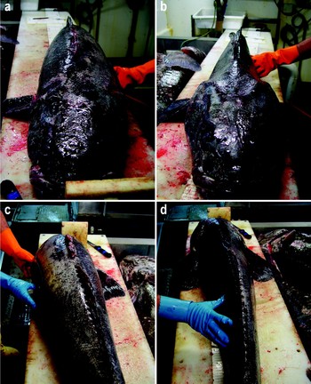
Poorly conditioned D. mawsoni are often referred to as “axe handles” by crew of the fishing vessels. These fish appear shrunken behind the head, with a notably thinner trunk than fish having “normal” length to weight ratios. This morphology is shown in Fig. 2.
a. & b. Anterior, and c. & d. posterior views of specimens of Dissostichus mawsoni in a. & c. good, and b. & d. poor physical condition. The specimen shown in b and d is an example of an “axe handle” with a low condition factor (K). Note the narrowness and shrunken appearance of the trunk. Photos from Fenaughty (Reference Fenaughty2006) with permission of the Commission for the Conservation of Antarctic Marine Living Resources, North Hobart, Tasmania, Australia.

What level of the condition factor (K) defines an axe handle?
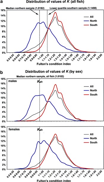
In order to examine the distribution of poorly conditioned D. mawsoni it is necessary to define a level of K associated with this condition. Figure 3a provides values of K grouped in bins of 0.05 and plotted as percentages of the total sample for the northern and southern areas. Sample numbers for each area are shown in Table I and examination of this data indicates that the upper quartile value of K for D. mawsoni from the northern sample is very close to the lower quartile value of K from the southern data - 1.1487 and 1.1498, respectively. The median value of K for the northern sample is 1.0182 and this is less than the lower quartile value of the southern fish. Dashed vertical lines in Fig. 3b indicate this value. Thus we have designated 1.0182, the median value of K for the northern sample, as the break point to define “axe handle” fish in this paper. For simplicity this point is referred to as Kah.
Analysis of condition factor (K) values in Dissostichus mawsoni from northern and southern areas of CCAMLR Subarea 88.1. Data are presented as percentages of the total sample for each area. a. Distribution of the values of K in the sample and delineation of the K value for axe handle specimens. b. Distribution of the values of K by sex. The median value for K of all axe handle (Kah) specimens is shown for reference.

Is there a difference in condition factor (K) between the two areas?
As shown by the curves in Fig. 3a, D. mawsoni in the north are generally in much poorer condition than those found in the south. Using the median value of the northern sample 1.0182 to define axe-handle fish (Kah), only 4.9% of the southern fish lie below this value compared with 50% in the north.
Is the geographical difference in condition factor common to both sexes?
The distribution of values of K for each sex is shown in Fig. 3b. For reference the median value (Kah) for all fish sampled in the northern area is shown in this paper as the index defining poor condition or “axe-handles”. Although there are greater numbers of poorly conditioned males, females are also well represented among axe handle specimens. In the northern sample 55.4% of the males sampled were less than Kah compared with 43.2% of the northern females. In the southern region 5.3% of the males were less than Kah compared with 4.5% of the females.
Are the differences in K size related?
Using 100 cm L T to define sexually mature D. mawsoni, 55.5% of males and 43.1% of females in the north were below Kah. This is very similar to the overall figures for all sizes in this area (previous paragraph) as there have been very few fish smaller than 100 cm L T caught north of 70°S. Only 25 males and 15 females below 100 cm L T were present in the northern sample of 9104 fish. In the south, however, the numbers of sexually mature fish having values less than Kah amounted to only 1.98% of the males and 1.67% of the females. Comparisons with the overall values for the south indicate that most of the poorly conditioned fish in that area were pre-recruits. Thus differences in the proportion of the sample below Kah are size related but size is also related to locality since larger sexually mature specimens predominate in the north (Fenaughty Reference Fenaughty2006). More details concerning length-frequency distributions by sex and area are provided by Fenaughty (Reference Fenaughty2006).
Are axe handle specimens reproductively spent adult fish?
To answer this and the following question we must first consider how reproductive maturity is assessed in D. mawsoni. The macroscopic gonadal maturity stage is thought to be of limited use in this species (Fenaughty Reference Fenaughty2006). This is because vitellogenesis in many species of Antarctic fish is a prolonged process (Everson Reference Everson1977, Kock Reference Kock1992), meaning that sexually mature female fish will have ovaries with yolked oocytes at all times of the year (Everson et al. Reference Everson, Kock and Parkes1996). In the case of D. mawsoni, ovarian sections from specimens taken in the Ross Sea during autumn 2001 at the onset of the spawning season often show two size groups of vitellogenic oocytes (Patchell Reference Patchell2002, New Zealand National Institute of Water and Atmosphere, personal communication). The macroscopic gonadal maturity stage does not distinguish between developing and resting states; and the difference between “developed/ripe” (stage 3) and “running ripe” (stage 4) is subjective. If D. mawsoni generates multiple batches of eggs and spawns in episodes over an extended period, as may be the case, the distinction between the developed/ripe and the spent stages may be obscured. However, if the spent stages have been accurately identified using the macroscopic gonadal maturity stage (Table II), less than 0.2% of D. mawsoni of either sex were reproductively spent at the time of sampling, and thus axe handles are not simply reproductively spent adults.
Summary of data on macroscopic gonadal maturity stages with reference to sex and region of CCAMLR Subarea 88.1.
What is the distribution of K with respect to reproductive maturity for northern and southern samples?
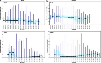
Because of the ambiguity of the macroscopic gonadal maturity stage, we have weighed gonads from sampled D. mawsoni since the 2002–03 season. The gonadosomatic index (GSI) provides an additional objective tool for use as a proxy for reproductive maturity. We analysed the GSI using grouped values of K in order to assess the relationship between the condition factor and the reproductive state of adult fish. We grouped values of K in bins of 0.05 and calculated and plotted maximum and minimum values, medians, and interquartile ranges of GSI. The analysis was restricted to fish ≥ 100 cm LT, our benchmark for sexually mature fish, and the sample numbered 21 751 specimens (see Materials and methods). As the question to be answered in relation to axe handle fish relates to the somatic body weight, we calculated an additional value of K, the Ksomatic, based on the somatic weight of the fish (wet body weight in grams minus the weight of the gonads in grams). We used this value as the basis for Fig. 4a & b as it negates any potential systematic bias in fish condition factor that could be caused by incrementally increasing gonad weight. We treated grouped values of Ksomatic as described for K above. Analyses of both K (not shown) and Ksomatic show only minor differences in the trends (although there will be an obvious reduction in weight) obtained using grouped values of K and Ksomatic. In examining the GSI in Fig. 4a & b, it should be kept in mind that the only published data for running ripe females of D. mawsoni is for two specimens with values of 26–30% (Fenaughty Reference Fenaughty2006).
a. Gonadosomatic indices for male Dissostichus mawsoni ordered and grouped by values of Ksomatic (somatic condition factor) and by area - north and south. The bar shows the median, the box the inter-quartile range and the whisker the range of values recorded for each interval. b. Gonadosomatic indices for female Dissostichus mawsoni ordered and grouped by values of Ksomatic (somatic condition factor) and by area - north and south. The bar shows the median, the box the inter-quartile range and the whisker the range of values recorded for each interval.

Figure 4a & b show the association between grouped values of Ksomatic and the GSI and indicate that low values of Ksomatic (and K) are generally associated with the higher GSI of more reproductively mature fish. The trend is more obvious in females, a reflection of the comparatively greater proportional size of the gonads. For both sexes the southern samples show high GSI values at the low end of the fish condition range followed by a lower but steadily increasing GSI with increasing values of Ksomatic. There is variability in the data for both geographical areas as shown by the maximum values - small numbers of reproductively advanced fish are present in both areas throughout the range of Ksomatic.
What causes the axe handle morphology?
The metabolic loss of muscular and subcutaneous lipid stores is the most parsimonious explanation for at least a significant component of the emaciated axe handle appearance. It is also possible that muscle proteins are metabolized.
Where are lipid stores located and what is their microscopic appearance in large specimens of Dissostichus mawsoni?
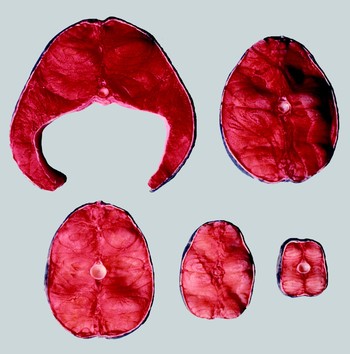
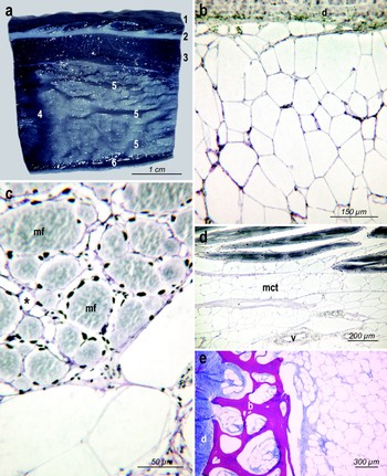
The primary lipid stores in D. mawsoni are muscular and subcutaneous and we emphasize that this description applies to specimens greater than or equal to 100 cm L T in good condition. The white axial musculature is 50.6% of body weight and its contained lipid is 4.8% of body weight or 23% on a dry weight basis (Eastman & DeVries Reference Eastman and DeVries1981). Figure 5 shows sequential cross sections of the trunk as stained by oil red O. The intensity of lipid staining is greatest anteriorly, especially near the centre of mass in the pectoral region, and tapers off caudally where myoseptal deposits become more prominent than muscular deposits. When a fillet is dissected free and placed in seawater it floats, with the anterior one-half remaining at the surface and the posterior portion sinking. The ventral body wall in the abdominal region is especially fatty with grossly visible “marbling” of the muscle by the perimysial deposits (Fig. 6a). A subperitoneal lipid layer is also present in this area (Fig. 6a). The muscular lipid deposit is contained in adipocytes around the individual fibres, around fasciculi (groups of fibres), and in the myosepta. Small adipocytes (25–45 µm) are adjacent to the endomysium of individual fibres and large (100–150 µm) adipocytes occupy the perimysial connective tissue around fasciculi (Fig. 6c). A longitudinal section shows the continuity of the perimysial and adjacent myoseptal adipose tissue (Fig. 6d). The blood supply to the white muscle and associated adipose tissue of D. mawsoni is sparse although larger branches of the segmental vessels are prominent in the myosepta (Fig. 6d).
Oil red O-stained cross sections of the trunk of large neutrally buoyant specimen of Dissostichus mawsoni in good condition showing gross distribution of lipid in white muscle. Sections are arranged from anterior (top row, left) to posterior (bottom row, right). Intensity of red staining reflects the amount of lipid present, with content in muscle decreasing posteriorly to the extent that myoseptal lipid is more prominent in the most posterior section. Sections range in diameter from c. 25 cm (top left) to 4 cm (bottom right).

Gross and microscopic appearance of lipid deposits in large neutrally buoyant specimens of Dissostichus mawsoni. These samples are from normal (non-axe handle) fish in good condition. a. Longitudinal section of the ventral body wall lightly stained with Sudan black B to show extensive subcutaneous and muscular lipid deposits. 1. epidermis and scales, 2. dermis, 3. subcutaneous lipid, 4. myoseptal lipid, 5. perimysial lipid, 6. subperitoneal lipid. x2.3. b. Part of the subcutaneous lipid layer from ventral body wall showing that it consists almost mostly of relatively large adipocytes. Most adipocytes in the field are 100–150 µm in diameter. d = dermis. Bodian's Protargol stain. x135. c. Cross section of a fascicle of dorsal white muscle showing multinucleate muscle fibres containing contractile proteins or myofilaments (mf) that could also be used as a metabolic source of energy. Small adipocytes surround the endomysium of each muscle fibre. Long axis of small adipocyte indicated by the asterisk is 25 µm. Large adipocytes dominate the perimysial connective tissue around muscle fasciculi. Bodian's Protargol stain. x370. d. Longitudinal section of ventral white muscle adjacent to a myoseptum. Myoseptal connective tissue (mct) consists mostly of large adipocytes with a few blood vessels (v). Bodian's Protargol stain. x60. e. Cross section of dermis (d), bone (b) and subcutaneous adipose tissue of supraorbital region of skull. Bone is cancellous with cavities containing small adipoctyes and is underlain by an adipose deposit composed of larger adipocytes. Gomori's trichrome stain. x30.

The subcutaneous adipose tissue, usually 2–8 mm thick and amounting to 4.7% of the body weight (Eastman & DeVries Reference Eastman and DeVries1981), is situated immediately beneath the dermis of the skin (Fig. 6a). Lipid content is 74% of wet weight (Clarke et al. Reference Clarke, Doherty, DeVries and Eastman1984). This layer consists of a mesh of large polygonal adipocytes 100–150 µm in diameter (Fig. 6b). Adipocytes consist of a flattened nucleus and a thin rim of cytoplasm surrounding a single lipid droplet. There is little connective tissue and few capillaries.
Although their loss would not contribute to the thinning of the trunk and axe handle appearance, there are other lipid stores in D. mawsoni. As is the case in most notothenioids, there is relatively little visceral lipid. However, the skull bones in D. mawsoni are thin and cancellous and there is a 1–2.5 mm thick layer of adipose tissue between lamina of bone as well as adipocytes within cavities in the bone (Fig. 6e).
We found no evidence of extracellular lipid in D. mawsoni and all lipid from the stores we documented here is contained in adipocytes typical of those in other vertebrates except for their larger size (100–150 µm). Thus, in theory, this lipid is available for metabolic purposes.
Discussion
Our sample of D. mawsoni from CCAMLR Subarea 88.1 shows substantial differences in the condition factor, especially as represented by the axe handle morphology, between specimens from northern and southern regions. Based on specimens taken from the stomachs of sperm whales captured in the oceanic waters of the northern region, Yukhov (Reference Yukhov1971, p. 13) identified “emaciated” fish with “no internal fat” and with K as low as 0.81, specimens that we are here referring to as “axe handles”. Yukhov (Reference Yukhov1970, Reference Yukhov1982) also suspected that D. mawsoni from this area had undertaken lengthy migrations from shelf waters. While there are large scale tagging programmes currently underway (e.g. by New Zealand), there is little tagging data, especially from larger fish, to support long distance migration. Evidence for migration is presented below. Our discussion will focus on the evolutionary and metabolic aspects of migration, especially with reference to specimens of low condition factor.
Migration is a feature of phyletically basal notothenioid clades that persists in Dissostichus mawsoni
Sustained locomotion, and migration in particular, is not a typical life history trait for the five families of Antarctic notothenioids that dominate the cold waters of the Antarctic shelf. In fact water temperatures constantly ≤ 0°C have produced an evolutionary minimization of baseline energy costs as reflected in a reduced scope for activity (Pörtner Reference Pörtner2006). Many notothenioids are inactive most of the time. For example, the nototheniid Notothenia coriiceps Richardson 1844 spends only 1.7% of each day engaged in locomotion (North Reference North1996) and artedidraconids of the genus Pogonophryne exhibit periods of up to 22 hours without significant movement (Zimmermann & Hubold Reference Zimmermann, Hubold, di Prisco, Pisano and Clarke1998). However, the life cycles of the phyletically basal non-Antarctic Pseudaphritis urvillii (Valenciennes 1832) and Eleginops maclovinus (Cuvier 1830) involve migration (Eastman Reference Eastman1993). Eleginops, from southern South America and the Falkland Islands, is the sister species of the Antarctic clade (Near et al. Reference Near, Pesavento and Cheng2004), and this suggests that the capacity for migration may be a life history trait present in the most recent common ancestor of Eleginops and the Antarctic notothenioids. Eleginops has a relatively larger mass of red pectoral musculature and a greater capacity for sustained labriform swimming than other notothenioids (Fernández et al. Reference Fernández, Calvo and Johnston1999). Some populations of Eleginops are catadromous, spending most of their life in fresh or brackish water and migrating as adults to spawn at sea or in the mouths of estuaries. For example, in the Falkland Islands juvenile Eleginops reside in smaller creeks and adults forage in larger creeks but migrate to spawn in shelf waters 30–100 m deep, occasionally reaching the shelf break at 250 m (Brickle et al. Reference Brickle, Arkhipkin and Shcherbich2005a, Reference Brickle, Laptikhovsky and Arkhipkin2005b).
Brain morphology, specifically the size of the corpus cerebellum, also reflects movement, migratory ability and complexity of the life cycle in teleosts. The corpus cerebellum is an important integrative centre receiving visual, mechanosensory lateral line, and somatosensory inputs (Meek & Nieuwenhuys Reference Meek, Nieuwenhuys, Nieuwenhuys, ten Donkelaar and Nicholson1998, p. 769, 835–836, Wullimann Reference Wullimann and Evans1998) as well as being involved in motor learning and coordination (Wullimann Reference Wullimann and Evans1998, p. 266). Among notothenioids, the corpus cerebellum is one of the most variable regions of the brain (Eastman & Lannoo Reference Eastman and Lannoo1995). It is large, with a caudally directed lobe, in Eleginops maclovinus (Eastman & Lannoo Reference Eastman and Lannoo2008) and some nototheniids, especially the two species of Dissostichus (Eastman Reference Eastman1993). On the other hand, the corpus cerebellum is small in the closely related Pleuragramma antarcticum (Eastman & Lannoo Reference Eastman and Lannoo1995, Reference Eastman and Lannoo2008), an inactive member of the neutrally buoyant clade, and in artedidraconids such as Pogonophryne (Eastman & Lannoo Reference Eastman and Lannoo1995, Reference Eastman and Lannoo2003).
Long distance migrations can be documented by mark and recapture studies. Unfortunately most tagging to data has been confined to smaller specimens of D. mawsoni, although New Zealand agencies have begun tagging larger fish. Records, summarized by Dunn & Hanchet (Reference Dunn and Hanchet2006), document movements of 800–2300 km for five recaptured specimens of D. mawsoni (size unreported) originally tagged in McMurdo Sound (≈ 78°S). However, the long distance migratory propensities of the genus Dissostichus are substantiated by the ≈ 10 000 km journey of a vagrant D. eleginoides from the South Atlantic to Greenland (Møller et al. Reference Møller, Nielsen and Fossen2003), by a tagged D. eleginoides that moved over 1900 km in the Indian Ocean sector (Williams et al. Reference Williams, Tuck, Constable and Lamb2002) and by the Falkland Islands population of D. eleginoides that migrates 1000 km from feeding to spawning grounds (Laptikhovsky et al. Reference Laptikhovsky, Arkhipkin and Brickle2006).
Historical perspective on migration of Dissostichus mawsoni as related to productivity, feeding and reproduction
As inferred from molecular sequence data, the split between Eleginops and the Antarctic clade occurred 40 million years ago and the divergence of D. mawsoni and D. eleginoides is estimated at 14.5 million years ago during the middle Miocene (Near Reference Near2004). During its historical association with the Ross Sea, D. mawsoni has experienced many glacial cycles and periods when ice sheets advanced onto the continental shelf and rendered much of the area uninhabitable. For example during the middle Miocene ≈16–10 million years ago, there were at least five shelf-wide grounding events of the West Antarctic Ice sheet on the Ross Sea outer continental shelf (Chow & Bart Reference Chow and Bart2003). At the Last Glacial Maximum ≈ 20 000 years ago, models indicate that the grounding line of the Antarctic ice sheet extended to the shelf break almost everywhere (Huybrechts Reference Huybrechts2002). More specifically, the West Antarctic Ice Sheet was grounded on the western Ross Sea shelf as far north as Coulman Island (Conway et al. Reference Conway, Hall, Denton, Gades and Waddington1999) and grounded near the shelf break in the central and eastern Ross Sea (Anderson et al. Reference Anderson, Shipp, Lowe, Wellner and Mosola2002).
Since it has the largest shelf and is the most productive region in the Southern Ocean (Arrigo et al. Reference Arrigo, Worthen, Schnell and Lizotte1998), the Ross Sea is a key feeding ground for vertebrate predators including D. mawsoni. Its importance is attributable to the biotic interactions in a food web where Euphausia crystallorophias and Pleuragramma antarcticum are grazers at lower trophic levels and available as food for higher levels to the extent that Pleuragramma may dominate energy transfer in the water column in areas where E. crystallorophias is absent (Smith et al. Reference Smith, Ainley and Cattaneo-Vietti2007). The productivity of the shelf is reflected in the diet of specimens captured at various latitudes in Subarea 88.1. In the vicinity of McMurdo Sound (≈ 78°S) in the southern Ross Sea, fish are 85% of the diet overall with Pleuragramma 71% by frequency of occurrence and 89% by dry weight (Eastman Reference Eastman, Siegfried, Condy and Laws1985). As the second most fatty notothenioid fish next to Aethotaxis mitopteryx (Friedrich & Hagen Reference Friedrich and Hagen1994, Hagen et al. Reference Hagen, Kattner and Friedrich2000), Pleuragramma have a high nutritional value. At 60%, mysids are the second most frequently occurring item. In deeper waters of the northern Ross Sea slope D. mawsoni also consume primarily fish (80–86%), but these are less lipid-rich fish, icefishes of the notothenioid family Channichthyidae (54%) and the macrourid grenadier Macrourus whitsoni (37%) (Fenaughty et al. Reference Fenaughty, Stevens and Hanchet2003). Here squid are the second most common item at 14%. Farther north in less productive oceanic waters (Smetacek & Nicol Reference Smetacek and Nicol2005), their principal food source is squid with mantle lengths of 20–33 cm (Yukhov Reference Yukhov1971, Reference Yukhov1982). Since the southern extent of the historical range of D. mawsoni in the Ross Sea may have waxed and waned with glacial advances and retreats, the shelf may be a fortuitous feeding ground during periods of deglaciation.
While the southern shelf is a prime feeding ground for D. mawsoni, it is not a breeding ground. The only literature records for running ripe females of this species are from the northern part of Subarea 88.1 (Fenaughty Reference Fenaughty2006). Sexually mature fish from the southern Ross Sea shelf may undertake a spawning migration to seamounts north of the Ross Sea, a distance of 1000 km or more (Fenaughty Reference Fenaughty2006). In this regard D. mawsoni may retain fidelity to its historical spawning grounds near the ridges and seamounts north of the shelf break in Subarea 88.1. If this aspect of its biology is substantiated, it will be similar to its sister species D. eleginoides in spawning on the slope in water > 1000 m deep near isolated islands, seamounts and banks (Evseenko et al. Reference Evseenko, Kock and Nevinsky1995, Laptikhovsky et al. Reference Laptikhovsky, Arkhipkin and Brickle2006). This may be a phyletically persistent feature of the biology of Dissostichus.
In light of our comments in the previous two paragraphs we should note that, although water < 1750 m deep is limited, longline catches of D. mawsoni are sometimes very large on northern ridges (Fenaughty, personal observation). High population density is not generally associated with low productivity, and it is unlikely that these areas support large resident populations of D. mawsoni. However, there is habitat heterogeneity within deep-water ecosystems elsewhere to the extent that certain ridges and seamounts are, compared with surrounding oceanic areas, sites of high density and biomass representing reproductive aggregations of fish and invertebrates (Drazen et al. Reference Drazen, Goffredi, Schlining and Stakes2003). Such areas could be considered transient reproductive hot spots that facilitate mate location thereby increasing reproductive success (Domeier & Colin Reference Domeier and Colin1997, Drazen et al. Reference Drazen, Goffredi, Schlining and Stakes2003). Finally we should note that we formulated our hypothesized migratory life cycle for D. mawsoni with respect to only the Ross Sea population.
Axe handle specimens and the energy metabolism of Dissostichus mawsoni
On the basis of mitochondrial DNA, population genetic studies of D. mawsoni within Subarea 88.1 suggest a single genetically homogeneous stock of this species (Smith & Gaffney Reference Smith and Gaffney2005). This information coupled with the observation of marked morphological differences, typified by axe handle specimens, between southern and northern sampling regions of Subarea 88.1, led Fenaughty (Reference Fenaughty2006) to propose movement of reproductively mature fish of a single population from the south to northern areas for purposes of spawning. Fenaughty (Reference Fenaughty2006) also indicated that the effects of migration to the north and the lower productivity in these waters might contribute to the poor condition of many individuals. During this period their endogenous caloric stores are drawn upon to support the combined energetic demands of migration and gametogenesis as they move to the ridges and seamounts of the northern region for spawning. The trends shown in Figs 4a & b are consistent with this hypothesis.
Although we cannot definitively validate the above interpretation of the life cycle of D. mawsoni, other marine fishes display similar morphological changes as a result of migration and reproduction. Lipids, specifically the fatty acid components, are the most important energy reserve for reproduction and swimming (Cowey & Sargent Reference Cowey, Sargent, Hoar, Randall and Brett1979, Tocher Reference Tocher2003). However, in some species, after lipid reserves are mostly depleted, muscle proteins are metabolized (Mommsen Reference Mommsen and Evans1998). Probably the most widely known examples are the Pacific salmonids of the genus Oncorhynchus. Migration and spawning of these animals is fuelled initially by primary mobilization of lipid stores, followed by a very significant involution of white axial muscle mass (Mommsen et al. Reference Mommsen, French and Hochachka1980, Kiessling et al. Reference Kiessling, Lindahl-Kiessling and Kiessling2004). Free amino acids released by breakdown of protein in the axial mass are presumably carried by the circulation to the liver for subsequent deamination and use of carbon skeletons in gluconeogenic production of glucose, which is then conveyed by the blood to support metabolism of other tissues. This pattern of fuel utilization is not restricted to semelparous or anadromous species. For example, Atlantic cod (Gadus morhua), a marine iteroparous species, show clearly similar reductions in somatic energy reserves and condition factor as a function of reproductive effort (Lambert & Dutil Reference Lambert and Dutil2000). This temporal hierarchy of metabolic fuel use is also the typical pattern displayed by fishes in response to starvation (reviewed by Love Reference Love1980, Navarro & Gutierrez Reference Navarro, Gutierrez, Hochachka and Mommsen1995). Subsequent to food deprivation, liver glycogen is rapidly depleted, followed next by mobilization of fatty acids from triglyceride stores in adipose tissues and, in later stages by breakdown of soluble and myofibrillar proteins from white axial muscle. Protein complement of oxidative (red) muscle tissue is typically spared, presumably to preserve ability for sustained locomotion and because it typically represents such a small fraction of body mass. In all probability, a similar series of physiological events could lead to the significant wasting of the axial muscle mass in axe handle specimens of D. mawsoni. If this interpretation is correct, it speaks directly to one of the significant outstanding questions about lipid stores in many notothenioid fishes: Is a single store of lipid in these animals used for both buoyancy purposes and as a metabolic reserve? Our observations suggest that, if the lipid is stored in typical adipocytes, the answer to this question is “yes”. Another implication of this answer is that neutral buoyancy in sexually mature D. mawsoni may be ephemeral, with lipid stores and buoyancy fluctuating over the course of the life cycle in response to migration, food supply and reproductive state. In this sense, storage of lipid in adipocytes in D. mawsoni is less specialized than in Pleuragramma where much of the lipid is stored in large, presumably less metabolically active sacs (Eastman & DeVries Reference Eastman and DeVries1989), allowing this species to maintain a relatively constant density/buoyancy.
Regression relationships between length and body mass of D. mawsoni specimens collected in southern and northern sectors of Subarea 88.1 were reported for five years of captures by Fenaughty (Reference Fenaughty2006, table 6). If one assumes 140 cm L T for a typical sexually mature fish, the mean calculated mass from regressions for all fish over the five year period yields estimates of 35.863 kg for the southern sector and 28.724 kg for the northern sector, c. 19.9% reduction of body mass in the latter group. Eastman & DeVries (Reference Eastman and DeVries1981) estimate that intramuscular lipid stores of well-nourished D. mawsoni collected in the southern sector near McMurdo Sound account for c. 4.8% of body mass while subcutaneous lipid stores account for 4.7% of body mass, i.e. a total of ~9.5% of body mass can be accounted for in the major triglyceride stores of the axial body. Given the morphology of axe handle specimens and in light of the above calculations, two conclusions appear to be warranted. First, in all probability, most of the axial lipid stores of northern axe handles have, in fact, been mobilized for energetic and/or gametogenic purposes. This conclusion is based upon the usual pattern of mobilization of caloric stores described above and the fact that these extensive lipid stores are contained in adipocytes, which typically contain hormone-sensitive triacylglycerol lipase, an activity that has been demonstrated in other notothenioids (Sidell & Hazel Reference Sidell and Hazel2002, Hazel & Sidell Reference Hazel and Sidell2004). Second, even complete mobilization of axial lipid stores can account for less than 50% of the observed differential in body mass between southern sector and axe handle specimens. A significant wasting of the lean (protein) portion of the axial musculature is probably involved in the morphological differences reported here since axe handle specimens with soft and watery axial muscle, called “jelly fish” in the industry, have been observed (Fenaughty, personal observation). This conclusion becomes even more evident when considering the widely reported observation that loss of lipid mass from axial musculature during starvation of fishes is generally matched by an identical increase in water content (Jobling Reference Jobling1994).
All indications are that both lipid and protein stores of the axial portion of D. mawsoni are metabolically volatile and that neutral buoyancy of this species is, indeed, an ephemeral phenomenon that is gained and lost cyclically in sexually mature adults. Once feeding is resumed and forage species are abundant, there is every reason to expect that both lipid stores and muscular protein could be restored rapidly. There is widespread documentation of “compensatory” growth of fish after spawning or food deprivation such that animals with the poorest condition factor grow most rapidly (Jobling Reference Jobling1994). In the case of D. mawsoni from Subarea 88.1, one of the most pressing biological questions would seem to be whether feeding recommences in the northern sector or whether these animals must rely upon their already depleted caloric reserves to return to the productive southern foraging grounds.
The above interpretation of the axe handle morphology is speculative. However, if it is correct, it leads to several hypotheses that could be tested if suitable tissue samples from axe handle specimens were to become available. Samples prepared for histology should be able to resolve whether intramuscular and subcutaneous lipid stores have been depleted. Protein breakdown in the axial musculature of fishes usually occurs in soluble and myofibrillar protein fractions while the scaffolding of connective tissue is spared (Love Reference Love1980). Consequently, greater proportions of proline, hydroxyproline and glycine (which dominate the composition of collagen) would be expected in axe handles compared to well-nourished southern sector specimens. Finally, if the pattern of preferential utilization of monoenoic and polyunsaturated fatty acids that has been described for other notothenioid species is shared by D. mawsoni, the fatty acyl composition of dramatically depleted fat stores in axe handle specimens should be particularly dominated by saturated fatty acyl residues, such as myristate (Hazel & Sidell Reference Hazel and Sidell2004).
Acknowledgements
The assistance of Sanford Limited in data collection and provision is gratefully acknowledged. This work was also supported by NSF grants ANT 04-36190 to JTE and ANT 04-37887 to BDS. Danette Pratt assisted with some of the figures. We thank Dr Mario La Mesa and an anonymous reviewer for their comments on the manuscript.