Introduction
The inner ear, or labyrinth, is well protected from infection because of its deep location, surrounded by the hard otic capsule. When infection occurs, it is known as labyrinthitis, and may present with hearing loss or balance disturbance. Viral causes are the most common, although the exact incidence and prevalence is unknown.1 Prior to the development of antibiotic therapy, bacterial infection in this area frequently resulted in complications, including permanent sensorineural deafness, vestibular dysfunction, cranial nerve palsies, intracerebral abscess and death.
Infection may spread to the inner ear via three main routes: from the middle ear (tympanogenic labyrinthitis), via the meninges or from the blood.Reference Glasscock, Shambaugh, Glasscock and Shambaugh2 The resulting labyrinthine infection may be serous or suppurative, acute or chronic in nature.Reference Sugiura and Paparella3 Since the development of antibiotics, acute bacterial infections of the labyrinth can usually be treated quickly and effectively with a combination of antibiotics and steroids. Occasionally, in the presence of an erosive pathology such as cholesteatoma, chronic labyrinthine infections may become established.Reference Sugiura and Paparella3–Reference Paparella and Sugiura5 The pathology in these cases is usually that of ‘labyrinthine ossificans’, a process of ossification and fibrosis of the labyrinth that represents healing and resolution of the infection.Reference Sugiura and Paparella3 Rarely, labyrinthine infection does not resolve, and a sequestrum of bone is formed, leading to chronicity, recurrence and progression of symptoms. This is known as labyrinthine sequestrum.Reference Paparella and Sugiura5–Reference Thomas Prasannaraj, De and Narasimham8
We present two cases whereby middle and inner-ear surgery provided a tympanogenic access route for infection of the labyrinth. The patients concerned both had auditory implants that preceded the onset of their symptoms. Here, we highlight the clinical and radiological features of labyrinthine infection following middle and inner-ear surgery. We review the literature on tympanogenic labyrinthitis and challenge the perception that aggressive surgical intervention is the only management option in these patients. In an era where an increasing number of auditory implantations are being performed, it is important that all ENT surgeons are aware of this devastating complication and how to manage it.
Case reports
Case one
A 53-year-old man underwent non-eventful right stapedectomy, with excellent closure of the air–bone gap. Three weeks post-operatively, he developed otorrhoea and was treated with topical antibiotics. Microbiology swabs grew Pseudomonas aeruginosa. Three months post-operatively, he developed sudden onset sensorineural hearing loss and suffered an acute vertigo attack.
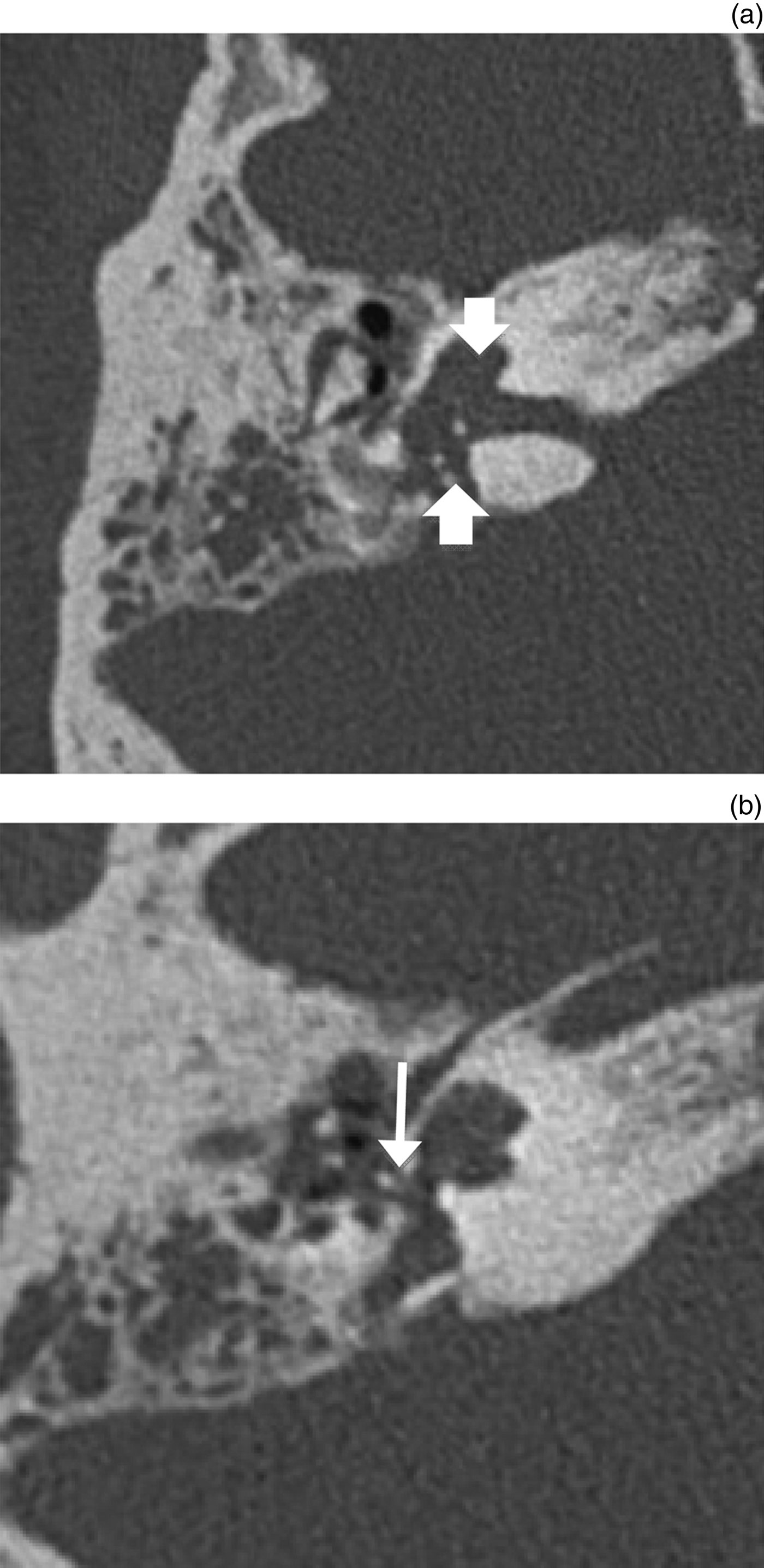
A computed tomography (CT) scan was performed, which showed diffuse erosion of the otic capsule. This resulted in the bony labyrinth having an amorphous appearance (Figures 1a and 1b). Bony sequestrum could be seen adjacent to the cochlea.
Axial computed tomography scans of the temporal bone with bone windows (right side). (a) Arrows show diffuse expansion of the otic capsule with loss of the internal architecture of the cochlea (top arrow), with no interscalar septae or modiolus evident. There are calcific opacities probably representing bony sequestrations (bottom arrow) in the vestibule ossicles. There is also mastoid and middle-ear opacification. (b) The stapes prosthesis is surrounded by soft tissue in the oval window area, and may have dislodged (arrow). Note the ill-defined peri-labyrinthine lucency adjacent to the expanded posterior semicircular canal and vestibular aqueduct.

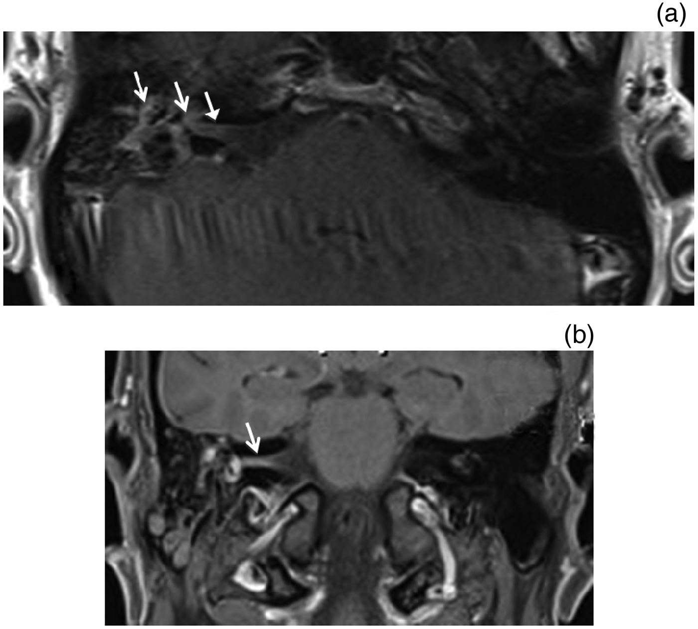
A gadolinium-enhanced magnetic resonance imaging (MRI) scan revealed enhancement extending from the right middle ear to the internal auditory meatus and cochlea (Figures 2a and 2b). There was no enhancement on the left.
Axial (a) and coronal (b), post-gadolinium, fat-saturated magnetic resonance images at the level of the internal auditory meatus. There is significant enhancement in the internal auditory canal, inner ear and middle ear (arrows), suggesting an ongoing inflammatory process. No enhancement was identified on the left side.
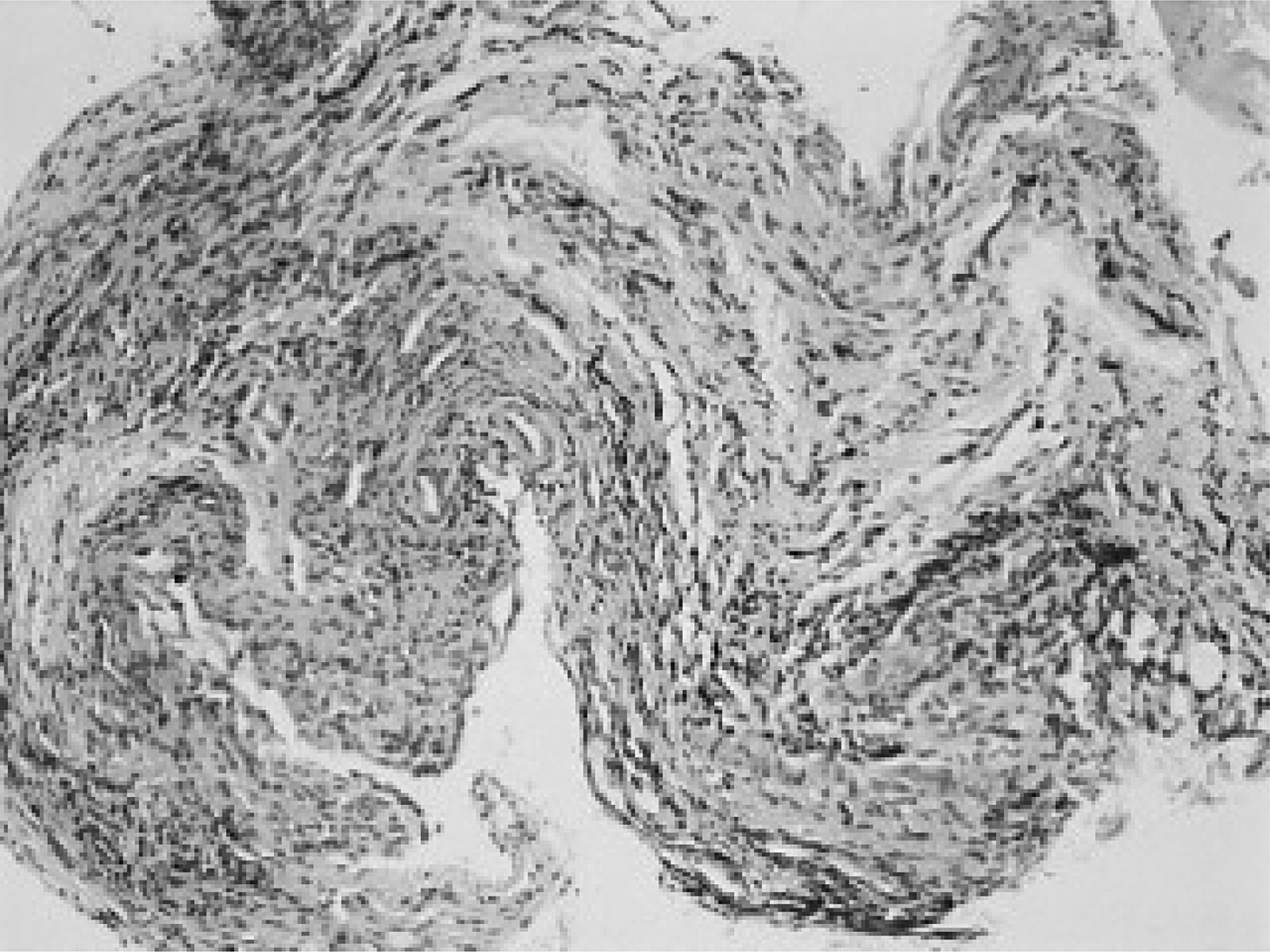
Surgical exploration revealed that the stapes prosthesis was dislodged, and the ossicles were partially eroded and surrounded by soft tissue. Tissue samples were sent to microbiology, but no bacterial or viral organisms were detected. Histopathological analysis revealed non-specific features of chronic inflammation (Figure 3).
Histopathology showing evidence of chronic inflammation (inflammatory cells are dark in the image). (H&E; ×10)
Case two
A 75-year-old man with bilateral profound hearing loss underwent successful right cochlear implantation. He was diabetic and had previously undergone a heart transplant following severe ischaemic damage. Four years post-cochlear implantation, he developed recurrent right otorrhoea, which was treated with antibiotics and steroid ear drops. Seven years post-implantation, he developed partial right facial palsy in association with right otorrhoea. This was treated with prednisolone and acyclovir, in addition to topical and systemic antibiotics.
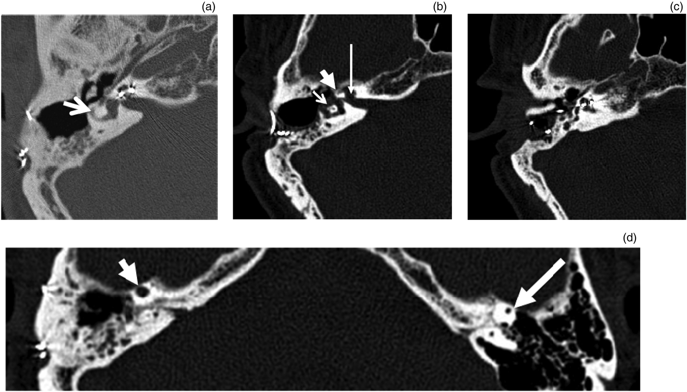
A petrous temporal bone CT scan demonstrated that the cochlear implant electrode array was in situ (Figure 4). There was expansion of the otic capsule and the labyrinthine portion of the facial nerve canal. In addition, there was erosion of the bony covering of the lateral semicircular canal.
Axial, high-definition computed tomography scans of the temporal bone (right side). (a) In 2008, the lateral semicircular canal (arrow) was largely normal. (b) In January 2012, there was diffuse erosion of the otic capsule, resulting in an amorphous appearance to the semicircular canals (thin, short arrow) and the labyrinthine portion of facial nerve canal (thin, long arrow), with a speck of calcification present indicating sequestration (wide arrow). (c) January 2012: there was bony change of the cochlea with the electrodes still in situ. There was lucency adjacent to the posterior semicircular canal. (d) January 2012: there was right-sided sequestration and enlargement of the anterior limb of the superior semicircular canal (short, wide arrow) compared to the normal left side (long, wide arrow).
The patient was not fit for surgery because of his significant co-morbidities. He was therefore treated with an extended course of antibiotics and interval scans. He had a repeat CT scan 12 months later to review progress. Figure 4 shows the developing pathology. He had a largely normal lateral semicircular canal in 2008 (Figure 4a), prior to the development of vestibular symptoms. By January 2012, the superior border of the lateral semicircular canal had been eroded and sequestered bone was visible (Figure 4b).
Following 12 months of extended antibiotic treatment, the right semicircular canal had ossified, whilst appearances remained unchanged on the left side (Figure 5).
Axial, high-definition computed tomography scans of the temporal bone from December 2012. (a) After 12 months of conservative treatment, the right lateral semicircular canal had ossified (arrow). (b) The right superior semicircular canal had ossified (long arrow) compared with the normal left side (short arrow).
Discussion
Labyrinthitis is an inner-ear infection. It causes the deep inner-ear structures to become inflamed, resulting in vertigo and hearing loss. Tympanogenic labyrinthitis is a potential consequence of otitis media. It has also been reported to occur post-operatively in up to 2 per cent of middle-ear procedures performed for indications including otosclerosis, meningitis and cancer surgery (patients undergoing cochlear implantation were excluded from that study).Reference Lao, Sha, Chen, Dai, Huang and Cheng7, Reference Vincent, Sperling, Oates and Jindal9
Tympanogenic labyrinthitis occurs either through the passage of inflammatory mediators and bacterial toxins into the labyrinth (serous labyrinthitis), or from direct invasion (suppurative labyrinthitis).Reference Sugiura and Paparella3, Reference Paparella and Sugiura5 Fortunately, both the structure of the inner-ear labyrinth and the timely use of antibiotics have meant that bacterial causes have become rare in the post-antibiotic era, with viral causes being much more common.1 When bacterial infection of the labyrinth does occur, it usually leads to fibrosis and to ossification of the labyrinthine structures.Reference Sugiura and Paparella3 There have only been seven reported cases where a sequestrum has formed instead; the patients concerned presented with recurrent, progressive symptoms of inner-ear disease.Reference Matthews6–Reference Thomas Prasannaraj, De and Narasimham8
The authors of the previous studies advocated aggressive surgical management for these patients because of the high risk of intracranial complications and difficultly of antibiotic penetration. Our second patient was deemed unfit for surgical intervention; in that case, we demonstrated success with aggressive antibiotic therapy alone, which can be used as an alternative to surgery in such patients.
Role of auditory implantation
The presence of prosthetic material is known to make infection more difficult to clear.Reference Zimmerli, Lew and Waldvogel10 Implantation of a stapes prosthesis and cochlear implant provided the route for infection to enter. In addition, the presence of prosthetic material provided an ongoing focus for chronic infection to become established and a sequestrum to form.
Role of immunocompromise and causative pathogens
We could not find a particular role for patient factors, either ones shared amongst the two cases reported here or in the published literature. Whilst the patient in case two was immunocompromised from both immunosuppressive drugs following heart transplantation and type II diabetes, our patient in case one was young and fit, without co-morbid disease. Similarly, in the literature, whilst there was a case of an adult with a prior scarlet fever infection in childhood developing the pathology, the other patients were not immunocompromised.Reference Sheehy, Brachmann, Graham, Hattori, Sade and Abramson4, Reference Matthews6–Reference Thomas Prasannaraj, De and Narasimham8
The pathogens isolated in the reported cases were those typical for aural disease: Pseudomonas spp, Staphylococcus aureus and Streptococcus spp. Perhaps because of previous antibiotic treatment, the surgical specimens in case one and swabs in case two never grew a significant organism.
Imaging
Computed tomography can reliably identify the characteristic features of a sequestered labyrinth. It remains the ideal imaging modality for this condition because of its excellent bone resolution. In case one, MRI was performed to further evaluate soft tissue invasion and to exclude cholesteatoma as a cause of the appearances. In case two, MRI imaging was contraindicated because of the patient's cochlear implant, but CT scans provided satisfactory evidence for the labyrinthine sequestration.
Treatment recommendation
Antibiotics may temporarily limit progression of the disease. However, optimal treatment of this condition involves removing the infected bony tissue and the prosthesis. Without this treatment, infection can be very difficult to clear.Reference Zimmerli, Lew and Waldvogel10 Even in the absence of a prosthesis, Lao et al. warn of fatal encephalopathic complications if vestibular sequestration is not treated surgically.Reference Lao, Sha, Chen, Dai, Huang and Cheng7
Although removing the infected prosthesis may be the best option, sometimes patients may not be fit for surgery. Our second case had extensive co-morbidities and was not deemed anaesthetically fit for the procedure. He was treated conservatively with an extended course of antibiotics for this reason. Interestingly, serial CT scans showed evidence of healing and resolution of the labyrinthine infection over 12 months.
It is imperative that this destructive pathology is prevented from occurring. The consequences are devastating, and include profound hearing loss, vestibular hypofunction and facial nerve palsy. No real trends in age, or predisposing conditions could be found in the published literature. This makes the creation of criteria for exclusion from auditory implantation or other surgical programmes problematic. Arguably, immunosuppression (either in the form of co-morbid diabetes or organ transplantation), previous brain abscess treatment or extremes of age may make this pathology more likely.Reference Matthews6 Several previous reports of patients who developed the pathology did not fall into these categories however. Those cases concerned young adults who were previously healthy, with no predisposing co-morbidities, as in case one and in the case series published by Lao et al.Reference Lao, Sha, Chen, Dai, Huang and Cheng7
On balance, the authors recommend that in high-risk cases such as immunosuppressed or elderly patients, it may be judicious to use antibiotics at induction, and a course of broad-spectrum oral antibiotics should be considered post-operatively. However, given the rarity of the complication, and the potential for harm from unchecked antibiotic use, the authors do not propose that a full course of antibiotics should be given to every patient following every auditory implant surgical procedure.
• Labyrinthitis can occur due to viruses, bacteria and fungi, and autoimmune diseases; symptoms include hearing loss and vertigo
• The membranous labyrinth is usually protected from infection by the otic capsule, but occasionally this capsule is breached
• Labyrinth infection can seed from the bloodstream via adjacent structures such as the meninges or through the middle-ear cavity
• If the labyrinthine infection is not cleared quickly, or there is an ongoing nidus of infection, chronic infection may occur
• A complication of chronic bony infection is sequestrum formation
• This paper reports the first two cases where bony sequestrum formed following auditory implantation
Otologists should be astutely aware of this complication, and respond rapidly with aggressive early antibiotic treatment if persistent otorrhoea develops after auditory implantation. Surgical removal of an implanted prosthesis should be considered if the infection does not respond to antibiotic therapy, in order to remove any ongoing nidus for infection and allow resolution of the pathology. However, our second case demonstrates that contrary to previous literature advocating radical surgical approaches in all instances, a conservative approach can be successfully employed when necessitated by the circumstances.Reference Lao, Sha, Chen, Dai, Huang and Cheng7