Introduction
Pathological changes that characterize Alzheimer's disease (AD) can begin two or more decades prior to the emergence of clinical symptoms (Sperling et al., Reference Sperling2011). Recent developments in neuroimaging and fluid biomarkers for Aβ amyloid have improved understanding of clinical pathological associations in adults without dementia at risk for AD. Abnormally high Aβ amyloid levels, as defined by some validated criterion (e.g. standardized uptake value ratio (SUVR) of greater than 1.5 on positron emission tomography (PET) neuroimaging with 11C-labelled Pittsburgh Compound-B (PiB); Jack et al., Reference Jack2008; Rowe et al., Reference Rowe2010), have been observed to provide excellent sensitivity for classifying clinically diagnosed AD. Consequently, there is now great interest in determining the clinical consequences and theoretical meaning of Aβ amyloid in approximately 50–60% of individuals who meet criteria for mild cognitive impairment (MCI; Jack et al., Reference Jack2008; Villemagne et al., Reference Villemagne2011; Doraiswamy et al., Reference Doraiswamy2012) and 20–30% of cognitively healthy older adults (HA) who show high Aβ amyloid (Aizenstein et al., Reference Aizenstein2008; Jack et al., Reference Jack2008).
The presence of high Aβ amyloid in healthy older adults or those with MCI has been proposed to be indicative of incipient AD, while MCI with low Aβ amyloid may reflect other neurodegenerative or psychiatric processes (Dubois et al., Reference Dubois2010; Albert et al., Reference Albert2011). Individuals with MCI and high Aβ amyloid have been observed to have worse performance on episodic memory measures than those with low Aβ amyloid, with the magnitude of this difference in performance moderate to large by convention (d = 0.12–1.28; e.g. Pike et al., Reference Pike2007; Jack et al., Reference Jack2008; Doraiswamy et al., Reference Doraiswamy2012). In contrast, most cross-sectional comparisons of cognitive performance between healthy older adults with high and low Aβ amyloid typically observe no differences in the cognitive function of these groups (Pike et al., Reference Pike2007; Jack et al., Reference Jack2008; Aizenstein et al., Reference Aizenstein2008; Amariglio et al., Reference Amariglio2012; Doraiswamy et al., Reference Doraiswamy2012). However, some studies have observed worse cognitive function in healthy adults with high Aβ amyloid (e.g. on Logical Memory (d = 0.74) and Alzheimer's Disease Assessment Scale – Cognitive subscale (ADAS-Cog, d = 0.77); Sperling et al., Reference Sperling2013) and in a combined group of healthy adults and MCI with high Aβ amyloid (e.g. on Animal Fluency (d = 0.36) and Trail Making A (d = 0.37); Mathis et al., Reference Mathis2013). Similarly, where investigations have considered the strength of associations between the Aβ amyloid level and memory or cognition, rather than define abnormality categorically, the results have generally shown only very small or no association to be present (e.g. r = 0.08–0.12; Hedden et al., Reference Hedden, Oh, Younger and Patel2013). However, for the majority of studies conducted to date in healthy adults, the sample sizes have been uniformly small and therefore studies may not have had sufficient power to render even small effects as statistically significant (Hedden et al., Reference Hedden, Oh, Younger and Patel2013). Thus, in healthy adults the meaning of high Aβ amyloid remains unclear.
In addition to small samples, the inconsistent evidence for a relationship between cognitive function and Aβ amyloid in both MCI and healthy older adults, could reflect the use of small test batteries or even individual neuropsychological tests to define cognitive function. The sensitivity of neuropsychological test batteries to Aβ amyloid related cognitive impairment, and even disease stage, may be improved through the use of composite cognitive measures derived from multiple tests of the same neuropsychological construct (Nuechterlein et al., Reference Nuechterlein2008). By reducing several neuropsychological variables into a single composite measure, the impact of measurement error or idiosyncratic responses for an individual test is reduced, which often improves the reliability of composite cognitive outcomes over that of individual tests (Gibbons et al., Reference Gibbons2012; Johnson et al., Reference Johnson, Gross, Pa, Mclaren, Park and Manly2012). Studies using composite measures to define cognitive domains report that these are generally equally or more sensitive to MCI- and AD-related cognitive impairment and more strongly associated with AD biomarkers than the individual tests that comprise them (Crane et al., Reference Crane2012; Gibbons et al., Reference Gibbons2012; Johnson et al., Reference Johnson, Gross, Pa, Mclaren, Park and Manly2012). In addition, the use of composite measures minimizes the number of statistical comparisons required to assess cognitive outcomes, therefore reducing the probability of Type I error (Crane et al., Reference Crane2012).
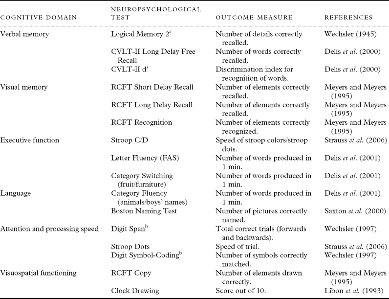
The Australian Imaging Biomarkers and Lifestyle (AIBL) study of aging is a prospective study that incorporates an extensive neuropsychological test battery to detect the cognitive impairment that characterizes very early AD (Ellis et al., Reference Ellis2009, see also Table 1). Performance on each of the AIBL neuropsychological tests is impaired in MCI (Ellis et al., Reference Ellis2009), although, as yet, the nature of the impairment has not been characterized completely, as the magnitude of impairment in different cognitive domains has not been quantified. Importantly, a large subgroup of the AIBL cohort has undergone Pittsburgh compound B (PiB)–PET neuroimaging to determine Aβ amyloid levels, which will enable investigations of the relationship between cognition, AD risk factors, and AD-related pathology.
Summary of the Australian imaging biomarkers and lifestyle study neuropsychological test battery and composite structure

Note: CVLT-II = California Verbal Learning Test – Second Edition; RCFT = Rey Complex Figure Test.
aStory A only, Subtest of the Wechsler Memory Scale, bSubtest of the Wechsler Adult Intelligence Scale – Third Edition (WAIS-III).
The aim of this study was to use performance on cognitive domain-specific composite measures to describe the nature and magnitude of cognitive impairment in MCI and then to determine whether cognitive impairment on these measures was associated with Aβ amyloid in older adults without dementia. The first hypothesis was that, while cognitive domains of memory, executive function, language, and visuospatial ability would be impaired in MCI, the magnitude of impairment would be greatest for memory. In contrast, there would be no impairment in the domains of attention and psychomotor function. As the MCI group was recruited from memory clinics that specialize in AD and all group members had undergone extensive assessment to exclude other pathologies that might affect their cognition prior to entry into the AIBL study, we expected this particular cognitive profile in the MCI group. The second hypothesis was that in both MCI and healthy older adults, individuals with high Aβ amyloid levels would show cognitive impairment on the composite measures when compared with individuals with low Aβ amyloid levels.
Methods
Participants
Complete sample
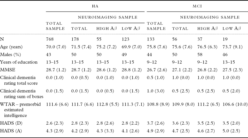
The cohort assessed was the AIBL baseline cohort that consisted of 768 healthy older adults and 133 adults who met criteria for MCI, all of which were included in the present study. These groups differed in age, premorbid intelligence, functional ability, and levels of anxiety and depression (Table 2). The HA group was recruited to the AIBL study through a media campaign, while the MCI group were recruited by referrals from AD clinicians and AD specialist clinics, as well as some volunteers who were identified as meeting criteria for MCI following baseline assessment. Prior to enrolment, the AIBL exclusion criteria were applied to all participants, and included history of non-AD dementia, schizophrenia, bipolar disorder, significant current depression (the Geriatric Depression Scale score greater than 5/15), Parkinson's disease, cancer (other than basal cell skin carcinoma) within the last two years, symptomatic stroke but not transient ischaemic attack, uncontrolled diabetes, or current regular alcohol use exceeding two standard drinks per day for women or four per day for men. These exclusion criteria ensured that individuals with frank pathology that might cause cognitive impairment, other than AD, were not recruited. The study was approved by and complied with the regulations of three institutional research and ethics committees. All participants provided written informed consent prior to participating in the study.
Demographic means (SD) for age, MMSE, WTAR and HADS scores, and median (range) CDR scores for each group
Note: MMSE = Mini-Mental State Examination, WTAR = Wechsler Test of Adult Reading, HADS (D) = Hospital Anxiety and Depression Scale, Depression Subscale, HADS (A) = Hospital Anxiety and Depression Scale, Anxiety Subscale.
In the total sample HA and MCI groups differed significantly on age, MMSE, Clinical Dementia Rating Total Score, Clinical Dementia Rating Sum of Boxes, WTAR, HADS (D), and HADS (A), p < 0.05.
In the neuroimaging sample, HA and MCI groups differed significantly on age, MMSE, Clinical Dementia Rating Total Score, Clinical Dementia Rating Sum of Boxes, and HADS (D), p < 0.05.
In the HA group High Aβ and Low Aβ differed significantly on age, p < 0.05. In the MCI group High Aβ and Low Aβ differed significantly on WTAR, p < 0.05.
A clinical review panel, consisting of old age psychiatrists, neurologists, geriatricians, and neuropsychologists reviewed all available data following assessment to confirm the clinical status (MCI or HA) of each participant. Classification of MCI was based on internationally agreed criteria (Winblad et al., Reference Winblad2004) and has been described in detail in Ellis et al. (Reference Ellis2009). Briefly, classification of MCI required that performance on two cognitive measures be at least 1.5 standard deviations below relevant published normative data for that measure; however, if an individual was referred to the study with a clinical diagnosis of MCI, then they need demonstrate impaired cognitive performance on only a single cognitive measure. In addition, all individuals presented with a subjective memory complaint (either by self or informant) and with substantially intact activities of daily living, and social and occupational functioning.
Neuroimaging sample
A subgroup of the AIBL sample, consisting of 178 healthy older adults and 56 MCI, underwent PiB–PET neuroimaging. Selection for neuroimaging from the HA group was controlled to obtain a wide age range, and an over representation APOE ε4 carriers; MCI participants were selected opportunistically. Groups differed in age, premorbid intelligence, functional ability, and level of depression symptoms (Table 2).
Measures
Demographic and clinical characteristics
Depression and anxiety levels were assessed using the Hospital Anxiety and Depression Scale (HADS). The Wechsler Test of Adult Reading (WTAR) was used to estimate the premorbid intelligence of participants. Functional ability was rated using the Clinical Dementia Rating (CDR) scale on the basis of information obtained from cognitive testing, direct questioning of the participant, and an informant/treating clinician for those with a diagnosis of MCI.
An 80-mL blood sample was also taken from each participant, 0.5 mL of which was forwarded for APOE genotyping at a clinical pathology laboratory.
Neuropsychological assessment
The AIBL neuropsychological test battery has been described in detail previously (Ellis et al., Reference Ellis2009) and is summarized in Table 1. Neuropsychological assessments were conducted in a single 90-min session by trained research assistants under supervision of a neuropsychologist. All measures were administered and scored according to standard protocols.
PiB–PET neuroimaging
The PiB–PET neuroimaging has been described in detail previously (Rowe et al., Reference Rowe2010). Briefly, standardized uptake value (SUV) data were acquired 40–70 min post-injection with the PiB compound. Data were summed and normalized to the cerebellar cortex SUV, giving the region-to-cerebellar ratio, termed as SUV ratio (SUVR; Rowe et al., Reference Rowe2010).
Data coding
The data for each neuropsychological test were assigned to one of the six cognitive domains on the basis of consensus among neuropsychologists, psychologists, and neurologists involved in the AIBL study (Table 1). To compute each composite score, the raw scores for each neuropsychological test were converted to a z-score using the mean and standard deviation of the total HA group, and then averaged to create the single composite score for that domain. This process was repeated for the neuroimaging sample, using the mean and standard deviation of the HA group from this sample only to calculate the z-scores. SUVR was coded as a dichotomous categorical variable using the value of 1.5 as a cut-off for high/low Aβ amyloid level in accordance with established criteria (Jack et al., Reference Jack2008; Villemagne et al., Reference Villemagne2011). APOE genotype was coded as a dichotomous categorical variable according to the carriage of an ε4 allele.
Data analysis
Comparison of cognitive impairment in complete and neuroimaging samples
Means and standard deviations for each group were calculated for each of the demographic and mood variables. Where data were not continuous (CDR total and sum of boxes), median and range were calculated. Demographic and clinical characteristics were compared between groups using t-tests or χ 2 analysis.
To determine the magnitude of impairment in the MCI group on each composite measure, a series of one-way analyses of covariance (ANCOVA) were conducted, in which the scores on the cognitive composite measures were treated as the dependent variable and the group (HA, MCI) as the factor. As the HA and MCI groups differed in age, estimated premorbid intelligence, and levels of anxiety and depressive symptoms (Table 2), these demographic variables were included as covariates in the model.
For each composite measure, the magnitude of the difference between ANCOVA-adjusted means and standard deviations for the HA and MCI groups was estimated using Hedges’ g. As Hedges’ g expresses the magnitude of the difference between group means in terms of the control group standard deviation units (Zakzanis, Reference Zakzanis2001), this effect size is consistent with the classification of abnormal performance against a normative data range, as occurs in neuropsychological evaluation of MCI (e.g. Albert et al., Reference Albert2011). This same analysis was then repeated in the neuroimaging sample. The 95% confidence interval (CI) for each measure of effect size was compared between the total sample and the neuroimaging sample. For the effect size of each composite measure, significant differences between the complete and neuroimaging samples were inferred to be present when there was no overlap between the respective 95% CI.
Effect of Aβ amyloid on cognitive function in healthy adults and mci
Data from the neuroimaging sample were submitted to two sets of planned comparisons constructed within a one-way ANCOVA, with age, premorbid intelligence, and anxiety and depressive symptoms as covariates. Group (HA and MCI), Aβ amyloid level (high, low), and Group × Aβ amyloid interaction were entered as fixed factors. To determine the nature and magnitude of Aβ-related cognitive impairment in healthy and MCI groups, the first set of planned comparisons compared the performance of the high and low Aβ amyloid subgroups within the MCI and HA groups separately. Differences between the high and low Aβ amyloid subgroups were expressed using Hedges’ g. To determine the extent to which impairment in healthy adults and MCI was influenced by disease classification and Aβ amyloid status, the second set of planned comparisons compared the performance of HA high Aβ amyloid, MCI low Aβ amyloid, and the MCI high Aβ amyloid subgroups to performance of the HA low Aβ amyloid subgroup. For all these comparisons the magnitude of impairment relative to the HA low Aβ amyloid group was expressed using Hedges’ g.
Results
Comparison of cognitive impairment in complete and neuroimaging samples
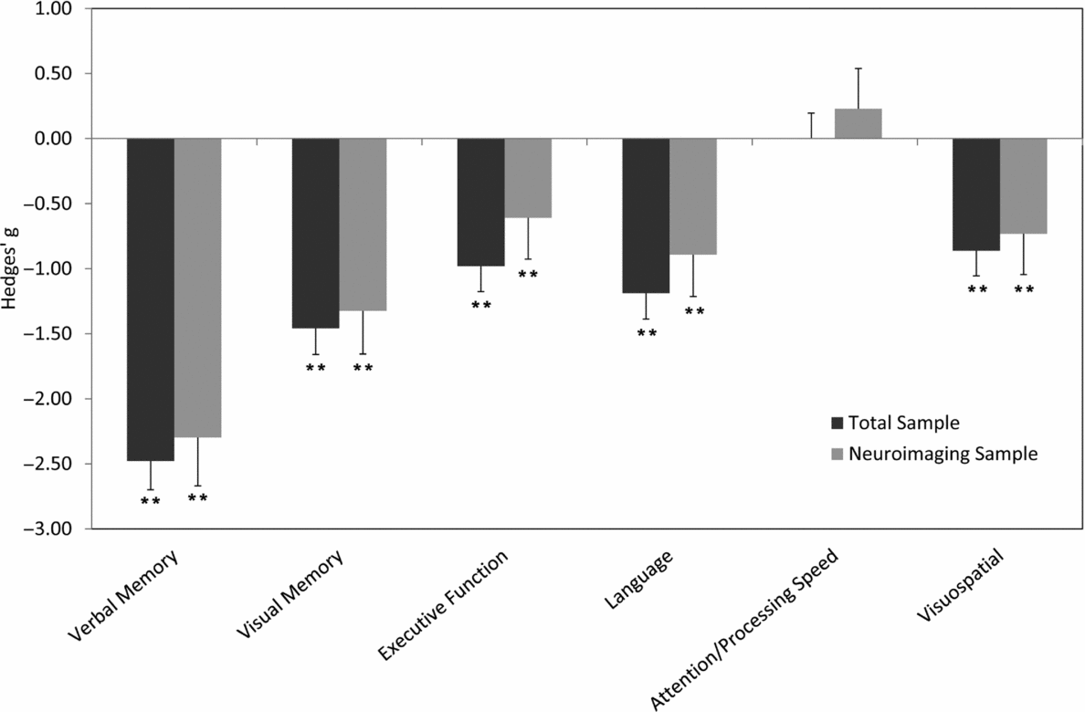
The magnitude of impairment (Hedges’ g) in the MCI group, from the total sample and the neuroimaging sample, relative to their respective HA groups are shown in Figure 1. For each composite, except attention and processing speed, the ANCOVA was statistically significant and the differences between the groups were moderate to large in magnitude. This was the case for both samples, as indicated by the substantial overlap of the 95% CI of the effect sizes reflecting MCI-related impairment on each cognitive composite measure between samples.
Effect sizes (Hedges’ g) with 95% CI for the performance of MCI group, relative to HA group on cognitive composite measures when controlling for age, premorbid intelligence, anxiety, and depression. **Significant difference (p < 0.01) between HA and MCI groups.
Effect of Aβ amyloid on cognitive function in healthy adults and MCI
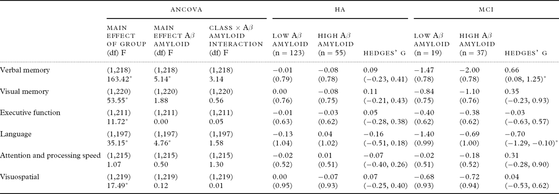
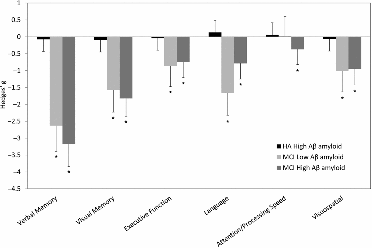
Outcomes for the main effects of the Group (HA, MCI) and Aβ amyloid level (high and low), and the interaction of Group × Aβ amyloid from the one-way ANCOVA are summarized in Table 3. Group estimated marginal means and standard deviations for high and low Aβ amyloid subgroups within the HA and MCI groups separately are also shown in Table 3. The first set of planned comparisons indicated that within the HA group, there were no statistically significant differences between high and low Aβ amyloid subgroups on any cognitive composite measure. Within the MCI group, the high Aβ amyloid subgroup performed significantly worse than the low Aβ amyloid subgroup on the verbal memory composite and this difference was moderate in magnitude. Furthermore, the low Aβ amyloid subgroup performed significantly worse than the high Aβ amyloid subgroup on the language composite and this difference was moderate in magnitude.
ANCOVA results and estimated marginal mean (SD) of HA high and low Aβ amyloid subgroups, and MCI high and low Aβ amyloid subgroups, and Hedges’ g (95% CI) of difference between groups
Note: *p < 0.05.
The second set of planned comparisons indicated that, when compared to the HA low Aβ amyloid subgroup, the impairment in the MCI high Aβ amyloid subgroup on each of the memory composite measures was statistically significant and large in magnitude (see Figure 2). For the executive function, language, and visuospatial composites, the impairment in the MCI high Aβ amyloid subgroup was of moderate to large magnitude, and was also statistically significant. The MCI high Aβ amyloid subgroup did not differ significantly from the HA low Aβ amyloid subgroup on the attention and processing speed composite. Furthermore, the MCI low Aβ amyloid subgroup also showed large and statistically significant impairment, when compared to the HA low Aβ amyloid subgroup, on each of the memory composites. Moderate to large impairments, which were statistically significant, were also observed for the executive function, language, and visuospatial composites in this subgroup. The MCI low Aβ amyloid subgroup did not differ significantly from the HA low Aβ amyloid subgroup on the attention and processing speed composite.
Effect sizes (Hedges’ g), with 95% CI, comparing performance of HA high Aβ amyloid, MCI high Aβ amyloid, and MCI low Aβ amyloid subgroups with HA low Aβ amyloid subgroup on cognitive composite measures. *Significant difference (p < 0.05) from the HA low Aβ amyloid group.
Discussion
The first hypothesis that memory, executive function, language, and visuospatial ability would be impaired in MCI, and that there would be no impairment in attention and psychomotor function, was supported. As expected, impairment in verbal memory was very large in the MCI group (i.e. performance ~2 standard deviation below healthy adults) even after statistical control of the effects of age, premorbid intelligence, and anxiety and depressive symptoms (Figure 1). For visual memory, impairment in the MCI group was large, although somewhat smaller than that observed in verbal memory. Impairment was also observed for executive function, language, and visuospatial domains, albeit at a reduced magnitude (i.e. performance ~1 standard deviation below healthy adults (Figure 1)). No MCI-related impairment in attention or psychomotor function was observed. This pattern of impairment is consistent with that defined in current neuropsychological models of MCI (Albert et al., Reference Albert2011) and can be considered to be independent of the effects of age, premorbid intelligence, and anxiety and depressive symptoms as these were controlled in the analysis.
The second hypothesis that in both MCI and healthy older adults, individuals with high Aβ amyloid levels would show cognitive impairment on composite measures when compared with individuals with low Aβ amyloid levels was supported partially. Individuals with high Aβ amyloid did show poorer memory performance in MCI, although no differences between groups with high and low Aβ amyloid levels were observed in healthy adults (Table 3). In the AIBL study, patients with MCI were recruited from AD memory clinics and were classified as meeting clinical criteria by expert clinicians. Despite a very exacting workup, neuroimaging indicated that Aβ amyloid was abnormally high in only 68.5% of these patients. This characteristic of MCI samples has been described previously (Jack et al., Reference Jack2008; Villemagne et al., Reference Villemagne2011; Doraiswamy et al., Reference Doraiswamy2012) and has given rise to speculation that MCI without high Aβ amyloid does not reflect early or prodromal AD (Dubois and Albert, Reference Dubois and Albert2004; Albert et al., Reference Albert2011). In fact, recent research criteria for MCI reflecting early AD now emphasize that the clinically classified syndrome be accompanied by positive AD biomarker (Albert et al., Reference Albert2011).
From a neuropsychological perspective, MCI with high Aβ amyloid was characterized by greater impairment in episodic memory and less impairment in language when compared with MCI with low Aβ amyloid (Table 3). Although confirmation of amnestic MCI classification at enrolment to the AIBL study required individuals to show impaired performance on the tests of memory from the AIBL battery, the AIBL clinical panel did not have access to information about levels of Aβ amyloid when neuropsychological assessments were made or when clinical classifications were confirmed. Thus, the greater impairment of memory in MCI with high Aβ amyloid is highly unlikely to reflect any classification bias. Furthermore, as high and low Aβ amyloid samples were well matched on demographic characteristics (see Table 2), and any effects of depressive symptoms, anxiety symptoms, age, or premorbid intelligence were controlled statistically in the analyses, this difference is unlikely to reflect any characteristics of the individuals themselves. While the large impairment in memory is to be expected in the MCI group generally, the increased memory impairment in MCI with high Aβ amyloid reinforces the predilection of AD pathology for medial temporal lobe structures and memory function (Dubois and Albert, Reference Dubois and Albert2004; Dickerson and Sperling, Reference Dickerson and Sperling2008). The finding of increased impairment in language in the MCI group with low Aβ amyloid reinforces the notion that impairment in the two MCI groups may reflect different pathological processes (Dubois and Albert, Reference Dubois and Albert2004; Pike et al., Reference Pike2007; Rowe et al., Reference Rowe2010; Albert et al., Reference Albert2011). As such, further investigation into the specific profile of language impairment and potential underlying pathologies within this group is warranted.
There were no statistically significant differences in cognitive performance between healthy adults with high and low Aβ amyloid levels. The relatively large sample sizes (n = 174) assessed in the present study provided good statistical power to detect even small differences had they existed. These data therefore suggest that in healthy older adults, high Aβ amyloid level does not manifest as any cognitive impairment. This conclusion is consistent with the results of other studies that have also found that elevated Aβ amyloid, detected from neuroimaging and cerebrospinal fluid (CSF) analysis, is not associated, or only weakly associated, with increased cognitive impairment in healthy adults (Pike, et al., Reference Pike2007; Aizenstein, et al., Reference Aizenstein2008; Jack, et al., Reference Jack2008). However, one recent study reported significant differences of a moderate to large magnitude (d = 0.67–0.82) between healthy older adults with high and low Aβ amyloid levels on a test of episodic memory (e.g. Logical Memory test), but not on tests of language, executive function, or attention (e.g. verbal fluency and digit symbol substitution; Sperling et al., Reference Sperling2013). Importantly, the sample in this report (i.e. total n = 78 with 18 amyloid positive) was much smaller than that in the current study (n = 178 with 55 amyloid positive), where no Aβ amyloid-related memory impairment was observed in healthy adults. One possible explanation for the difference in study outcomes is that a proportion of the high Aβ amyloid group in the Sperling et al. (Reference Sperling2013) sample may actually have met criteria for MCI. This possibility is evident in the scatter plots of individual scores on the Logical Memory test for immediate and delayed recall, where for the high Aβ amyloid group, at least 11% of the sample had memory scores that would be classified as significantly impaired given the sample's mean education (based on the ADNI education corrected cut-off scores for WMS-R Logical Memory subtest). As the individuals in this study will have undergone careful diagnoses, it is likely that despite having this memory impairment the individuals or their spouses did not complain about their memory, as such a complaint would have likely resulted in them meeting clinical criteria for MCI. Hence, the finding of a difference between healthy individuals with high and low Aβ amyloid levels may have been due to the high Aβ amyloid group containing a proportion of individuals with objective memory impairment similar to that of MCI. Further studies and meta-analyses will be necessary to determine whether healthy older adults with high Aβ amyloid do show cognitive impairment, although at the present time we believe that the weight of evidence, based on sample size and number of negative studies, suggest that they do not.
The current results have some important implications for neuropsychological assessment of AD-related cognitive impairment. Given the strong relationship between Aβ amyloid and AD, in addition to the recent approval by the Food and Drug Administration for use of one neuroimaging measure to determine Aβ amyloid levels in humans suspected of having AD (Yang et al., Reference Yang, Rieves and Ganley2012), it is likely that demands for assessment of Aβ amyloid levels will increase. However, the procedure of Aβ amyloid neuroimaging remains expensive and is not widely available; hence in MCI, the recognition of a cognitive profile that suggests increased risk for cerebral Aβ amyloid may help optimize health resource utilization. As such the identification of a cognitive profile that can distinguish MCI with high Aβ amyloid from MCI with low Aβ amyloid should be sought. In the current study, high Aβ amyloid was best predicted by a specific impairment in memory. It is unlikely that sampling bias in the current study would have influenced the difference in the profiles between MCI groups with high and low Aβ amyloid, given that individuals in this sample had been extensively screened for other neurological, psychiatric, and systemic diseases. However, with broader inclusion criteria it is likely that differences in cognitive profiles determined from neuropsychological assessment may become even greater.
Importantly, the findings of this study highlight the inability to identify cognitive impairment in healthy older adults with high Aβ amyloid. This may require more sensitive tests than the composite measures used here. Alternatively, the development of algorithms that include other known risk factors for high Aβ amyloid (e.g. age, APOE ε4 allele carriage) may result in increased sensitivity and thereby predictive validity. This is an important challenge as there is a growing consensus that anti-amyloid therapies may be most useful in the very early or asymptomatic stages of the disease (Sperling et al., Reference Sperling2011); however, widespread screening for Aβ amyloid remains impractical and expensive.
Some important limitations of this study warrant consideration as to the extent to which they affect the conclusions drawn. First, the conclusions drawn here were based on cross-sectional comparisons between groups and assumed that high Aβ amyloid is a valid proxy for early AD. Given that the hallmark of AD is progressive cognitive decline, ultimately the importance of Aβ amyloid and the extent to which its effects can be detected with neuropsychological instruments will require prospective studies. There are currently several large prospective studies, including the AIBL study, underway that will enable future investigations of these relationships to compare groups longitudinally. Second, the AIBL cohort is not an epidemiological sample and as such the extent to which the findings extend to general neuropsychological settings must still be determined. Nevertheless, despite these limitations, the current findings provide further evidence of the relationship between Aβ amyloid and cognition in both healthy older adults and those who meet criteria for MCI, with impairments to memory function being the primary symptom of the underlying disease during the preclinical and prodromal phases of AD.
Conflict of interest
None.
Description of authors’ roles
Karra D. Harrington, Paul Maruff, Yen Ying Lim, and Kathryn A. Ellis designed the study. David Darby, Ralph N. Martins, Colin L. Masters, Paul Maruff, Kathryn A. Ellis, Victor L. Villemagne, Cassandra Szoeke, Greg Savage, and Christopher Rowe are responsible for the design and conduct of the AIBL study. Christopher Rowe and Victor L. Villemagne collected and analyzed all neuroimaging data. Karra D. Harrington, Yen Ying Lim, Paul Maruff, Kathryn A. Ellis, Michael Weinborn, David Darby, and Greg Savage developed the composite structure. Karra D. Harrington and Paul Maruff conducted all statistical analyses and interpretation of the data. Karra D. Harrington, Yen Ying Lim, and Paul Maruff prepared the manuscript. Kathryn A. Ellis, Carly Copolov, David Darby, Michael Weinborn, David Ames, Ralph N. Martins, Greg Savage, Cassandra Szoeke, Christopher Rowe, Victor L. Villemagne, Colin L. Masters drafted and revised the manuscript.
Acknowledgments
Core funding for the AIBL study was provided by the Commonwealth Scientific and Industrial Research Organisation (CSIRO), which was supplemented by “in kind” contributions from the study partners (see http://www.aibl.csiro.au/). The research was also supported by the Science Industry and Endowment Fund (see www.sief.org.au), and the National Health and Medical Research Council (NHMRC) via the Dementia Collaborative Research Centres program (DCRC2). Mental Health Research Institute (MHRI) acknowledges the funding support from the Victorian Government's Operational Infrastructure Support program. Pfizer International has contributed financial support to assist with the analysis of blood samples and to further the AIBL research program. The McCusker Foundation has contributed financial and in-kind support to AIBL. The authors acknowledge the financial support of the Cooperative Research Centre (CRC) for Mental Health program.
The AIBL investigators thank Richard Head (CSIRO) for initiating and facilitating the AIBL collaboration. Alzheimer's Australia (of Victoria and Western Australia) assisted with the promotion of the study and screening of telephone calls from volunteers. The AIBL team thanks the clinicians who referred AD patients to the study, including Associate Professor Brian Chambers, Professor Edmond Chiu, Drs. Roger Clarnette, Mary Davison, John Drago, Peter Drysdale, Jacqueline Gilbert, Kwang Lim, Professor Nicola Lautenschlager, Drs. Dina LoGiudice, Peter McCardle, Steve McFarlane, Alastair Mander, John Merory, Professor Daniel O’Connor, Drs. Ron Scholes, Mathew Samuel, Darshan Trivedi, and Associate Professor Michael Woodward. We thank all those who participated in the study for their commitment and dedication to helping advance research into the early detection and cause of AD.