

Many food-grade microorganisms, including lactic acid bacteria (LAB), are known to synthesise exopolysaccharides (EPS) which have been widely used in the dairy industry, mainly when produced in situ during fermentation, to improve attributes such as thickening properties, syneresis reduction and rheology augmentation (Folkenberg et al. Reference Folkenberg, Dejmek, Skriver, Guldager and Ipsen2006). Apart from the technological relevance, there is increasing evidence that suggests a role of EPS from LAB and bifidobacteria in the interaction between the producing bacteria and the host, exerting an impact on human health. It was suggested that hetero-EPS exhibits immune stimulating (Fanning et al. Reference Fanning, Hall, Cronin, Zomer, MacSharry, Goulding, O'Connell Motherway, Shanahan, Nally, Dougan and van Sinderen2012; Salazar et al. Reference Salazar, López, Garrido, Moran, Cabello, Gueimonde, Suárez, González, de los Reyes-Gavilán and Ruas-Madiedo2014), antitumor and cholesterol-lowering activities (Ruas-Madiedo et al. Reference Ruas-Madiedo, Hugenholtz and Zoon2002a; Wang et al. Reference Wang, Li, Rui, Chen, Jiang and Dong2014). Additionally, they may act as protective agents against pathogens (Nagai et al. Reference Nagai, Makino, Ikegami, Itoh and Yamada2011; Fanning et al. Reference Fanning, Hall, Cronin, Zomer, MacSharry, Goulding, O'Connell Motherway, Shanahan, Nally, Dougan and van Sinderen2012; Maruo et al. Reference Maruo, Gotoh, Nishimura, Ohashi, Toda and Takahashi2012) and stomach ulcers (Rodríguez et al. Reference Rodríguez, Medici, Rodríguez, Mozzi and de Valdez2009), as prebiotic factors (Salazar et al. Reference Salazar, Gueimonde, Hernandez-Barranco, Ruas-Madiedo and de los Reyes-Gavilan2008, Reference Salazar, Binetti, Gueimonde, Alonso, Garrido, Del Rey, González, Ruas-Madiedo and de los Reyes- Gavilán2011) or by improving the intestinal functional barrier (Del Piano et al. Reference Del Piano, Balzarini, Carmagnola, Pagliarulo, Tari, Nicola, Deidda and Pane2014). Despite these apparent beneficial aspects for health, the low yield of hetero-EPS from LAB (80 to 600 mg/l for L. lactis subsp. cremoris; Cerning, Reference Cerning1995; Ruas-Madiedo et al. Reference Ruas-Madiedo, Tuinier, Kanning and Zoon2002b) prevents their commercial use, in contrast to homo-EPS (10–25 g/l; Becker et al. Reference Becker, Katzen, Puhler and Ielpi1998).
Lactobacillus fermentum Lf2 is a strain which was isolated as a contaminant culture in cheese manufacture. The particular interest in this strain resides in the high levels (approximately 1000 mg/l) of EPS when grown in well-defined conditions. The strains of this bacterial species which have been studied in relation to the EPS production are L. fermentum TDS030603 (Fukuda et al. Reference Fukuda, Shi, Nagami, Leo, Nakamura, Yasuda, Senda, Motoshima and Urashima2010) and an isolate from Burkina Faso fermented milk (Savadogo et al. Reference Savadogo, Ouattara, Savadogo, Barro, Ouattara and Traoré2004). To our knowledge, at this moment there are no studies related to the functional properties of EPS from L. fermentum or based on the application of EPS as food additives in dairy products. This fact, combined with the increasing demand for natural and functional food ingredients, were the main factors to address the isolation and functional characterization (immunological role and protection against Salmonella Thyphimurium) of the EPS produced by L. fermentum Lf2, focusing on its final application as a food additive in a functional yogurt.
Materials and methods
Organisms and growth conditions
L. fermentum Lf2 was isolated as a non-starter culture in Cremoso cheese, and stored at −80 °C (INLAIN Collection) in MRS (de Man, Rogosa and Sharpe, Biokar, Beauvais, France) broth plus 150 ml/l glycerol. It was routinely grown in MRS broth at 37 °C and was identified by sequencing of its 16SrDNA (Edwards et al. Reference Edwards, Rogall, Blöcker, Emde and Böttger1989).
For yogurt production, the commercial strains Streptococcus thermophilus SC42 and Lactobacillus delbrueckii subsp. lactis 254 were used as starter cultures. They were selected based on their inability (visual test) to produce EPS in milk. They were routinely grown in 100 g/l reconstituted skimmed milk (RSM) at 43 °C and stored at −80 °C in the same medium.
For in vivo trials, a human isolate of Salmonella enteritidis serovar Typhimurium was kindly provided by the INEI-ANLIS ‘Dr Carlos Malbrán’ Institute (Buenos Aires, Argentina). The strain was maintained at −80 °C in BHI (Brain-Heart Infusion, Britania, Buenos Aires, Argentina) broth with 150 ml/l glycerol. Overnight cultures (16 h, 37 °C, aerobiosis) obtained immediately after thawing were diluted and used to infect animals.
Fermentations and EPS isolation
Fermentations were performed in a 2-l fermentor (Sartorius Biostat A plus)® in a semi-defined broth (SDM; Kimmel & Roberts, Reference Kimmel and Roberts1998). L. fermentum Lf2 was inoculated from an overnight culture (1 ml/l) and incubations were made at 30 °C for 72 h, with an agitation rate of 5 g and sparging with CO2 at 0·2 l/min. The pH was kept automatically at 6·0 with sterile 8 m NaOH. After incubation, bacteria were removed by centrifugation (19 630 g , 30 min, 5 °C) and EPS was precipitated at 4 °C for 48 h by adding 2 volumes of chilled absolute ethanol (Cicarelli, Buenos Aires, Argentina). The precipitate was collected by centrifugation (4053 g , 30 min, 5 °C), dissolved in ultrapure water and dialyzed against distilled water (12–14 kDa MWCO membranes, Sigma- Aldrich) for 3 d, at 4 °C with a daily change of water, and finally freeze-dried (Chris Alpha 1–4 LD Plus, Tokyo, Japan) to obtain the EPS fraction. The total protein concentration of EPS extract was determined with the Bio-Rad (Hercules, CA, USA) protein assay (Bradford, Reference Bradford1976).
Functional properties of the EPS extract
Manufacture of yogurts
Yogurt was produced in cylindrical bottles with 100 ml RSM inoculated with S. thermophilus SC42 (106 cfu/ml) and L. delbrueckii subsp. lactis 254 (105 cfu/ml), added with 0 (control) and 300 mg/l of EPS extract, with incubation at 43 °C until a final pH value of 4·6, and immediately cooled-down and stored overnight at 4 °C. The dose of EPS added was selected based on previous assays performed by our group, demonstrating that yogurt with 300 mg/l of EPS extract showed rheological and sensory differences when compared with control yogurts.
Animals and feeding procedures
For in vivo trials, 6-week-old male BALB/c mice weighing 19–21 g were obtained from the random-bred colony of the Centro de Experimentaciones Biológicas y Bioterio, Facultad de Ciencias Veterinarias (UNL, Esperanza, Santa Fe, Argentina). All the procedures were approved by the Ethical Committee for Animal Experimentation of the Facultad de Ciencias Veterinarias (FCV-UNL, Esperanza, Santa Fe, Argentina), and were made following the recommendations of the INLAIN animal facility (Zacarías et al. Reference Zacarías, Reinheimer, Forzani, Grangette and Vinderola2014).
Immunostimulatory ability: determination of s-IgA and cytokines in small and large intestines
Thirty two mice were clustered in 4 groups (8 mice/group) and received, during 15 consecutive days and by gavage, 300 µl/d of any of the following samples: (a) RSM (M), (b) RSM + 300 mg EPS/l (equivalent to 4·5 mg/kg/d) (M-EPS), (c) yogurt (Y), (d) yogurt + 300 mg EPS/l (Y-EPS). All animals simultaneously and ad libitum received sterile tap water and a conventional balanced diet (Cooperación, Buenos Aires, Argentina).
After the feeding period, animals were anaesthetised and sacrificed by cervical dislocation. Liver was removed and homogenized in 5 ml of sterile PBS and pour plated onto ABRV agar (37 °C for 24 h in aerobiosis) in order to evaluate translocation of enterobacteria to liver. The small intestine was recovered and flushed with 5 ml of cold PBS buffer containing 10 g/l of a protease inhibitor cocktail (Sigma-Aldrich). This fluid was recovered, centrifuged (10 000 g , 10 min, 4 °C) and the supernatant was stored at −80 °C for secretory IgA quantification by ELISA (Martins et al. 2009). Portions of the distal small intestine (jejunum and ileum) and whole large intestine were removed for homogenate preparations. The tissues were kept frozen (−80 °C) immediately after extractions and then resuspended (1 ml/100 mg of tissue) in extraction buffer (100 ml PBS, 0·293 g EDTA and 50 µl Tween 20) containing a cocktail of protease inhibitor (10 ml/l), homogenized (Ultra-turrax T8, IKA Labortechnik, Staufen, Germany) and centrifuged (9600 g , 10 min, 4 °C). Cytokines IL-10, IL-6 and IFN-γ were determined in the supernatants using the corresponding mouse ELISA Set (BD OptEIA, BD, Biosciences PharMingen, San Diego, CA, USA).
Protective capacity of EPS against an infection with S. enteritidis serovar Typhimurium
A completely randomized design was chosen for this study including 64 animals which were clustered in 2 groups (32 animals/group). Each mouse received, during 15 consecutive days and by gavage, 300 µl/d of any of the following samples: (a) 600 mg/l of EPS in RSM (equivalent to 9 mg/kg/d) and (b) RSM (control group). This dose was selected based on a previous assay with 10 animals/group, where 4·5 and 9·0 mg/kg/d of EPS extract were administered, evidencing that the lower dose (4·5 mg/kg) caused no significant effect on the survival of treated mice. After the feeding period, all animals were challenged with a single infective dose of a fresh culture (16 h incubation, 37 °C, aerobiosis) of S. enteritidis serovar Typhimurium (106 cfu/mouse) according to a previous study of the optimal dose able to infect 40–50% of mice. The body weight and number of surviving animals were registered daily for 25 d post-infection and results were expressed as the percentage of animals that survived the infection.
Statistical analysis
SPSS software (SPSS Inc., Chicago, IL, USA) was used to carry out the statistical analysis of the data. T-test was performed to compare control groups with treated groups, and the Mann-Whitney U test was applied when the distribution was not normal (Shapiro-Wilk test). Data were considered significantly different when P < 0·05.
The proportions of survivors at the end of the challenge with Salmonella Typhimurium were compared with Barnard's exact test using the software R (R Core Team, 2015), as implemented in the ‘Exact’ library. The difference in death risk between the treated and control groups was estimated with Cox's model of Proportional Hazards, calculated with the ‘survival’ R library (Calhoun, Reference Calhoun2015; Therneau, Reference Therneau2015). The P-value of the estimation of the risk ratio in this model was estimated using a permutation test of group membership (100 000 repetitions), while confidence intervals were estimated using bootstrap (100 000 repetitions).
Results
Fermentation, EPS isolation and manufacture of yogurt
At controlled pH (6·0), 30 °C, and in SDM broth, the best EPS production (1090·9 mg/l) was reached after 72 h of fermentation, corresponding with the late stationary phase of growth of L. fermentum Lf2. It is important to highlight that the EPS extract had 0·9% protein (Bradford method), a value lower than the limit recommended for EPS when added to food matrices (<3%, De Vuyst et al. Reference De Vuyst, Zamfir, Mozzi, Adriany, Marshall, Degeest and Vaningelgem2003). The EPS that was released in SDM at pH 6·0, after its acid hydrolysis in 2 mol/l TFA (120 °C for 2 h, Ruas-Madiedo et al. Reference Ruas-Madiedo, Hugenholtz and Zoon2002a) consisted of glucose and galactose, in a relation 2 : 1 approximately (data not shown).
Functional properties of the EPS extract
Immunostimulatory ability: determination of s-IgA and cytokines in small and large intestines
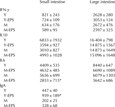
The chosen dose of EPS extract was appropriate, since no translocation was observed in mice from the groups M-EPS and Y-EPS (data not shown). The effects of EPS administration on small intestine s-IgA are shown in Table 1. The levels of s-IgA were increased when the exopolysaccharide extract was added in yogurt (Y-EPS P < 0·05) relative to the yogurt control group. When the EPS was administered in milk, the stimulation of immune response mediated by IgA was markedly lower (approximately 2·6 times) than that observed in yogurt, evidencing only a slight and non-significant (P > 0·05) enhancement.
Effects of EPS extract from L. fermentum Lf2 on the small intestine fluid IgA and small and large intestines tissues cytokines IFN-γ, IL-10 and IL-6, when suspended (4·5 mg/kg body weight) in yogurt (Y-EPS) or milk (M-EPS) after 15 d of administration, compared to control mice (Y and M, respectively)

Asterisks indicate statistically significant (P < 0·05) difference from the corresponding control value. Results are expressed as mean values ±sem
The cytokines measured in the small and large intestine tissues at the end of the administration were comparable to their respective controls (Table 1), with the exception of IL-6 from small intestine in mice that received the EPS extract in milk (M-EPS), in which the lowest production (P < 0·05) of this cytokine was detected.
Protection capacity of the EPS against an infection with S. enteritidis serovar Typhimurium
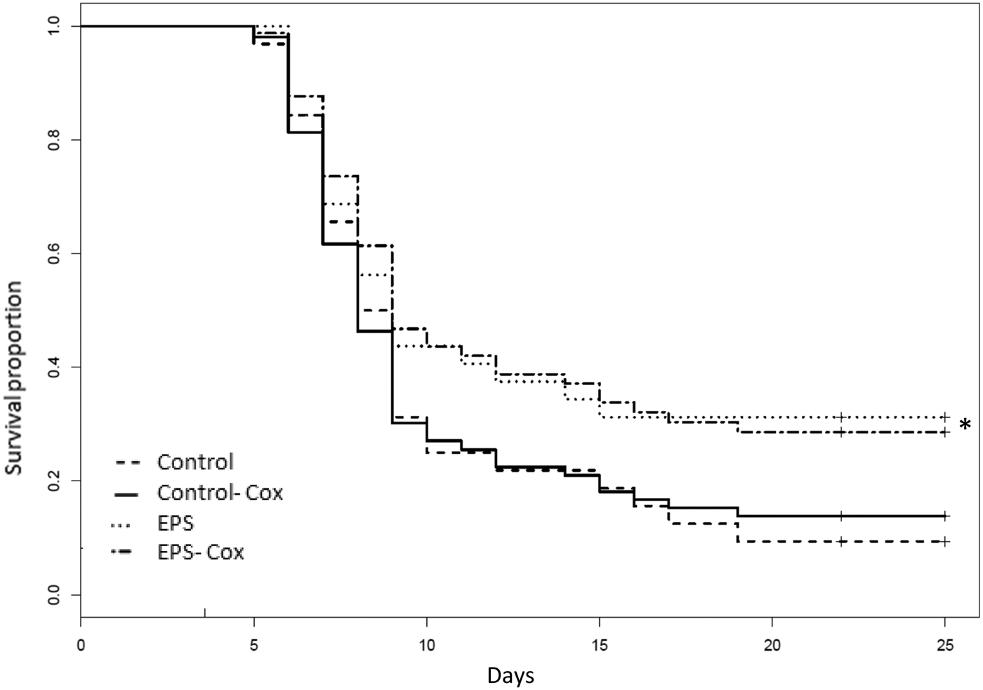
The survival proportion of mice that received EPS was 31%. Significant differences (unilateral P-value of 0·016, exact Barnard test of proportions for a 95% confidence level) were observed when compared with the control group which evidenced 9% survival (Fig. 1 shows the graphics of survival applying the Kaplan-Meier estimation of distribution for this EPS dose). The estimated power of this test was 87·3% (assuming 95% confidence level and, true proportions equal to those observed, for 2 groups of 32 mice/group), which is the probability of detecting a significant difference when the difference does exist. The difference ratio in the instantaneous death risk among groups was estimated by the Cox Proportional Hazards model. The instantaneous death risk for the EPS group calculated by permutation test was 0·63 of that for the control group (unilateral P-value of 0·053, 95% confidence level: [0·35, 1·09]), indicating that no significant difference was detected among groups when this statistical treatment was applied.
Effect of the EPS extract from L. fermentum Lf2 on the survival rate of mice infected with Salmonella enteritidis serovar Typhimurium applying the Kaplan-Meier estimation of distribution. BALB/c mice (32 animals/group) were administered orally with sterile RSM (Control) or EPS suspended in sterile RSM (9 mg/kg body weight). Mice were infected with S. Typhimurium (106 cfu) and the survival rates were observed for 25 d. The curves corresponding with the estimated Cox Proportional Hazards model are specified (Control-Cox and EPS-Cox). Asterisk indicates a statistically significant difference (P < 0·05, exact Barnard test of proportions) from the control group.

Discussion
Our study was addressed to the functional characterization of milk and yogurt supplemented with an EPS extract (300 and 600 mg/l) from L. fermentum Lf2. In previous experiments, these concentrations of EPS were suitable to produce beneficial rheological and sensory changes in yogurt, improving the consistency without unpleasant defects, when compared with controls (unpublished).
This study is the first to report the effect on the s-IgA and cytokines (IL-6, IL-10 and INF-γ) in small and large intestines following the administration, at a dose of 4·5 mg/kg of an exopolysaccharide produced by L. fermentum when used as food additive in milk and yogurt. It seems that the EPS extract when suspended in milk showed a mild in vivo immune suppressive profile by reducing the proinflammatory cytokine IL-6 in small intestine. Similar results were reported by Salazar et al. (Reference Salazar, López, Garrido, Moran, Cabello, Gueimonde, Suárez, González, de los Reyes-Gavilán and Ruas-Madiedo2014) for an EPS-producing B. animalis subsp. lactis strain in healthy rats, evidencing an immune protective profile by a suppression of IL-6 and a promotion of the synthesis of the regulatory cytokine TGF-β. When yogurt was used as matrix, we observed an increased (P < 0·05) level of s-IgA for the EPS extract, again in small intestine. As expected (Gill, Reference Gill1998; Simon et al. Reference Simon, Vahjen and Scharek2003), live microorganisms delivered via yogurt seemed to improve the gut mucosal immune system by increasing the level of secretory IgA, since the effect of this food matrix was significantly higher than those detected in milk. In the gut, secretory IgA is the main immunoglobulin whose key role is to exert an immune exclusion by intimate cooperation with the innate non-specific protection mechanisms (Brandtzaeg et al. Reference Brandtzaeg, Bjerke, Kett, Kvale, Rognum, Scott, Sollid and Valnes1987). Various authors described antibody production at the systemic level after the oral administration of exopolysaccharides or their producing strains (Willers et al. Reference Willers, Weiler and Kolb1995; Vinderola et al. Reference Vinderola, Perdigón, Duarte, Farnworth and Matar2006; Fanning et al. Reference Fanning, Hall, Cronin, Zomer, MacSharry, Goulding, O'Connell Motherway, Shanahan, Nally, Dougan and van Sinderen2012; Salazar et al. Reference Salazar, López, Garrido, Moran, Cabello, Gueimonde, Suárez, González, de los Reyes-Gavilán and Ruas-Madiedo2014). In general, these studies refer to administration in higher doses than those evaluated in the present work, which are more representative of a real concentration of any EPS in a food matrix. For example, Vinderola et al. (Reference Vinderola, Perdigón, Duarte, Farnworth and Matar2006) have reported the administration of 100 mg/kg of kefiran (a dose 22 times higher than the used in our study), evidencing an immunomodulation (mediated by IgA, Il-2, IL-6, IL-10, TNF-α) at the level of large intestine and blood, enhancing the IgA production at both the small and large intestine level.
More recently, diverse reports indicate that not only the presence/absence of the polymer, but also the intrinsic characteristics of each EPS, mainly charge and molecular weight, are relevant for their capability to induce an immune response. López et al. (Reference López, Monteserin, Gueimonde, de los Reyes-Gavilán, Margolles, Suarez and Ruas-Madiedo2012) have demonstrated that different bifidobacterial EPS in vitro induced a variable cytokine production pattern by human peripheral blood mononuclear cells. In general, it was proposed that EPS having negative charge and/or small molecular weight (lower than 106 Da) could act as mild stimulators of immune cells, whereas those polymers non-charged and with a large size could elicit a lower production of any cytokine, evidencing a suppressive profile (Hidalgo-Cantabrana et al. Reference Hidalgo-Cantabrana, Nikolic, López, Suárez, Miljkovic, Kojic, Margolles, Golic and Ruas-Madiedo2014; Salazar et al. Reference Salazar, López, Garrido, Moran, Cabello, Gueimonde, Suárez, González, de los Reyes-Gavilán and Ruas-Madiedo2014; Ryan et al. Reference Ryan, Ross, Fitzgerald, Caplice and Stanton2015). Therefore, based on preliminary evidence indicating a relatively low MW (lower than 105 Da) of our EPS, we could justify the observed mild stimulation of immune cells when administered to mice.
Our experimental model was performed with standard, naïve (not challenged) healthy BALB/c mice. Thus, this could be the main reason why most cytokines tested were not significantly modified by the ingestion of the EPS extract. In this regard, Fanning et al. (Reference Fanning, Hall, Cronin, Zomer, MacSharry, Goulding, O'Connell Motherway, Shanahan, Nally, Dougan and van Sinderen2012) have demonstrated in a naïve murine model that the EPS-producing Bifidobacterium breve UCC2003 strain failed to elicit a strong immune response in comparison to its EPS-deficient variant strains, evading the B-cell response. The reason why the two matrices (milk and yogurt) evidenced different responses on s-IgA and IL-6 remains unclear, and would require additional studies to elucidate (by increasing the number of cytokines evaluated, for example).
In general, it is desirable for a pathogen challenge to cause 40–50% mortality in the control group, in order to evaluate the protective effect of the treatment in a moderately hostile model. We based our pathogen dose on our previous studies with the same strain of S. Typhimurium, which indicated a dose of 106 cfu to allow the survival of 40% of mice. In our assay, only 9% of the control group survived after the challenge, which turned the assay into a very demanding one. The reason for this variability remains unknown, but it could be related to the fact that Salmonella infection in mice is considered dynamic and dependent on multiple host variables (Watson & Holden, Reference Watson and Holden2010). Despite the challenging conditions of our assay, the results indicated that the daily administration of EPS extract, at the dose of 9 mg/kg before a challenge with Salmonella provided enhanced protection against the infection. Similar survival rates were observed by Nagai et al. (Reference Nagai, Makino, Ikegami, Itoh and Yamada2011) when evaluating the effect of an EPS from a strain of Lactobacillus delbrueckii subsp. bulgaricus on the influenza virus infection after 21 d of administration of yogurts. Although they evaluated the capacity of EPS against a virus, the survival of mice ranged from 0% for the control groups to 38·9% for diverse EPS-fractions. Similar results were reported by an EPS producing strain of Lactococcus lactis subsp. cremoris (Nagai et al. Reference Nagai, Makino, Ikegami, Itoh and Yamada2011). The role of polysaccharides in oral tolerance and in balancing the immune status associated with some infections or inflammatory disorders has been well described (Hidalgo-Cantabrana et al. Reference Hidalgo-Cantabrana, López, Gueimonde, de los Reyes-Gavilán, Suárez, Margolles and Ruas-Madiedo2012). Fanning et al. (Reference Fanning, Hall, Cronin, Zomer, MacSharry, Goulding, O'Connell Motherway, Shanahan, Nally, Dougan and van Sinderen2012) demonstrated the effects of an EPS+ Bifidobacterium breve strain on the immunostimulation and pathogen protection, since EPS presence was associated to the evasion of adaptive B-cell responses and to the reduction on the colonisation levels of Citrobacter rodentium (murine model). These authors proposed that the EPS covers the ephitelial host cell surfaces by formation of a biofilm, leaving C. rodentium with fewer receptors on which to attach to successfully.
Conclusions
Our study highlights the relevance of evaluating the functional properties of EPS from L. fermentum Lf2 when carried in dairy matrices as additive (and not as an isolated component) and considers a reasonable concentration to be applied in real cases without overestimating its potential. This fraction contributed to functional properties and, considering that it may also enhance the rheological characteristics of the yogurt, it could function as a food additive with both, technological and functional roles, making possible the manufacture of a new functional dairy product.
This work was financed by the Secretaría de Ciencia, Tecnología e Innovación de Santa Fe (SECTeI) and by Universidad Nacional del Litoral (UNL) through the projects 2010-194-12 and CAID 2013 (PI 501 201101 00043 LI), respectively. E. Ale and M. Perezlindo thank CONICET and Consejo Interuniversitario Nacional (CIN) for their Doctoral and Student grants, respectively.