INTRODUCTION
Microglia and astrocytes play central roles in maintaining brain homeostasis (Graeber and Streit, Reference Graeber and Streit2010; Halassa and Haydon, Reference Halassa and Haydon2010). Both types of glial cells secrete a myriad of bioactive substances, and regulate the activity of neurons by physical and humoral factors (Banati et al., Reference Banati, Gehrmann, Schubert and Kreutzberg1993; Raivich et al., Reference Raivich, Bohatschek, Kloss, Werner, Jones and Kreutzberg1999; Schwartz et al, Reference Schwartz, Butovsky, Brück and Hanisch2006). For instance, astrocytes provide structural and metabolic support for neurons and control synaptic transmission (Haydon, Reference Haydon2001; Horner and Palmer, Reference Horner and Palmer2003; Perea et al., Reference Perea, Navarrete and Araque2009). Microglial cells continuously survey the local microenvironment and work as housekeepers in the brain (Davalos et al., Reference Davalos, Grutzendler, Yang, Kim, Zuo and Jung2005; Nimmerjahn et al., Reference Nimmerjahn, Kirchhoff and Helmchen2005). In addition to these beneficial functions, glial cells mediate detrimental effects on neurons under specific circumstances. Namely, reactive astrocytes impede neuronal survival and regeneration by forming scar tissue and synthesizing neurotoxic molecules, i.e., nitric oxide and arachidonic acid metabolites (Marchetti and Abbracchio, Reference Marchetti and Abbracchio2005). Activated microglia kill and phagocytose damaged neurons following excessive and uncontrolled adverse stimulation (van Rossum and Hanisch, Reference van Rossum and Hanisch2004; Cardona et al., Reference Cardona, Pioro, Sasse, Kostenko, Cardona and Dijkstra2006). Altogether, the evidence to date suggests that glial cells may act as a double-edged sword with protective features predominating in the healthy nervous system and destructive properties in certain pathological conditions (Biber et al., Reference Biber, Neumann, Inoue and Boddeke2007).
The axotomy paradigm offers systematic and detailed study of glial reactions and neuronal cell death in the central nervous system. Because there is no disruption of the blood–brain barrier or infiltration of macrophages, the axotomy model has the advantage of selectively stimulating the reactions of resident glial cells (Moran and Graeber, Reference Moran and Graeber2004). The changes in neuron–glia interactions are one of the most extensively studied events occurring after axotomy. The seminal work by Blinzinger and Kreutzberg (Reference Blinzinger and Kreutzberg1968) has indicated that perineuronal microglia might actively engage in the displacement of synaptic boutons from the surface of regenerating motoneurons in the axotomized rat facial nucleus. This phenomenon is referred to as synaptic stripping (Graeber et al., Reference Graeber, Bise and Mehraein1993), which has been considered to promote survival and regeneration of axotomized motoneurons. Although a number of studies have aimed to elucidate the mechanisms underlying synaptic stripping following axotomy, the roles of glial cells in this phenomenon are still under debate.
Here we suggest that species and strain differences that are often ignored in previous papers might be related to the current controversies surrounding the interpretation of axotomy studies. For instance, the survival ratios of axotomized neurons were higher in rats than in mice (Kiryu-Seo et al., Reference Kiryu-Seo, Hirayama, Kato and Kiyama2005). However, the majority of studies were devoted only to glial reactions and synaptic stripping, without accounting for differences in neuronal survival between species. In the first part of this article, we summarize and discuss the current knowledge on species and strain differences in neuronal survival, glial augmentation and synaptic stripping. In the second part, we focus on our recent studies using rats and mice, which unveil the differential involvement of microglia and astrocytes in synaptic stripping and neuronal survival after axotomy (Yamada et al., Reference Yamada, Hayashi, Jinno, Wu, Inoue and Kohsaka2008, Reference Yamada, Nakanishi and Jinno2011; Yamada and Jinno, Reference Yamada and Jinno2011). Ultimately, we consider the species and strain differences seen in the axotomy paradigm to offer novel and important insights into the glial involvement in synaptic stripping, neuronal regeneration and apoptotic cell death.
SPECIES AND STRAIN DIFFERENCES IN THE AXOTOMY PARADIGM: AN OVERVIEW OF PAST AND PRESENT STUDIES
Neuronal viability
Retrograde changes in neuronal cell bodies after peripheral nerve transection were first recognized more than 100 years ago (Nissl, Reference Nissl1894). After peripheral nerve injury, affected neuronal somata undergo a series of metabolic and structural changes (Snider et al., Reference Snider, Elliott and Yan1992). Central chromatolysis occurs almost exclusively in response to axon injury and is present in many but not all axotomized neurons, though transiently and acutely (Barron, Reference Barron2004). It is marked by rounding and enlargement of the soma and nucleus, disaggregation and peripheral dispersion of the somal ribonucleoprotein aggregates and clearing of the cytocentrum. The basic degenerative and regenerative responses of axotomized neurons were well documented at the light microscopic level by the early part of the 20th century (Cajal, Reference Cajal, DeFilipe and Jones1928; Sofroniew, Reference Sofroniew, Tuszynski and Kordower1999). In the late 20th century, several studies examined the responses of neurons to axotomy by electron microscopy. For instance, Hornung et al. (Reference Hornung, Koppel and Clarke1989) observed the formation of cytoplasmic vacuoles in the soma and dendrites of dying neurons in the developing chick isthmo-optic nucleus after mimicking axotomy via colchicine injections. Intravascularly injected horseradish peroxidase labeled vacuoles but not the free cytoplasm, indicating that the uptake occurred by endocytosis rather than by leakage through holes in the membrane.
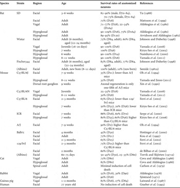
Up to the present time, a number of studies have reported the fate of axotomized neurons (Table 1). At 10–12 weeks after facial nerve axotomy of adult Sprague–Dawley (SD) rats, the survival ratios of motoneurons were 80% in males and 70% in females (Yu, Reference Yu1988). In the dorsal motor nucleus of the vagus (DMV) and in the hypoglossal nucleus of the SD rat, 70 and 100% of neurons survived 28 days after axotomy (D28), respectively (Aldskogius et al., Reference Aldskogius, Barron and Regal1980). At D28 and 2 months after hypoglossal nerve injury, almost all motoneurons survived in the Wistar rat (Kiryu-Seo et al., Reference Kiryu-Seo, Hirayama, Kato and Kiyama2005; Yamada et al., Reference Yamada, Hayashi, Jinno, Wu, Inoue and Kohsaka2008). Age dependency in the neuronal survival after axotomy has also been reported. After crush lesion of the facial nerve in the albino rat, the survival ratio of motoneurons was about 100% in adult animals whereas the ratio was 10% in newborn animals (Søreide, Reference Søreide1981a). On the other hand, axotomy-induced cell death is probably more severe in mice than in rats. Oh et al. (Reference Oh, Murray, Bhattacharya, Holland and Tatton1994) reported that only 35% of axotomized facial motoneurons survived at D21 in C57BL/6J mice. At 8 weeks after the hypoglossal nerve axotomy, 20% of motoneurons persisted in C57BL/6 mice (Kiryu-Seo et al., Reference Kiryu-Seo, Hirayama, Kato and Kiyama2005). In the DMV, 25–35% of lesioned motoneurons survived at D28 in C57BL/6N mice (Yamada et al., Reference Yamada, Hayashi, Jinno, Wu, Inoue and Kohsaka2008). Several strain differences in neuronal viability were found in mice. At D35 of hypoglossal nerve axotomy, the survival ratio of motoneurons in ICR mice was 80%, whereas that in C57BL/6 mice was 50% (Kiryu-Seo et al., Reference Kiryu-Seo, Gamo, Tachibana, Tanaka and Kiyama2006). Facial nerve axotomy in 5-month-old Balb/C mice led to a severe loss of motoneurons, with only 40% of the neurons surviving compared with the nonlesioned side at D30 (Hottinger et al., Reference Hottinger, Azzouz, Déglon, Aebischer and Zurn2000). It might also be noted that the axonal regeneration after dorsal root crush in C57BL/6J mice was only one-fifth of that in the A/J mouse (Xin et al., Reference Xin, Richardson, Gervais and Skamene1990). Variable degrees of motoneuron death following axotomy have also been reported in other species. For instance, in the adult cat DMV and hypoglossal nucleus, 72 and 85% of lesioned neurons survived, respectively, at D80 (Cova and Aldskogius, Reference Cova and Aldskogius1986). In the adult cat spinal cord, axotomized motor neurons survived for long term after peripheral nerve injury (Carlson et al., Reference Carlson, Lais and Dyck1979). In the adult rabbit, 90% of injured hypoglossal neurons survived at D28 (Sjöstrand, Reference Sjöstrand1971), whereas 65% of DMV neurons disappeared 18 days following unilateral cervical vagotomy (Aldskogius, Reference Aldskogius1978). In the axotomized human facial nucleus, there was no indication of cell death (Graeber et al., Reference Graeber, Bise and Mehraein1993). These results might indicate an important aspect of the axotomy paradigm: experiments using some regions in specific species and strains will work as models for neuronal regeneration, but other animal models will work best for studying neuronal cell death.
Species and strain differences in survival rates of neurons after axotomy.

Glial augmentation
Franz Nissl (Reference Nissl1894) was the first to report augmentation of glial cell numbers in the rabbit facial nerve nucleus following peripheral axonal injury. The origin and structural characteristics of proliferating cells after axotomy has been an object of study for a long time (Sjöstrand, Reference Sjöstrand1971). Incorporation of isotope-labeled thymidine and thymidine analogs into newly synthesized DNA has been used to identify proliferating cells. In the axotomized hypoglossal nucleus of adult A/J mice, 3H-thymidine-labeled cells were considered to be microglia by the ultrastructural features (Adrian et al., Reference Adrian, Williams and George1978). Schelper and Adrian (Reference Schelper and Adrian1980) examined whether axotomy-induced proliferating cells exhibited the diffuse cytoplasmic activity of sodium fluoride (NaF)-sensitive non-specific esterase (NSE), an enzyme characteristic for monocytes. The authors observed that no 3H-thymidine-labeled cells showed NaF-sensitive NSE activity in the axotomized hypoglossal nucleus of A/J mice. They thus concluded that the origin of proliferating cells were intrinsic microglia in the hypoglossal nucleus but not hematogenous monocytes. The identity of 3H-thymidine-labeled mitotic cells as microglia was also confirmed by electron microscopy in the lesioned facial nerve nucleus of Wistar rats (Graeber et al., Reference Graeber, Tetzlaff, Streit and Kreutzberg1988).
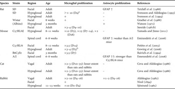
To date, expansion of intrinsic microglial population after axotomy is widely accepted, and the degree of proliferation has been estimated in several species and strains (Table 2). In the SD rat, the number of microglial cells dramatically increased (more than ten times) by hypoglossal axotomy at D4 (Svensson and Aldskogius, Reference Svensson and Aldskogius1993). Approximately three- to four-fold increase in microglial cells occurred after facial nerve axotomy in the albino rats (Søreide, Reference Søreide1981b). In the rat hypoglossal nucleus, microglial hyperplasia occurred within a few days after axotomy, but it waned rapidly (Barron et al., Reference Barron, Marciano, Amundson and Mankes1990). Comparable degrees of axotomy-induced increase in microglial cells were found in mice. For instance, the number of microglial cells drastically increased (about 15 times) by hypoglossal axotomy at D7 in the C57BL/6J mouse (Yamada and Jinno, Reference Yamada and Jinno2011). In the lesioned facial nucleus of B6C3Fe mice, microglia showed a 3.5-fold increase at D7 (Raivich et al., Reference Raivich, Moreno-Flores, Möller and Kreutzberg1994). Similarly, in the injured DMV and hypoglossal nucleus of adult cats, the microglial cell densities increased by three- to four-fold at D35 (Cova and Aldskogius, Reference Cova and Aldskogius1986). In the lesioned DMV of adult rabbits, the numbers of microglia showed two- to ten-fold increase at D3–30 (Aldskogius, Reference Aldskogius1982). Interestingly, activated microglial cells were located more remotely from the neuronal cell bodies in rabbits, whereas these microglia were closely associated with lesioned neurons in rats and mice (Sjöstrand, Reference Sjöstrand1971). At any rate, the mitotic responses of microglia to peripheral nerve injury seem to commonly occur in rats, mice, cats and rabbits, and the degrees of proliferation are rather similar among these species.
Species and strain differences in microglial and astrocyte proliferation after axotomy.

aThe data were calculated from Fig. 2A of the article by Svensson and Aldskogius (Reference Svensson and Aldskogius1993).
bThe data were calculated from Fig. 2G of the article by Gowing et al. (Reference Gowing, Vallières and Julien2006).
It has been reported that astrocytes proliferate in response to brain damage, such as stab wounds and ibotenic acid injection (Latov et al., Reference Latov, Nilaver, Zimmerman, Johnson, Silverman and Defendini1979; Smith et al., Reference Smith, Gibbs, Forno and Eng1987). However, some studies have demonstrated that peripheral nerve injury does not lead to mitotic response by astrocytes. For instance, there was no increase in the number of astrocytes in the SD rat following hypoglossal nerve transection (Svensson and Aldskogius, Reference Svensson and Aldskogius1993). In the facial nucleus of Wistar rat, no increase of astrocytes was observed after axotomy either (Graeber et al., Reference Graeber, Tetzlaff, Streit and Kreutzberg1988). Counter to these studies, a small degree of astrocytic hyperplasia was seen during D1–10 of hypoglossal axotomy in the albino rat (Watson, Reference Watson1972). Enhanced expression of glial fibrillary acidic protein (GFAP) was also seen in the rat facial nucleus after axotomy (Tetzlaff et al., Reference Tetzlaff, Graeber, Bisby and Kreutzberg1988). GFAP is a major constituent of glial intermediate filament proteins, and the upregulation of GFAP by astrocytes is a well-established hallmark of reactive astrocytes and gliosis (Pekny and Nilsson, Reference Pekny and Nilsson2005). When axon regeneration was prevented or delayed by placing a metal clip on the proximal nerve stump, increased GFAP expression lasted longer (up to 1 year) in the axotomized facial nucleus in CD rats (Laskawi and Wolff, Reference Laskawi and Wolff1996). Mouse inbred strain differences in astrocytic response were reported in the spinal cord after sciatic nerve transection (Emirandetti et al., Reference Emirandetti, Graciele Zanon, Sabha and de Oliveira2006). The degree of GFAP increase after axotomy was significantly higher in A/J mice than in C57BL/6J mice. In the adult cat, the density of astrocytes was not affected by hypoglossal or DMV nerve transection (Cova and Aldskogius, Reference Cova and Aldskogius1986). In the adult rabbit, astrocytes increased to some extent at a later postoperative stage of DMV axotomy (Aldskogius, Reference Aldskogius1982). The author suggested that astrocytic hyperplasia was likely accompanying a necrobiotic neuronal response. Altogether, the mitotic response of astrocytes to the peripheral nerve transection seems weaker than that of microglia. Future comparative studies are required to determine the degree of proliferation of astrocyte after axotomy.
Synaptic stripping
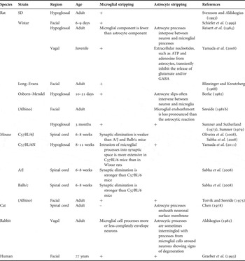
The intrusion of glial processes into synaptic space, so-called synaptic stripping, has been considered to be involved in the protection of lesioned neurons via physical denervation. For decades, many neuroscientists have been attracted to this phenomenon (Table 3). Early work using electron microscopy provided essential structural information on what happens during synaptic stripping. For instance, Sumner and Sutherland (Reference Sumner and Sutherland1973) described the time course of synaptic stripping after rat hypoglossal axotomy. At D1, no microglial wrapping was seen in the hypoglossal nuclei. At D7, microglial wrapping of neurons and dendrites had begun. At 2 weeks after the transection, neuron perikarya were completely surrounded by microglial processes. Similar synaptic detachments were seen after facial nerve lesion in adult albino mice (Torvik and Søreide, Reference Torvik and Søreide1975). Ultrastructural measurements of synaptic elimination after axotomy were performed in the sciatic motor nuclei using three isogenic mouse strains (Sabha et al., Reference Sabha, Emirandetti, Cullheim and De Oliveira2008). The authors showed that C57BL/6J mice exhibited a significantly larger preservation of synaptic boutons when compared to A/J mice and Balb/cJ mice. By contrast, an apparent separation of synaptic boutons by microglia was not observed in the lesioned spinal cord of adult cats (Chen, Reference Chen1978). These results indicate the possible existence of species and strain differences in the involvement of microglial cells in synaptic stripping.
Species and strain differences in microglial and astrocytic synaptic stripping after axotomy.

Recent studies emphasize that astrocytes play various roles in the stabilization and maintenance of synapse by providing a proper microenvironment surrounding the active synapses (Araque and Perea, Reference Araque and Perea2004), limiting the diffusion of ions and neurotransmitters, and releasing neurotransmitters (Walz, Reference Walz1989; Castonguay et al., Reference Castonguay, Lévesque and Robitaille2001). In addition to microglial cells, astrocytes are also involved in synaptic stripping. In close association with microglial process, lamellar astrocytic processes were observed around the axotomized neurons in the hypoglossal nucleus of albino rats (Sumner and Sutherland, Reference Sumner and Sutherland1973). Thin layers of astrocyte processes were often located between microglial process and neuron perikarya, or between microglial process and boutons in the lesioned hypoglossal nucleus of Wistar rats (Reisert et al., Reference Reisert, Wildemann, Grab and Pilgrim1984). In comparison with the volume and surface contribution of astrocytic processes, the presumed microglial component was minimal. The authors thus hypothesized that the microglial contribution to the regeneration of hypoglossal nerves might be negligible in rats. As opposed to the rat model, astrocytes did not show any clear tendency to enclose or encircle degenerating nerve cells in the mouse (Torvik and Skjörten, Reference Torvik and Skjörten1971b). In the rabbit DMV, astrocytic process intrusion appeared only at a later postoperative stage (Aldskogius, Reference Aldskogius1982). Around neurons showing signs of degeneration, astroglial cell processes were sometimes intermingled with process from microglial cells. In the adult cat, many layers of astrocytic processes ran parallel to each other and ensheathed the motoneuronal surface membrane at D15 after the transection of ventral root (Chen, Reference Chen1978). The astrocytic responses coincided with the loss of synapses, whereas microglial cell processes covered only a small portion of lesioned neuronal surface in adult cats. In the human facial nucleus, wrapping of a number of neurons by newly formed GFAP-positive astrocytic cell process were identified in the regenerating facial motor nucleus (Graeber et al., Reference Graeber, Bise and Mehraein1993).
To date, several studies have suggested plausible molecular mechanisms behind the different synaptic stripping responses in different animal strains/species after axotomy. For instance, MHC class I molecules, which traditionally have been linked with immunological mechanisms, are crucial for a selective maintenance of synapses during the synaptic removal process in the axotomy paradigm (Oliveira et al., Reference Oliveira, Thams, Lidman, Piehl, Hökfelt and Kärre2004). After lesion of peripheral nerves of motoneurons, MHC I was upregulated in microglia surrounding axotomized motoneurons (Lindå et al., Reference Lindå, Hammarberg, Cullheim, Levinovitz, Khademi and Olsson1998; Lidman et al., Reference Lidman, Olsson and Piehl1999), and might be induced in astrocytes both in vitro and in vivo (Jarosinski and Massa, Reference Jarosinski and Massa2002). The strain differences in MHC I expression level after the sciatic nerve axotomy were examined in inbred mice (Sabha et al., Reference Sabha, Emirandetti, Cullheim and De Oliveira2008). C57BL/6J mice, that had a comparatively poor regenerative potential, displayed a lower upregulation of MHC I, coupled with a slower synaptic stripping. On the other hand, A/J mice, which had a stronger axonal regrowth potential, showed a clear upregulation of MHC I and a sharp acute loss of afferents. These findings indicate that upregulation of MHC I may positively influence the regenerative outcome associated with a more effective axonal regrowth.
Neurotransmission
A single motoneuron is contacted by as many as 100,000 synaptic boutons containing a variety of different neurotransmitters (Ulfhake and Cullheim, Reference Ulfhake and Cullheim1988). Among numerous substances, glutamate is one of the most important excitatory neurotransmitters. In the adult rat superior cervical ganglion cells, the excitatory postsynaptic potentials (EPSPs) recorded in vitro with intracellular microelectrodes showed a decline to about 50% of their control values at D7–10 of axotomy (Sánchez-Vives and Gallego, Reference Sánchez-Vives and Gallego1993). In the lesioned adult cat dorsal root motoneurons, the amplitude of unit EPSPs recorded in vivo was significantly reduced (Kuno and Llinás, Reference Kuno and Llinás1970). The authors have further suggested a lack of the excitatory synaptic input to the cell body and the proximal dendrites of the axotomized spinal motor neurons on the basis of Rall's compartment model (Rall, Reference Rall and Reiss1964). Within 72 h of axon interruption of superior cervical ganglion cells in the adult guinea pig, the amplitude of EPSPs recorded in vitro in response to maximal preganglionic stimulation were reduced by more than 70% (Purves, Reference Purves1975). Such reduction of EPSPs is presumed to promote neuronal survival. For instance, blockade of the N-methyl-d-aspartate (NMDA)-type glutamate receptor rescued spinal motoneurons after peripheral nerve transection in the neonatal rat (Mentis et al., Reference Mentis, Greensmith and Vrbová1993; Greensmith et al., Reference Greensmith, Mentis and Vrbová1994). Intramedullary axotomy resulted in a preferential elimination of glutamatergic inputs to lesioned motoneurons in the adult cat, which may support the survival of motoneurons through reducing glutamate excitotoxicity on these neurons (Lindå et al., Reference Lindå, Shupliakov, Ornung, Ottersen, Storm-Mathisen and Risling2000). In summary, excitatory synaptic transmission might be modulated in response to peripheral nerve injury, although the mechanisms underlying such alteration remain to be determined. On the other hand, few studies have electrophysiologically examined the species and strain differences in alterations of synaptic transmission following axotomy. Novel hints about the structural and humoral factors regulating synaptic transmission after axotomy have been gained from our recent comparative studies on the vagal and hypoglossal motoneurons between Wistar rats and C57BL/6N mice at juvenile and adult ages (see the next section).
DIFFERENTIAL INVOLVEMENT OF ASTROCYTES AND MICROGLIA IN THE AXOTOMY PARADIGM: FROM OUR RECENT FINDINGS
Alterations in neurotransmission and synaptic stripping in the DMV after axotomy in rats and mice
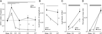
We examined whether alterations in synaptic transmission after nerve injury were correlated with synaptic detachment from injured DMV neurons using Wistar rats and C57BL/6N mice at juvenile ages (Yamada et al., Reference Yamada, Hayashi, Jinno, Wu, Inoue and Kohsaka2008). Nerve transection was performed on 16- to 20-day-old animals, and electrophysiological recordings were made in acute slice preparations from 17- to 30-day-old animals. The mean frequency of spontaneous EPSCs in injured DMV neurons in rats was significantly decreased to about one-third of that of control neurons at D2 (Fig. 1A). This significant decline in the mean frequency of spontaneous EPSCs in injured DMV neurons lasted until D7 in rats. There was no significant change in the amplitude of spontaneous EPSCs recorded from injured DMV neurons in rats. By contrast, the mean frequency of spontaneous EPSCs in injured DMV neurons in mice showed no significant decrease from D3 to D7, although we encountered a transient increase at D1. Similar to rats, there was no significant change in the amplitude of spontaneous EPSCs in mice. These findings indicate that the suppression of synaptic transmission in the injured DMV neurons might occur in rats but not in mice during the early phase after axotomy.
Species differences in synaptic transmission and microglial reaction after DMV nerve axotomy. (A) The spontaneous EPSCs recorded from the lesioned DMV neurons in rats (open circles) and in mice (filled circles). (B) The areal density of synaptophysin-positive puncta in the lesioned DMV in rats and in mice. (C) The areal density of Iba-1-positive microglial processes in the lesioned DMV in rats and in mice. (D) The microglial coverage ratio of perikarya in the lesioned DMV in rats and in mice. Horizontal bars (open bar = rat; filled bar = mouse) at the upper-level of each graph show statistical significances between sham-operated and axotomized animals. Asterisks show statistically significant differences between rats and mice, **P < 0.01. Modified and reproduced from Yamada et al. (Reference Yamada, Hayashi, Jinno, Wu, Inoue and Kohsaka2008), with permission of the publisher.

The possible contribution of synaptic detachment to suppression of synaptic transmission in the injured DMV neurons was comparatively estimated in rats and mice. In the rat injured DMV, the areal density of synaptophysin-positive puncta was significantly lower at D7 versus sham treatment (Fig. 1B), whereas no significant difference was found at D2. In the mouse injured DMV, the areal density of synaptophysin-positive puncta showed no significant decrease from control to D7. The synaptophysin density in the lesioned DMV was significantly higher in mice than in rats at D7. In contrast, the areal density of Iba1-positive microglia processes in the lesioned DMV markedly increased at D7 versus sham in both in rats and mice (Fig. 1C). In the rat DMV, there was no statistically significant difference in the Iba1 density between sham and D2 animals. The microglia coverage ratio (percentage of perimeter surface covered by microglia) was significantly increased at D7 in both species versus sham (Fig. 1D). There was no difference in the microglia coverage ratio between mice and rats. It is noteworthy that the decline in synaptic boutons and the increase in microglial coverage first become apparent at D7 in rats, although the suppression of spontaneous EPSCs in the injured DMV occurred at D2, as mentioned above. In addition, the species differences between rats and mice suggest that the reduction of synaptic boutons and the increase in microglial intrusion might occur independently in the injured DMV during the early phase after axotomy.
Involvement of astrocyte-derived nucleotides in neurotransmission in the DMV at early stage post-axotomy in rats
We then carried out additional pharmacological experiments to elucidate the mechanisms underlying decline in spontaneous EPSCs preceding synaptic stripping in the injured DMV of Wistar rats. All recordings were made in juvenile (17- to 30-day-old) animals. The main results are summarized as follows. (1) Stimulation with high-K+ solution, which enhances the release of neurotransmitters from the presynaptic terminals, restored the frequency of miniature EPSCs and inhibitory postsynaptic currents (IPSCs) of injured DMV neurons to the control levels. (2) Application of a non-selective P2 purinergic receptor antagonist, pyridoxal-phosphate-6-azophenyl-2′,4′-disulfonic acid (PPADS), recovered the frequency of miniature EPSCs recorded from injured DMV neurons on D2 to the control levels. (3) Application of a selective A1-adenosine receptor antagonist, 8-cyclopentyl-1,3-dipropylxanthine (DPCPX), recovered the frequency of both the miniature EPSCs and IPSCs recorded from injured DMV neurons on D2 to the control levels. (4) There was no significant change in either the miniature EPSCs or IPSCs recorded from mechanically dissociated DMV neurons on D3 and D7. (5) High-K+ stimulation failed to restore the mean frequency of either the miniature EPSCs or IPSCs of injured DMV neurons on D7. (6) Neither PPADS nor DPCPX was able to increase mean frequency of miniature EPSCs and IPSCs on D7.
Adenosine triphosphate (ATP), the energy currency of the cell, has been shown to control the synaptic activity in the central nervous system. For instance, ATP inhibits glutamate release from the presynaptic terminal through P2Y receptors (Koizumi and Inoue, Reference Koizumi and Inoue1997; Koizumi et al., Reference Koizumi, Fujishita, Tsuda, Shigemoto-Mogami and Inoue2003; Zhang et al., Reference Zhang, Wang, Ye, Ge, Chen and Jiang2003). Adenosine also suppresses the release of glutamate and/or GABA through A1 receptors (Oliet and Poulain, Reference Oliet and Poulain1999). Interestingly, the most likely source of ATP and adenosine is astrocytes (Koizumi et al., Reference Koizumi, Fujishita, Tsuda, Shigemoto-Mogami and Inoue2003; Martín, Reference Martín, Fernández, Perea, Pascual, Haydon and Araque2007). Recent studies show that astrocytic modulation of basal synaptic transmission is mediated by the release of purines and the activation of presynaptic P2A receptors by adenosine (Panatier et al., Reference Panatier, Vallée, Haber, Murai, Lacaille and Robitaille2011). Therefore, we hypothesize that extracellular nucleotides, such as ATP and adenosine from astrocytes, transiently inhibit the release of glutamate and/or GABA via activation of P2Y and A1 receptors of presynaptic terminals in the rat injured DMV during the early phase of axotomy (e.g. D2). After the stage of humoral suppression by astrocytes (e.g. D7), microglial cells could start suppression of neurotransmission through synaptic stripping.
Responses of perineuronal astrocytes and microglia in the hypoglossal nucleus after axotomy in rats and mice
It is generally accepted that reactive glial cells can be either detrimental or protective depending on the context (Vilhardt, Reference Vilhardt2005; Schwartz et al., Reference Schwartz, Butovsky, Brück and Hanisch2006; Biber et al., Reference Biber, Neumann, Inoue and Boddeke2007). However, the majority of axotomy studies have focused on the beneficial contribution of glial cells to neuronal survival, and little attention has been given to the possible multifaceted roles of glial cells. Consequently, we examined the structural alterations of perineuronal glial cells in the lesioned hypoglossal nucleus using Wistar rats and C57BL/6N mice to identify a possible unfavorable role of microglia and astrocytes in synaptic stripping (Yamada et al., Reference Yamada, Nakanishi and Jinno2011).
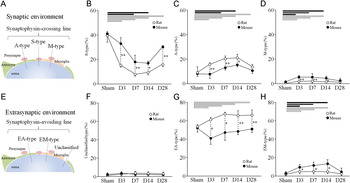
Gradual neuronal death occurred in C57BL/6N mice but not in Wistar rats at D28 after the hypoglossal transection. Based on immunofluorescent quadruple labeling for synaptophysin, Iba1, glutamate-aspartate transporter (GLAST; marker of astrocyte) and TOTO-3 (for counter staining), we classified the glial and synaptic coverage patterns of perikarya into three types (Fig. 2A): S-type, the profile closest to the soma was a synaptophysin-positive puncta; A-type, the closest profile was a GLAST-positive astrocytic process; and M-type, the closest profile was an Iba1-positive microglial process. A significant decline in S-type was observed after axotomy in both species (Fig. 2B). Axotomy-induced intrusion of astrocytic processes into the synaptic space (A-type) was significantly more extensive in rats than in mice, whereas microglial intrusion into the synaptic space (M-type) was significantly more severe in mice than in rats (Fig. 2C,D).
Species differences in glial reactions in the synaptic and extrasynaptic environment after hypoglossal nerve axotomy. (A) Synaptophysin crossing lines are classified into S-type, A-type and M-type. (B–D) Axotomy-induced alterations in the fractions of S-type (B), A-type (C) and M-type (D) in rats (open circles) and in mice (filled circles). (E) Synaptophysin-avoiding lines are classified into EA-type, EM-type and unclassified. (F–G) Axotomy-induced alterations in the fractions of unclassified type (F), EA-type (G) and EM-type (H) in rats and mice. Horizontal bars (open bar = rat; filled bar = mouse) at the upper-level of each graph show statistical significances between sham-operated and axotomized animals. Asterisks show statistically significant differences between rats and mice, *P < 0.05, **P < 0.01. Modified and reproduced from Yamada et al. (Reference Yamada, Nakanishi and Jinno2011), with permission of the publisher.

We also examined perisomal areas lacking synaptophysin-positive puncta, which were defined as the representation of the extrasynaptic environment. The patterns of coverage of perikarya in the extrasynaptic environment were classified into three categories (Fig. 2E): EA-type, the profile closest to the soma was an astrocytic process; EM-type, the closest profile was a microglial process; and unclassified, no apparent profiles were detected. The fractions of unclassified type were very low in both species (Fig. 2F). In the extrasynaptic environment, the prevalence of contact between astrocytic processes and lesioned motoneurons (EA-type) was significantly increased in rats, whereas no significant axotomy-induced alterations in astrocytic contact were observed in mice (Fig. 2G). Microglial intrusion into the extrasynaptic space (EM-type) was significantly more severe in mice than in rats (Fig. 2H).
Astrocyte and microglia may play distinct roles in the axotomy paradigm
The main findings of our second study can be summarized as follows: (1) In the healthy hypoglossal nucleus, motoneurons were mainly covered with synaptic terminals and astrocytic processes. (2) Axotomy caused slow motoneuron death in mice but not in rats. (3) In rats, axotomy induced the extensive astrocytic interposition and the removal of synaptic terminals. (4) By contrast, in mice, prominent microglial intrusion into synapse was induced after axotomy. Together, these observations give rise to a novel hypothesis regarding the double-edged sword nature of glial cells in the axotomy paradigm: the astrocytic interposition may primarily mediate anti-apoptotic effects through reduction of synaptic inputs, whereas the microglial intrusion might be involved in slow apoptotic cell death of motoneurons.
Our results also give some insight into the neuroprotective role of astrocytes via glutamates clearance in the extrasynaptic space. Glutamate transporter is abundantly expressed in astrocytes, which regulate synaptic and extrasynaptic glutamate by importing glutamate through transporters (Huang and Bergles, Reference Huang and Bergles2004). Inhibition of astrocytic glutamate transporters with pharmacological blockers, antisense oligonucleotides or by transgenic knockout, produce elevated extracellular glutamate levels and neuronal death. Recent studies showed that increased expression of astrocytic glutamate transporters rescued degradation of motoneurons in ALS animals (Lepore et al., Reference Lepore, Rauck, Dejea, Pardo, Rao and Rothstein2008; Blackburn et al., Reference Blackburn, Sargsyan, Monk and Shaw2009). Interestingly, the prevalence of contact between astrocytic processes and lesioned motoneurons in extrasynaptic sites was significantly increased in rats after axotomy, whereas there were no significant alterations in mice. We thus hypothesize that the enlargement of astrocytic processes in the extrasynaptic environment seen in rats might help neuronal survival via the increased uptake of excessive glutamate or suppression of synaptic inputs. This idea seems to be supported by several axotomy studies of different species. In the axotomized adult cat spinal cord, most areas of neuronal membrane bared of synaptic contact were occupied by astrocytic processes, whereas microglia covered only a small portion of it (Chen, Reference Chen1978). In line with our prediction, cat spinal motor neurons survived for long term after peripheral axotomy (Carlson et al., Reference Carlson, Lais and Dyck1979). In the rabbit DMV, rapid and remarkable nerve cell loss occurred after nerve transection (Aldskogius, Reference Aldskogius1978). Interestingly, in the rabbit DMV after vagal nerve lesion, microglial cell processes more or less completely enveloped neurons, whereas astrocytic processes were sometimes intermingled with processes from microglial cells around neurons showing signs of degeneration (Aldskogius, Reference Aldskogius1982). It should also be noted that, in the human facial nucleus, Graeber et al. (Reference Graeber, Bise and Mehraein1993) found no indication of cell death but wrapping of axotomized motoneurons by astrocytic processes in the facial nucleus.
CONCLUDING REMARKS
Despite a large amount of studies with the axotomy model, the mechanisms underlying synaptic stripping remain to be determined. We now know the results obtained from peripheral nerve injury vary substantially among species and strains. Here we suggest that using the comparative anatomy in the axotomy paradigm offers a unique experimental model to study glial involvement in synaptic stripping. Indeed, our recent studies using Wistar rats and C57BL/6N mice uncovered the differential roles of microglia and astrocytes in the axotomy paradigm. Contrary to past belief, which regarded microglia to be dominant player in synaptic stripping, the new hypothesis suggested by our findings regards astrocytes as an essential and major element in synaptic suppression and neuronal protection. Previous reports using other species, such as cats, rabbits and humans, could support the neuroprotective role of astrocytes after axotomy. Future studies utilizing the comparative anatomy in the axotomy paradigm will provide further important and unexpected discoveries on the multifaceted roles of microglia and astrocytes in injury and repair.
ACKNOWLEDGEMENTS
The authors wish to thank Dr. Hiroshi Nakanishi for his support to this work. The authors also thank Dr. Eric Bushong for his constructive comments and grammatical corrections on earlier versions of the manuscript. This study was partially supported by Grant-in-Aid for Research Activity Start-up (22890130 to J.Y.) and Grant-in-Aid for Research (C) (21500328 to S.J.) from the Ministry of Education, Science and Culture, Japan, Uehara Memorial Foundation, Kaibara Morikazu Medical Science Promotion Foundation, Takeda Science Foundation, and Kobayashi Magobe Memorial Medical Foundation.
Statement of interest
There is no conflict of interest.